Feeds:
Posts
Comments

Doppler E/A ratio reversal is probably the most reported abnormality in clinical echocardiography. We are also pleased to label it as a grade 1 diastolic dysfunction. Making a significant population who come for regular health checks anxious and worried.

Sharing a presentation from the Annual conference ECHO INDIA 2019, I participated in a symposium on Diastolic dysfunction.

Topic : Issues in diagnosing grade 1 diastolic dysfunction: Pearls and Perils

How did we get into this academic trap? Should we continue this practice?

The current ASE guidelines 2016 have a clear message. It has taken off the E/A ratio from the Initial screening for diastolic dysfunction.

Summary & Final message 

Are we ready for the change? By understanding a simple concept, one can reduce the incidence of indiscriminate diagnosis of grade 1 diastolic dysfunction.

  • E/A ratio apparently has a no role in diagnosing diastolic dysfunction in the normal population who have normal EF %.
  • Hence, never report E/A ratio in Isolation as grade 1 diastolic dysfunction.
  • However, in patients with HFrEF it does help in triaging diastolic dysfunction.
  • Always look for symptoms and 2D features  (Unexplained dyspnea, LA enlargement, LVH ) before considering diastolic dysfunction.

*For advanced readers and researchers grade 1 diastolic dysfunction does have a deep meaning and always continues to puzzle.

Patient corner

For all those anxious patients who ramble around with a report of grade 1 diastolic dysfunction, I can assure you this. Please realize, 9/10 times, this is just a decorative echocardiography abnormality meant to add some spice to the report  does not have any significance.

*Will post a PPT presentation shortly.

This is not a breaking news story. It’s the same old secret that was exposed in JUPITER  trial with Rosuvostatin 14 years ago. Yes, I am talking about the relationship between the usage of statin and the occurrence of diabetes. Now, we have this huge study on possible diabetes progression with statins. It’s not from a small journal to ignore. 

83 thousand patients data, the world’s largest series on link between statin therapy and diabetes.


This study has this to conclude

Diabetogenic statins

Something* happens as the statins antagonize the HMG COA  enzyme that resides within the delicate membranes of the endoplasmic reticulum inside the most specialized cells in our human body, called hepatocytes.

*What is that something?

Image source Umme Aiman et al Journal of Pharmacology and Pharmacotherapeutics 5(3):181-5DOI: 10.4103/0976-500X.136097

How to go about this issue?

With-holding statin in as many as possible is the best thing for such diabetic  (non-diabetic?) patients. But, the more pragmatic option is to ignore these negative studies, and instead intensify diabetes management if it worsens. After all, we can’t afford to lose the prodigious evidence-based cardio-vascular protective effects of statins and earn the wrath of our patients and peers you know!

Further Interest

1.Mansi IA, Chansard M, Lingvay I, Zhang S, Halm EA, Alvarez CA. Association of Statin Therapy Initiation With Diabetes ProgressionA Retrospective Matched-Cohort StudyJAMA Intern Med. Published online October 04, 2021. doi:10.1001/jamainternmed.2021.5714

2.Aiman U, Najmi A, Khan RA. Statin induced diabetes and its clinical implications. J Pharmacol Pharmacother. 2014 Jul;5(3):181-5. doi: 10.4103/0976-500X.136097. PMID: 25210397; PMCID: PMC4156828.


Atrial fibrillation has a direct effect on systemic blood pressure as stroke volume swings from beat to beat because of changing  RR Interval ( preload ). The variation in systolic pressure actually reflects not only the changing stroke volume but also the enhanced contractility of the ventricle to the preload( Frank-Starling principle ). The net effect is reflected in the pulse as an irregularly irregular pulse (Both rate and volume /Amplitude).* However, In dysfunctional ventricles or in acute AF* this variation in systolic  BP can be significant. Also realize, If the preload is changing every beat, there is a considerable dynamism in the afterload as well because of ventricular arterial coupling.

(*Acute effects on BP with the onset of AF : There can be transient hypotension with loss of atrial booster pump.This is not significant in otherwise healthy hearts. Inpatient with baseline LV or RV dysfunction, the onset of AF can be detrimental. The ventricular rate is also a determinant of blood pressure. At fast rates, there can be a fall in BP)

How to record BP during AF ? 

As you record the BP by cuff, the Phase 1 Korotkoff sound floats up and down with each beat. If the variation in RR interval is huge one may get a beat-to-beat variation even up to 40 mmHg.We also know, AF can cause pulse drop /deficit intermittently.

What happens to Korotkoff sound during pulse deficit?

Obviously, there will be a loss of these phase 1 sounds, though the other phases of sound may be heard, which are generated by the previous cardiac cycle. So, measuring blood pressure in AF is not a clinically pleasant task. That’s why we are asked to record 3 times and take an average.

Now, coming to diastolic BP in AF. It’s a real hemodynamic riddle. Traditional teaching is, systolic BP is determined by cardiac output and diastolic BP by peripheral vascular resistance. This is at best a gross understanding of circulatory physiology. Both systole and diastole are coupled together as blood flow across the system of varying resistance. In fact. The preceding systolic pressure head stores the elastic energy in large vessels that are thrown back as diastolic BP.

So what happens to BP during AF? What does the literature say? It doesn’t say much. So we decided to look for ourselves. Here is a tracing of femoral arterial pressure curves during atrial fibrillation. Note: the systolic BP shows considerable variations with changing RR interval with little change in diastolic blood pressure.


Final message

In Atrial fibrillation, the systolic blood pressure changes from beat to beat and it impacts the timing of the Korotkoff sounds. The diastolic blood pressure behaves the same, but it’s less in magnitude and difficult to detect by conventional sphygmo-manometry.

Further reading 

Clinical Implication for irregular BP of Atrial fibrillation  :https://drsvenkatesan.com/2021/05/04/cerebral-hemodynamics-in-af-irregularly-irregular-brain-perfusion-and-risk-of-dementia-cordis/

The effect of AF on pulmonary arterial pressure is an unexplored topic .Cardiology fellows please note.

Is there a solution?

As I understand, we don’t have any. Maybe, we can try this.  No way, I can prevent it from appearing ridiculous for the mainstream scientists.

Truths often lie silently  buried deep (many times intentionally). They definitely deserve an intellectual resuscitation beyond the dirty world of data and evidence. Further, why should experience be considered as enemy of evidence ?

Dr.Richard Asher,  a British physician from Sussex addressed a group of young passing out medical students way back in 1948 in London. The lecture was titled seven sins of medicine! We should thank the Lancet for having published this brief speech the subsequent year in its journal making it immortal medical teaching!

Seven sins of medicine lancet 1949

Seven sins of medicine

Though he was listing these sins among medical students, it is very relevant to every health professional.

1. Obscurity
Asher endorses the use of clear communication and plain language whether writing or speaking. Obscurity may be used to cloak one’s own ignorance, or due to an inability to communicate with those outside of the medical profession. “If you don’t know, don’t admit it. Instead, try to confuse your listeners.” is not uncommon. Regardless of the intention, whether to misdirect from incompetence or to foster a feeling of superiority, the patient and those surrounding them are often left confused and uncertainiy.
2. Cruelty
 This sin is perhaps one of the most commonly committed by doctors and medical students. Whether it be the physical thoughtlessness of a half-dozen students palpating a painful tumor mass, or loudly taking (or presenting) a patient’s history in a crowded room, one of the first things that is unlearnt by a medical professional is to treat the patient as they themselves would like to be treated.
3. Bad Manners
 Often overlooked, rudeness or poor taste in humour is condoned within the hospital setting. At the end of the day, many doctors and students are simply rude to patients that do not suit them. Whether it is a snapping at an uncooperative patient or making a cruel joke about them after leaving the room, the impact of these “coping mechanisms” (as they are considered to be by many) must be taken into account.
4. Over-Specialisation
 In a growing trend by the medical establishment, over-specialization and under-generalization is a growing problem in the wider medical community. Ignoring aspects of one’s education in favor of more interesting aspects is a behavior that is pathological and outright negligent in a student. Failure to diagnose or to treat a patient because “their signs and differential fall outside of my field, let’s turf them to another service” ought to be a seriously considered Supervisory & Training issue.
5. Love of the Rare
 (aka “If you hear hoof-beats, think horses. Not zebras”) The desire for rare and interesting diseases causes many medical students and young doctors to seek the bizarre rather than seeing a mundane diagnosis.
6. Common Stupidity
As well as the standard definition for this sin, the specific example of “using empirical procedures rather than tailoring for the patient” or the young doctor “flying on autopilot” must be mentioned. Ordering another test that is redundant, and for which the results may already be interpreted from the history, before starting treatment is such a situation. For example: requesting a hemoglobin count before beginning transfusion, despite the fact that the patient appears obviously anaemic.
7. Sloth
 Laziness. Also includes ordering excessive numbers of tests, rather than simply taking the time to take an adequate history

Final message

 It is astonishing, to note  Dr.Asher made this observation in the very early days in the evolution of modern medicine,(No critical care units, no HMOs, No industry nexus with research, & commodification of medicine  )  I wonder what Dr. Asher would have to write if he is alive in 2021.

Wish, every medical professional shall find their Asher score. Looking back on my career, I must confess my score would be 3 ( may be 3.5 !) out of 7.  Now, desperately trying to get rid of them. Mind you, the 4th (Overspecailisation)  and 6 th (common stupidity) is inherently built into the system. I think, very tough to avoid them.

There are about 30000 scientific journals and two million papers every year. Of which 5000 are in medicine (Ref : World university news) 

Now, take a deep breath and answer this query. What do you think is the most important aspect of any scientific or medical research in the current era ?

Final message

With due respect to all researchers, What do you think is the most important aspect of any scientific or medical research?  This query is very much relevant today. All components are equally important is an easy way out. But, that’s not the pathway that will take us to the truth.

Postamble  

Having answered the above question, no way, we can escape from this question –“Which could be the least important component “?

I guess you got it right. In the current scenario, my choice is striking and is sandwiched in the middle of the 7 responses..

Background

A 52-year-old diabetic woman who had undergone recent PCI with a DES developed a febrile illness which was diagnosed as Dengue fever. She has been taking DAPT (Dual antiplatelet) meticulously to maintain her stent. Now, her platelet count has dropped from 1.5 and subsequently to 1 lakh. She is asking now, whether to stop DAPT or not? What is the risk of stent occlusion if she stops? 

The D³ cube syndrome 

Infectious diseases rarely bother a cardiologist (maybe a few IE,  myocarditis, etc). Now, a unique situation is emerging.

*Dengue affects 50-100 million people worldwide every year and one billion are at risk. CAD affects 5 % of the population that amounts to  350 million. As we fight CAD, 2 million coronary stents are implanted annually and at least one-third of them may be on DAPT at any time.

When a  global population is at risk of an infection that targets human platelets and another chunk of the same population in whom platelets are targeted with drugs, what is the Incident risk of overlap between these two groups


If you look at these two maps, I think we will not hesitate to call both Dengue and DES a global epidemic affecting the platelet function. The top one depicts the world stent market and the bottom Dengue prone countries


Mechanism of thrombocytopenia in dengue

The mechanism of thrombocytopenia in dengue is not clear. Both production at the marrow level and destruction in the periphery is attributed. The antibody-mediated NS (nonstructural protein) is the original antigenic sin  (Click to know more)

Chiao-Hsuan Chao PLoS Pathog. 2019 Apr; 15(4): e1007625. https://www.ncbi.nlm.nih.gov/pmc/articles/PMC6497319/

Meanwhile, DAPT paralyzes the platelet by blocking  P2Y12 and COX. It is obvious Dengue virus amplifies the antiplatelet action and increases the bleeding risk at any point during the illness.M

Risky period

Bleeding periods is highly unpredictable. The late recovery phase seems to be critical for hemorrhagic risk.it can go up to  2 to 3 weeks or even a month. (When we don’t have data, only experience becomes data. )

How to manage Antiplatelet agents in post PCI patients with dengue? 

While we have guidelines to stop DAPT during the need for emergency surgeries, the same can not be adopted in Dengue.(Curiously, we can stop DAPT without much fear, after all, dengue antigenic responses take up the role of antiplatelet agent )

Presario from Brasil proposed a practical suggestion.Pesaro AE  (Dengue: cardiac manifestations and implications in antithrombotic treatment. Arq Bras Cardiol. 2007 Aug;89(2):e12-5)


Switching to other drugs like eptifibatide or NSAIDs is not an option. 

Post dengue prolonged platelet dysfunction

Though the platelet count returns to normal soon after recovery, long-term platelet defects are also reported. This has implications in resumption of DAPT. Surprisingly, dengue recovery phase thrombocytosis also happens for some unknown reason. Ref: Rebound thrombocytosis causing MI following dengue fever? (Roy A, et al  Indian Heart J. 2007 Jan-Feb;59(1):94)

Final message

When both Dengue &  post PCI epidemics are trying to match in numbers, I guess D³ syndrome (Dengue-DES-DAPT)  would soon become a significant clinical issue. 

Now going back to the title question, Should I stop DAPT or not? 

  • Never easy to answer this question. It is a fine balance between the risk of bleeding vs the risk of stent thrombosis.No amount of algorithm and guideline may clarify it.
  • On any given day, the risk of bleeding in vital spots is more dangerous than thrombus.
  • It is wise to withhold antiplatelet drugs in all febrile illnesses when the platelet count is actively falling below 1 lakh. It may not be quixotic thinking to expect dengue viremia to help the DES with its DAPT equivalent action ! in the intervening period.

Reference

1. Ehelepola NDB, Athurupana Bowatte , Dissanayake  Continuation of Dual Antiplatelet Therapy in a Patient with a Coronary Artery Stent with Dengue Hemorrhagic Fever: A Clinical Conundrum.The American Journal of Tropical Medicine and Hygiene, 01 Jan 2020, 102(1):17-19  

2.Wishnu Aditya et all Proceedings of Singapore Healthcare 2019


Further queries

What about Heparin, Oral anticoagulation (OAC), usage in suspected Dengue?

Go back to basics. Heparin and OAC don’t affect platelet function. It is 100 % safe to continue.

Really? do you think so? No, coagulation physiology is not that simple. Thrombin and antithrombin interactions happen right on the platelet surface. Any antithrombin drugs do have some antiplatelet action as well. Extreme caution is required again. Withhold them unless absolutely indicated.


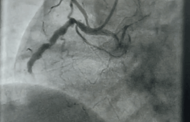
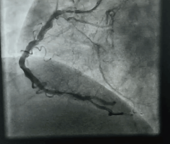
“It was severe double vessel disease &  turned out to be a complex angioplasty in LAD ” 

Why doctor? what happened?

It was a hard lesion, there was plenty of calcium deposits. It was not clearly visible in the angiogram. I had to do IVUS. Curiously, the calcium was clustered in all the three planes of the vessel ( intima the media and adventitia) and they projected into the lumen blocking the path.

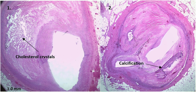
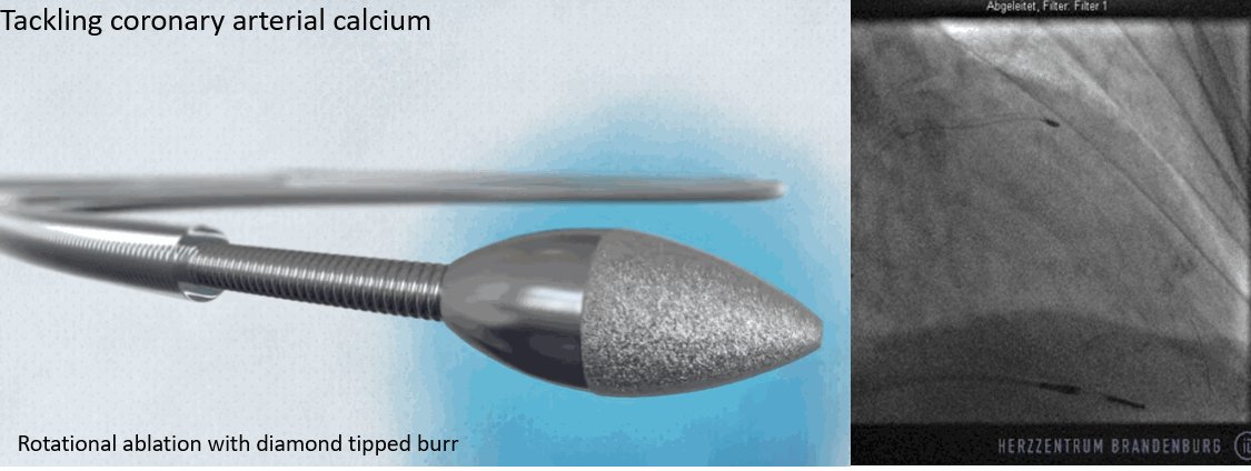
Image collage representation purpose

Thank you, doctor,  how did you manage to remove it,?

It was a real struggle. I had to break the calcium shell before deploying the stent. (What we refer to as lesion preparation). I thought Initially I can displace it with high-pressure balloon inflation, I went up to 40 ATM pressure,  finally needed a  rotational ablation with a burr.  ( IVL -Intravascular ultrasound was an option, but may not have helped either because of heavy Intimal load, Angiosculpts, and the cutting balloons (Wolverines) we don’t have)  

Was it a risky procedure?

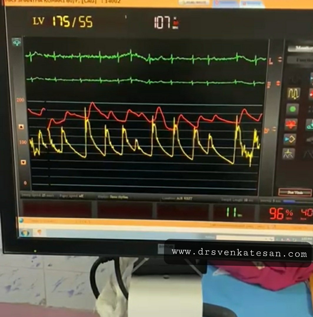
Yes, of course, see the sharp burr rotating 150 thousand resolution per second hugging the coronary artery without damaging it

By the way, why did he accumulate so much calcium inside doctor? Can it be due to his daily calcium supplement doctor?

Oh – my ? that is important new Info. I think that wasn’t noted in the case file. Tell me more, How long did he take it? 

I think he is taking it for long years. He is rather obsessed with it, consumes lots of calcium in his diet as well. He was also taking vitamin D as it once went down to 15 ng/ml. He needed to strengthen his bone which was porotic in the Dexa scan. Was that a problem now? But his serum calcium was always normal, Then, from where does, this calcium enter my dad’s coronary artery doctor?

I am sorry I  don’t know the answer so far. Now, your dad seems to teach me a lesson. It has to enter from the bloodstream or happen de nova due to degeneration. Calcium along with phosphate is a tightly regulated metabolism.They are closely linked to diet, bone, renal and endocrine glands function. We don’t understand how there can be a no-relationship between blood calcium and coronary arterial calcium. Again plaque calcium and serum calcium may or may not correlate. Understand our knowledge base with which we treat you. 

That’s ok doctor. But do you think,  should he stop calcium & vitamin D tablets from now on?  

Hmm, you keep asking tough questions. I will be a fool if I asked you to continue right!

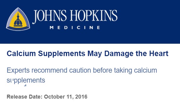
Please go through this article from Jhon Hopkins and decide ( & MESA study 10 year follow on calcium supplement Ref 2)

Final message

The human body is a wonderful machine. Calcium is a life-sustaining ion present in each of the 75 trillion cells we have. We are programmed to handle all elements except in a fraction when true pathology strikes. Never try to outsmart our natural homeostasis with unnecessary and unsolicited chemicals. Indiscriminate calcium supplements are definitely an unfriendly guest for the coronary artery.

Now, Is it just a coincidence?  the new epidemic of coronary microcalcification detected by CT scans(What is your Agatston’s score buddy ?) is matching with a steady increase in per capita consumption of calcium and vitamin D tablets.

Reference

1.Anderson JJB, Kruszka B, Delaney JAC, et al. Calcium intake from diet and supplements and the risk of coronary artery calcification and its progression among older adults: 10-year follow-up of the Multi-Ethnic Study of Atherosclerosis (MESA). J Am Heart Assoc. 2016;5:e003815.

2.Myung SK, Kim HB, Lee YJ, Choi YJ, Oh SW. Calcium Supplements and Risk of Cardiovascular Disease: A Meta-Analysis of Clinical Trials. Nutrients. 2021 Jan 26;13(2):368. doi: 10.3390/nu13020368. PMID: 33530332; PMCID: PMC7910980

For advanced readers

1. Does calcium stabilise a lesion or destabilize a lesion?

It does both .

2. Is CT calcium score correlate with serum calcium?

Yes, it does.Ref : Sanghoon Shin,  European Heart Journal, Volume 33, Issue 22, November 2012, Pages 2873–2881, https://doi.org/10.1093/eurheartj/ehs152

3. What are the various types of coronary calcium? Which is a more tricky lesion? 


Image courtesy -Abbot A calcific nodule is the tricky one, as it can be tiny still result in invisible stent malapposition inviting future problems.


Background

Yesterday, my fellow informed me about a frantic call for cardiac fitness for an emergency cesarian section in 24-year-old woman with hypertrophic cardiomyopathy, who is asymptomatic and has a 20mmhg gradient across LVOT.

“Was she in labor”?

“No, she is 36 weeks term.

“Why LSCS? Why emergency”?

“I don’t know sir. Let me discuss and come again”.

 HCM in pregnancy: An approach

Hypertrophic cardiomyopathy is a specific genetic disorder of myocyte (myosin and others) within the sarcomere. Though uncommon in pregnancy it raises considerable anxiety to the patient, family, and the obstetrician. 

Hemodynamics

Though we tend to worry more about dynamic LVOT obstruction, it is actually the restrictive physiology of LV myocardium that might cause more concern. Three key variables operate in this entity namely preload, afterload, and contractility that determine the cardiac hemodynamics and possibly the symptoms. We know the classical consequence of pregnancy is a fall in systemic vascular resistance( SVR) ie afterload.

In pregnancy, there is a complex interaction between these three parameters along with heart rate. Fortunately, the net effect ends up favorable for LV performance. This is made possible because a major compensation occurs by a 50% increase in blood volume that effectively counters the deleterious effect of fall in SVR on LVOT gradient. (If mitral regurgitation is significant, the fall in SVR actually may help reduce regurgitant fraction especially if its intrinsic defect )

Maternal outcome 

Is good (if not excellent). Maternal mortality reported in the literature, is gradually coming down (0 to .5% in various series)  However, about 15 % of HCM patients with gross LVH or obstruction, may develop pulmonary congestion in the third trimester. In some patients, VPDs, nonsustained VT, even AF can lead to some tense cardiac consultations but are usually innocuous. I am not sure about the sudden death in pregnancy. I guess it should be negligible, unlike the non-pregnant HCM. 

A mystery learning point

It is surprising  both fetal and maternal outcome is little related to the severity of LVOT gradient (Ref 2) 

Indication  for cesarian 

  • Most mothers can deliver per vaginalis without much hemodynamic challenge. 
  • Vey rarely cardiac indication for LSCS need to happen. (However, in the real world many land up in LSCS , since true indication can be blurred due to  cardio-obstetrical anxiety)
  • Spinal anesthesia to be avoided as hypotension is poorly tolerated 
  • Beta-blockers to be continued during pregnancy labor.(Need not start however if already not taking)

Fetal outcome

Premature birth, stillbirths, low weight are little more common than normal pregnancies. Fetal bradycardia due to beta-blockers has been noted but not troublesome.

What is the role of cardiologist?

The precise answer is “minuscule role”. I can vouch for this from a personal level. ( Consults are meant only for bringing some comfort to the obstetrical team). Active cardiac interventions are rarely required or rather desired. (Of course, patients who have significant symptoms, operated for HC, on OAC for AF, the rare ones with ICD needs expert care)

Final message 

  • Women with  HCM can safely become pregnant and deliver.
  • Best outcome is likely for both mother, and baby if basic precautions are taken.
  • LSCS is rarely required*.
  • Counseling about the condition needs to be gentle and just adequate. Dwelling deep into the pathology, hemodynamics, and statistics in totally asymptotic patients invites trouble to all stakeholders. 

*It is worthwhile to note other forms of severe  LVOT obstruction like valvular, supra valvular stenosis, and Aortic pathologies like Marfan, coarctation aorta are serious entities that deserve prompt cesarian sections.

Reference

1.Thaman R, Varnava A, Hamid MS, Firoozi S, Sachdev B, Condon M, Gimeno JR, Murphy R, Elliott PM, McKenna WJ. Pregnancy related complications in women with hypertrophic cardiomyopathy. Heart. 2003 Jul;89(7):752-6. doi: 10.1136/heart.89.7.752. PMID: 12807849; PMCID: PMC1767741.

2.

https://academic.oup.com/eurheartj/article/38/35/2683/3811991


3. ESC 2018 pregnancy heart disease guidelines 

Inferior STEMI, and see the first shot in RCA below. The patient was pain-free and hemodynamically stable at the time of the angiogram. (Don’t wonder how this is possible, defying the fundamental rules learned from  animal experiments after acute ligation of the coronary artery)


What needs to be done ?
  • Go ahead and do a primary PCI as we do in any other  IRA.
  • Be watchful, just pass on the wire, feel the lesion, and decide thereupon. Consider intracoronary lysis.
  • How about a long stent from proximal to distal RCA?
  • Kissing the lesion with DEB in the tightest segment (Not a funny option )


What was done? How is the patient?

Nothing was done & nothing happened to the patient as well. Just guidewire was crossed and few minutes of balloon touch-up work. Did the patient improve? Can’t say anything because he was fine even with this total occlusion. 

Lessons to be learned 

  • The art of leaving a lesion left unattended (rather unstented) in IRA without guilt.
  • TIMI zero flow in IRA need not be a death sentence for the distal myocardium, even in STEMI. 
  • Sometimes, a simple guidewire crossing can do the same job as a complex angioplasty in an IRA.(For acute salvage TIMI 2 or even TIMI 1 is good enough) Most IRA accidents happen when trying improve upon this in an ectatic vessel.
  • Risks of stenting in ectatic /Thrombotic segment is real
  • There can be a useful role for  STENTYS self-expanding stents in localized ectasia (Ref 1)
  • Long-term OAC (Soon NOACs) is a perfect remedy for protecting this type of coronary. 

* By the way, who are all bothered to know LAD anatomy in this patient.  Is it surprising the  RCA is sending collaterals to the left side in its hour of crisis? Yes. LAD had chronic sub-total lesion as well.

Reference

1.The Role of Self-expanding Stents in Patients with Atypical Coronary Anatomy | ICR Journal https://www.icrjournal.com/articles/role-self-expanding-stents-patients-atypical-coronary-anatomy


2.