Feeds:
Posts
Comments

If only  . . . we get  an  image like this , echo can help rule out most  left main disease with conviction .

Have a close look  at it ! One can get a good image of  coronary ostia in short axis view . But , here it is well visualized  in long axis .

left main

I tried to put color flow within left  main .

left  main color flow

What about pulsed  Doppler across left main ?

After all it needs 2mm sample volume and this left main was near 4.5mm . So keep trying !

In this complex world , simple innovations fail  . . . just because  they are simple !

Here is a new* PTCA  catheter which has a  two balloons  ,  the distal one dilates the lesion and the proximal one has a stent over it  . The stent is just deployed after the dilatation by the proximal balloon . The proximal balloon  not only help us prepare the lesion before stenting it also helps  in crossing difficult lesions  . Further   , it can be even used to post dilate the lesion . It can be a non compliant balloon  as well . It appears a  good  concept .

*Not really  new  I  believe  ,  Accuramed  owns the patent for  this twin balloon catheter over a decade now .(First twin balloon Gemini PTCA was used in 1988 )

gemini balloon catheter twin two ptca pci

I do not know why we haven’t adopted it yet ,  while many  dubious  innovations are making merry around the world !

GEMINI DOUBLE BALLON STENT ACCURAMED 2

GEMINI DOUBLE BALLON STENT ACCURAMED  3

The only downside could be ,   combinations of stent and balloon sizes are limited . But ,  it is not a major issue .The ability to  fine tune the stent apposition  moments  after the procedure , by h a simple pull back  is just amazing !

This catheter is  made by

A C C U R A
Medizintechnik GmbH
Max-Planck-Str. 33 61184 Karben Germany
Tel +49-6039-9201-0 Fax +49-6039-9201-22
E-Mail info@accura.info
http://www.accuramed.de

Final message

Two  balloons  over a  single catheter  is a  new development  .I wonder it can be  the standard of care in all PCIs . Hope somebody takes  this concept to the next level  for the benefit of our patients.

Link to the manufacture

http://www.accuramed.de/fileadmin/daten/en/Gemini_System.pdf


* Obfuscation:  hiding of intended meaning in communication, making communication confusing, wilfully ambiguous, and harder to interpret

This world can not be a perfect place and  it is foolish to expect the same !   What is published in medical literature is at best , an abstract thinking  of an unfinished agenda . Still public think  science is   . . . what doctors say ! They feel doctors can not simply watch a person  dying. They want us  act like  God. This is  how medical men became Demi-Gods  by default.

Here was a big opportunity . Who exploited it ? Obviously the greedy corporates  who embarked  on a dirty journey to en- cash this trust  and fill their coffers .This is the foundation  on which the  basics of medical market economy rides !

It is an un-pardonable on-going deceit among  modern human civilization . It has  spoiled  the trust between the patient and doctor and  probably  irreversibly  contaminated  in recent decades !

There are very few positives  though,  with occasional noble medical  souls (Like  BMJ,Lancet )   trying to keep the sinking ship afloat !

This sounding board article (Now we rarely  get to see )  from NEJM way back  in 1975  exposes a  shocking revelation  politely . Now, 40 years after ,  the importance of such article has grown  many fold . We are witnessing  every day ,  medical scientist break  stories ( Yes  . . . it is story )  in general media  with  absolute academic cowardice !

We expect more such  face bashing articles from NEJM . It would definitely  make   immense  good  for  our profession  which needs it  desperately !

Reference

I’m linking the original NEJM article ; Hope it does not violate copy right !

http://www.bumc.bu.edu/facdev-medicine/files/2011/03/Crichton_M_nejm1975_293_1257_medical-obfuscation_structure-function.pdf

6 minute walk test is the simplest of all functional testing in cardiac evaluation . Though  walking  is  a routine day to day motion ,  it is  essentially  a hemo-dynamic stress for the  heart ,  especially so for an ailing heart . Even though  it appears  simplest  of all investigation  there are strict guidelines  available for performing  this .

It is  surprising  American thoracic society  has come out  with a  specific guideline for this .Many of us  are not aware of  existence of such guideline   ,  hence this post  , with courtesy of ATS I am  giving a link.

    Guyatt G. H.,Sullivan M. J.,Thompson P. J.,(1985)  The six-minute walk: a new measure of exercise capacity in patients with chronic heart failure. Can. Med. Assoc. J. 132:919–923.

Butland R. J. A.,Pang J.,Gross E. R.,(1982) Two, six, and 12 minute walking tests in respiratory disease. B.M.J. 284:1607–1608.

History and genesis of the concept

It was originally used in COPD in 1976 with a 12 minute walk .Cardiologists abbreviated it to 6 mts for their convenience.

McGavin CR, Gupta SP, McHardy GJR. Twelve minute walking test  for assessing disability in chronic bronchitis. Br Med J 1976;i:822-3.

In modern times we have an Android application  for 6 minute walk test

This article   provides  you every thing  you  want to learn  about ASD device clsoure .The anatomy , the art of doing TEE in cath lab etc.Do not ever shy away from lesser known journals .It is simply amazing  to find  hidden treasures .Thanks to  Mr Tim BernersLee  invenor of the Internet !


mexican cardiology journal


Sample this arricle

 http://www.elsevier.pt/en/pdf/90140903/S300/


Even though cardiologists consider themselves master of ischemic heart disease , their collective clinical acumen is  put into  acute stress test   when they  confront  a patient with dilated LV and severe  LV dysfunction.This is not  a  rare situation  in clinical cardiology we stumble upon such instances often .Most of them are conferred a  tag  of DCM .

The differentiation from ischemic  vs idiopathic or primary muscular is not a  wasted academic exercise  , since   ischemic  DCM  may get reversed with revascularisation .We have  various  tests to differentiate  ischemic from idiopathic like CAG,MRI, 3D RTE, etc . Still common sense would tell us   95 % of times we can  differentiate ischemic DCM from non ischemic by asking  two critical questions  in the  bed side  echocardiogram

  1. Is there a regional wall motion defect ?
  2. Does all 4 chambers of the heart is enlarged ?

Idiopathic DCM is primary disease of muscle hence  the cardiac   muscle as a  whole  fails  ( We know they are a single  folded  muscle sheet )

Since  Ischemic DCM  primarily affect left ventricle and left atrium  RV,RA enlargement  are terminal events.

* Please note the traditional dependence on CAG to  diagnose  ischemic DCM is fraught with a risk of missing small vessels  induced  DCM,

*** If atrial fibrillation is present longstanding it can dilate both atrium but still RV will be normal  in sized in  ischemic DCM until very late stages

Here is a  20  second flow  chart  to differentiate ischemic  DCM  from idiopathic

ischemic verses idiopathic dcm

If you are a doctor  and  whatever  be your specialty   . . .  if you do  not read this article  and understand the nuances  probably you  will not become a   complete medical professional !

Master health check up futility general health check

master health check up geenral health checks bmj british medical journal cohcrane nordic


While  BMJ  struggles to  propagate  a vital truth , this  banner on a  Indian  high way   tempted the public  to go for 64 slice CT scan   . . .just like that !

Master health check up 2

Final message

Accruing medical knowledge and skills is only one aspect of medical profession. Applying it properly in our patient population is entirely  different ball game . Let us be disease curers and not disease hunters . This is important because disease  hunting is  a dangerous  game ,  where victims can be innocent  bystanders. This  is exactly same thing  Hippocrates  refered to as  Primum non nocere  1000 years ago.  It  has required  a huge statistical study to RE-INVENT  this universal fact !

The  major  issue of contention is fear of conversion of pure ischemic stroke into hemorrhagic stroke .

But here is a catch if you worry about that  . . . who will worry about recurrent emboli from heart ?

References

http://www.ncbi.nlm.nih.gov/pmc/articles/PMC2678170/pdf/nihms104070.pdf


anti coagulation following cardio embolic stroke

aha stroke guidelines 2007

aha stroke guidelines

Recommendation for heparin

aha antiplatlet agent

Recommendation for anti-platelet drugs

I think  the 2007   stroke guidelines are the latest .Even after going  through the guidelines  I am not really clear about the answer for the question posed in this article.

One more thing   I  (mis) understood was  ,  In acute stroke thrombolysis seems to be safe  . . . Heparin seems to be dangerous ?  Is that true ?  It defies logic for  me !

One possible explanation is thromolysis is a emergency single shot salvaging  process . While prolonged heparin will ooze blood into Infarct ! This is exactly is the reason  in   tPA   should not be   followed up with heparin  in acute strokes.(unlike STEMI  where a follow up heparin is a must )

Regarding prevention of recurrent emboli , we need to bother about whether it is predominately platelet rich or RBC rich

Readers may contribute to find the exact answer !

A 60 year old man with chest discomfort and severe breathlessness  and  blood pressure of 160/110 was  wheeled into CCU. A diagnosis of  acute anterior  STEMI was made and he  was about to be  thrombolysed . Since  his blood pressure was high they were waiting for it to come down with IV  Nitrglycerin

I was called to see this  patient  .Here is his ECG .

Q-LVH INCOMPLET LBBB STEMI DIFFERENTIAL DIAGNOSIS 2

Though   ECG  suggested anterior  STEMI  , I  was  fairly  convinced  it  was  in fact  LVH and  incomplete  LBBB.

I confirmed with the  patient  about the onset of symptoms . It was primarily  breathlessness and only a  vague discomfort .Meanwhile , the troponin came as positive and CPK MB    was  normal. The combined troponin  positivity  and ST elevation  almost confirmed the STEMI ,  and  the  urgency for  thrombolysis was  intensified . One resident suggested  an  emergency PCI.

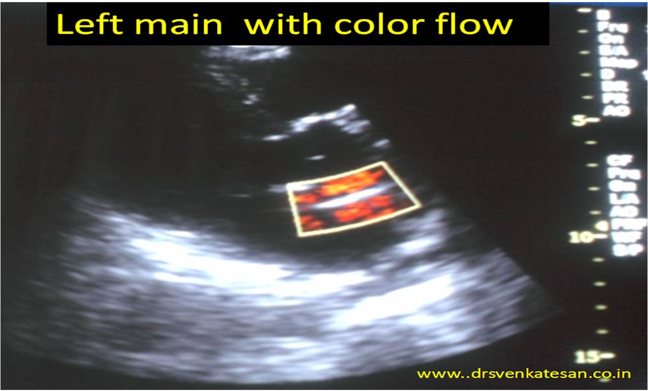
My self ,  in spite of  being a cardiologist was isolated among the physician team .  I  had to  urgently  prove to them it is indeed  not STEMI !  I did a bed side echo and showed  the  physician colleagues   a vigorously contracting  hypertrophied  left ventricle  with a EF of 68 % . There  was  negligible wall motion defect  . . .  if at all any !

They were still far from convinced ?  They  were  sort of  amused .There is   ST elevation ,  there is  troponin  positivity. . . what else you want  . . . they seemed to ask  ?

I asked them  . . . How can an  acute  extensive anterior   MI contract so well ,  without a trace of   wall motion defect ?

It took me considerable time and effort  to  convince them  that the whole thing was not a STEMI.  Finally they agreed .It was  a simple LVH with secondary ST elevation  due to incomplete  LBBB .  Troponin elevation  simply  represent minor myocardial  injury associated  with hypertensive  LVF . This  patient was discharged within 24 hours  in perfectly stable  condition . Since he had mild elevation of creatinine and was  sent for  nephrology  work up.

Final message

LVH with secondary  ST elevation in V1-V4 is a common situation that mimics  acute STEMI . Cardiac failure can result in non ischemic troponin  release .  Acute medicine is  an unique art . Some times it demands all your senses to be on alert mode . Realise ,  in the above case ,   in spite of the   the classical   triad of  chest pain ,    ST elevation , troponin positivity  it  almost led to a wrong diagnosis of  acute myocardial Infarction .

After thought : What  if they had thrombolysed this patient or taken for a PCI ?

When  the clinical suspicion is high and  circumstantial evidence  point to an ACS   ,   this error can be  justified . After all ,  5 % of   famous ISIS  study population were not suffering from STEMI  but got thrombolysis !

* One real possibility in this ECG is  old AWMI with re-infarction  or a dyskinetic septum lifting the ST segment .But both were excluded by the rapid bed side echo.


The popular clinical  entity Idiopathic dilated cardiomyopathy   is often a  dust-bin diagnosis” . The fact is the word   idiopathic simply reflects  our ignorance.

For God nothing is idiopathic . . . he knows how each and every cell  would   behave  .

so , when a patient presents with progressive dilatation and  heart failure refractory to all medical  therapy he is termed as idiopathic and posted for heart transplantation. And only later , we realize the whole thing is due  a  terrible form of reversible  DCM  . That is  pheochromocytoma  induced DCM , which recurred again in the   transplanted  heart.  What a  costly  Ignorance ?

pheochromocytoma and dilated cardiomyopathy reversible dcm tachycardic

Image courtesy and source http://www.dreamstime.com.

Is sub- clinical pheo like situations rampant ?

We know  that  high levels of both epi and nor- epinephrine circulate  in cardiac failure . We presume it  to be a secondary effect .

How can  we  so sure about it ?  There  is a distinct  possibility  of   adrenal gland hyperfunction  and hyperplasia in all DCMs (Idiopathic or ischemic ! )  The dramatic beneficial effects of beta blockers in cardiac failure  will vouch for it .

So , It remains a fertile filed for the youngsters to explore . . . the hyper  adrenergic mediated reversible component of any cardiomyopathy and cardiac failure .

Final message

The default  approach  in any  patient with progressive / refractory cardiac failure   should  be  ,  to consider  whether they fit into  any form of reversible myocardial disease  .  What is idiopathic in remote clinic of   your distant  country side  may be  well recognized secondary cardiomyopathy . The irony is , even sophisticated university hospitals many times miss the true etiology as in the above case report .

                                  So, the term Idiopathic  dilated  cardiomyopathy  (iDCM )  may  aptly be named as  Ignorant  forms  of  DCM  , with an  attractive  abbreviation    . . .   iDCM

Reference

1.J Surg Educ. 2009 Mar-Apr;66(2):96-101. doi: 10.1016/j.jsurg.2008.11.004. Pheochromocytoma presenting as acute severe congestive heart failure, dilated cardiomyopathy, and severe mitral valvular regurgitation: a case report and review of the literature.

2.Kelley SR, Goel TK, Smith JM.Prog Cardiovasc Nurs. 2005 Summer;20(3):117-9. Pheochromocytoma presenting as heart failure.

3.Pheochromocytoma   masquerading as a cardiomyopathy. Garcia R, Jennings JM.  Am J Cardiol. 1972 Apr;29(4):568-71.

4.  http://www.ncbi.nlm.nih.gov/pmc/articles/PMC1894695/pdf/20070600s00025p244.pdf

pheochromocytoma and dcm dilated cardiomyopathy .catecholamine induced dcm tachycardiac cardiomyopathy

5. http://downloads.hindawi.com/crim/medicine/2011/596354.pdf

pheochromocytoma and dcm dilated cardiomyopathy .catecholamine induced dcm tachycardiac adrenal cardiomyopathy