Feeds:
Posts
Comments

Background STEMI knowledge check : Evidence-based Ignorance

I think , It is unfortunate, In the management of STEMI , the two popular strategies of myocardial reperfusion is made to fight with each other as if they are perennial enemies for over two decades. Suddenly, someone with a rare coronary insight thought, why fight each other , they can have a friendly hug and work together. That brought the concept of pharmco -Invasive approach or strategy(PIA) backed up by STREAM, FAST-MI, and TRANSFER AMI studies.Yes, it appears to work well and devoid of all the early adverse events of pPCI. (Much to the dismay of ardent fans of Primary PCI )

*May I add one more shocker of a fact . Deep subset data mining from the above trials did show very early lysis may even act as a perfect stand-alone therapy negating the need for acutely one pharmaco Invasive PCI altogether.(Which was never published) Don’t get alarmed the concept is nothing but , the good old lysis , followed by leisure & elective Ischemia guided PCI in all uncomplicated STEMI.

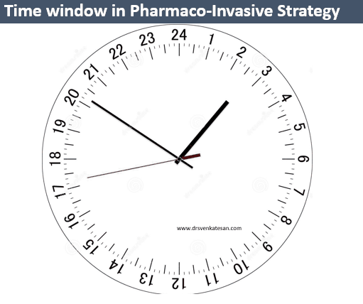
Now coming to the FAQ in Cardiology Boards: Why is the time window for PIA is 3 to 24 hrs ?

The simple answer for an uncomplicated fellow is “published studies have shown benefit only in this time window. If you do PCI early (,<3h) after lysis paradoxically both bleeding and pro-thrombotic complication over the stented lesions are more common. The upper limit is 24 hrs , since by that time we lose all the potential for myocardial salvage”

End-

Larger version of the answer

(Advanced readers who are willing to get confused, may read further)

1. Lysis and immediate PCI doesn’t go well at least in trial world. (FINESSE study, by Ellis et all NEJM 2008) Though cardiologists tend to blame lysis (effect of) to Interfere with their hand skills, it can very well be the opposite. The PCI undo the true benefit of lysis. For cardiologists to accrue maximum benefit in the early time window, they need to be too fast, in the process, they accelerate and fuse adverse events of both modalities.

2. The time window 3 to 24h could simply be evidence-based empiricism. In the major STREAM trial, invasive limb happened between 6 and 16 hours only. We stretched both in the top and bottom in the time clock and made it 3 to 24 hours with other trial data.

3. One realistic reason could be this. It requires a minimum of three hours for a patient to reach a place of coronary Invasion after lysis. So one may argue its time allowance for transport .It comes in handy at times.

4 .If the patient reaches earlier, we need to delay the PCI intentionally to please the evidence based medicine. Mind you, every minute delay increases the chance of no reflow as the microvasculature goes for edematous and porous death.

5. Please note, the time window for pharmaco Invasive strategy will go for a tail spin if the initial lysis is failed. Here, we have to rush I guess. Mind you, In this situation, the evidence based blaming that early PCI increases the adverse events immediately following lysis goes topsy turvy . This is where , we should recall old studies of routine rescue PCI (without clinical criteria) rarely succeeded to correct failed thrombolysis (SWIFT trial)

6.Now, why not PCI after 24hrs? The game can be played reversed if you document ongoing Ischemia in IRA or Non IRA, one may do it . The problem arises when the flawed thought process of a cardiologist could legally justify all PCI beyond 24 h /class 3 Indication after STEMI.The argument goes like this. I think this patient has residual silent Ischemia in- spite of severe LV dysfunction (Suspicion is the justification, to which ,unfortunately no one can dispute) It only suggests open artery hypothesis is still trying to raise from the graveyard more than a decade after its near burial.

Final message

To all those energetic, evidence-based cardiac physicians, we all know coronary care is all about time. In fact, we need to be blessed much more than a sense of time. There is something called medically( or spontaneously )stabilized ACS.  Please realise , “timely and safe intervention” for your patients could simply mean either playing the time button slow/ fast / slow or fast forward / pause or simply shutdown the cath lab, reach home early and enjoy some music or movie in your favorite streaming player.

Reference

1.Ellis SG, Tendera M, De Belder MA, FINESSE Investigators Facilitated PCI in patients with ST-elevation myocardial infarction. N Engl J Med. 2008;358(21):2205–2217. [PubMed]

2. Armstrong PW, Gershlick AH, Goldstein STREAM Investigative Team Fibrinolysis or primary PCI in ST-segment elevation myocardial infarction. N Engl J Med. 2013;368(15):1379–1387. [PubMed]

3. Danchin N, Puymirat E, Steg PG, T, on behalf of the FAST-MI 2005 investigators Five-year survival in patients with ST-segment-elevation myocardial infarction according to modalities of reperfusion therapy: the French Registry on Acute ST-Elevation and Non-ST-Elevation Myocardial Infarction (FAST-MI) 2005 Circulation. 2014;129(16):1629–1636. [PubMed]

4. Cantor WJ, Fitchett D, Borgundvaag B, TRANSFER-AMI Trial Investigators Routine early angioplasty after fibrinolysis for acute myocardial infarction. N Engl J Med. 2009;360(26):2705–2718.. [PubMed]
5.. Bonnefoy E, Steg PG, Boutitie F, , CAPTIM Investigators Comparison of primary angioplasty and pre-hospital fibrinolysis in acute myocardial infarction (CAPTIM) trial: a 5-year follow-up. Eur Heart J. 2009;30(13):1598–1606. . [PubMed]

Identifying the culprit after a criminal event may be easy for the police.For cadiologists investigating the crime scene after a coronary event, it is a different story. (Of course, localization of IRA after a STEMI may not be really difficult.) But , when a patient is having UA  and coronary artery shows multiple lesions, we do have real diagnostic issue. The general dictum could be, tightest lesion or the complex eccentric ones with thrombus is likely to be the culprit. This has important therapeutic Implication,  as we are argued to address the active lesions first. The following study was done in 2009 trying to find the ARA solely by ECG features.

The conclusion was

The following ECG findings were helpful in localizing Angina related artery . ST depression in V3- V5 correlated  with  LAD  angina .Global ST depression was highly correlated with proximal LAD or Left main disease ( 6/6 patients). ST depression in V1 –V3 was associated more commonly with dominant LCX/OM disease. ST depression in 2 ,3 , AVF , or I, AVL  had  no significant correlation with either RCA or LAD  system.However multiple culprit lesions or diffuse inflammatory CAD should always be thought off. One more possibility is , its simply a demand ischemia or micro vascular angina were there is no true epicardial culprit lesion. 

A revisit to my 2009 IHJ article.

http://indianheartjournal.com/ihj09/nov_dec_09/509-523.html


IDENTIFYING ANGINA RELATED ARTERY (ARA) IN UNSTABLE 
ANGINA /NSTEMI BY ADMISSION ECG AND ECHOCARDIOGRAPHY
S.Venkatesan C.Krishnakumar .G.Gnanavelu .R.Subramanian.Geetha Subramanian B.Ramamurthy.P.Arunachalam.M.Somsundram.V.E.Thandapani.M.A.Rajasekaran.
S.Murugan , Madupraphu doss ,P.Pachiappan.
Madras Medical College. Chennai

Unstable angina( UA /NSTEMI ) constitute a  heterogeneous  group of  patients with  lesions ranging from  normal coronary  artery  to severe multi vessel  disease. Even  though  multiple active plaques are documented ,  one  critical  lesion  would be   responsible  for  the  index  episode  of  angina..  Contrary to STEMI  there is no standard methodology   to identify  the  Angina  related artery.(ARA) in UA .We under took this  analysis  to find  whether  admission  ECG  with the help of echocardiography   could  predict  the ARA  in patients with UA

26  patients with  UA  admitted in  our  CCU  were  the  subjects of  study. Patients with   post  infarction angina,  CABG ,  PCI , old  MI , left ventricular  dysfunction  were  excluded. All patients  were treated  as per institutional protocol. Echocardiogrphic analysis   of  wall motion defects (WMD)  were  documented  between  2hrs  and  24hours of admission  .CAG  was  done  between  24 hrs and  7  days. The  coronary  lesion was considered angina related  if  the  WMD  detected   by  echocardiography matched with  the  myocardial  segments supplied by the  arterial territory  containing the lesion . After locating the ARA , the patient’s  admission ECG   was  compared  retrospectively   with  CAG  finding  to study  whether  it has  any  predictive  value  for identifying  ARA.  6 patients  who  had single vessel disease the ARA  localization  was straight forward. (LAD -4 , LCX -1 RCA-1 ). In 2  patients  there was  obvious  eccentric thrombus containing plaque indicating the culprit lesion . 18 had DVD or TVD with no clearcut culprit lesion.

The following ECG findings were helpful in localizing ARA.ST depression in V3- V5 correlated  with  LAD  angina .Global ST depression was highly correlated with proximal LAD or Left main disease ( 6/6 patients). ST depression in V1 –V3 was associated more commonly with dominant LCX/OM disease. ST depression in 2 ,3 , AVF , or I, AVL  had  no significant correlation with either RCA or LAD  system.

It  is  concluded  ARA  can be  identified  with  fair  degree  of accuracy   by admission  ST segment  profile. This  observation  differs with  the existing literature which  suggest little role for ECG to localize arterial lesion in UA. In patients with multivessel CAD  with  more than one  critical lesion  a  combination of ECG  and echo features  help  us to  fix the angina related artery and possibly the lesion. This has  important  therapeutic implication.

Keywords: Angina Related Artery, Unstable Angina/NSTEMI, ECG, Echocardiography.

Postample

I am reposting this abstract again because the same paper has been plagiarised in at least two occasions and got published in predatory journals. Now, we realise Journal article shopping and trading has become a scientific scam .

Reference

This paper  from Japan analysed this ARA concept in 1996 itself with SPECT Imaging


The mechanism of MR in ischemic /Functional is complex. Technically, pure ischemic MR should have a structurally normal leaflet and the subvalvular mechanism dominates But,the combination of the two is also prevalent. In fact, a degenerative component is added to this in many elderly patients.

Mechanism of Ischemic mitral regurgitation

Any of the following may contribute either alone or in in different combinations.

  • Global LV dilatation with or without annular dilatation
  • Spherical left ventricle
  • Altered inter papillary muscle distance (Degree and direction of  posterior vs apical displacement of pap muscle)
  • Chordal shortening /Lengthening/Abnormal tethering
  • Leaflet tenting distance and volume
  • Basal LV dysfunction and Local LV (Sub-mitral) remodeling

We have come a long way ,  since the days of  Carpentier and Duran who did pioneering work .It involved partial or complete mitral annular stabilization with surgical ring technology that  helped us to change the shape of the annulus. Advanced imaging, with 3 D printing will enable us to procure perfectly matched designer valve rings and (may be leaflets also) in the near future. Percutaneous mitral valve Interventions, with clips , valve, are going to dominate the mitral valve therapeutics.

Still, we are largely ignorant about Individual contribution from various components in the genesis of  ischemic /functional MR. This becomes important because the preservation of native valve is better on any day than replacing.  One thing is very clear, even though left ventricle forms part of mitral valve apparatus, the degree of LV dysfunction has no linear correlation with the severity of MR . Its a well-known fact, even severe LV dysfunction (Say < 25 % )may enjoy the company of a perfectly competent mitral valve. It’s interesting to note uniform global LV dysfunction cause more of central MR , while dispropotinate basalLV dysfunction especially the posteroinferior pap muscle cause eccentric jet. One more curiosity is mitral regurgitation improving with worsening disease as contractile force weakens.(Functional MR depends on LV function you know !)

We have witnessed at least two patients who had a significant MR following an inferior posterior MI which was managed medically, showed dramatic regression in the degree of MR  when he had anterior MI later*.The pleasant irony was apparently due balanced dysfunction of anterolateral pap muscle that happened in countering the original postero-medial pap muscle dysfunction.(*Allowing second MI to happen is of course a treatment failure !)

Image source -Christos G. Mihos  Journal of Thoracic disease Vol 8, No 1 (January 2016)

Mitral valve is essentially avascular structure, Still, ischemia affects this valve not by valve necrosis but by other sub valvular mechanisms .Note the MR here is due to poor motion of PML due to ischemic LV dysfunction.


Ischemic MR in early hours following STEMI (also NSTEMI) is still a nightmare. We realized in a harsh way, it’s rarely corrected fully even with a successful IRA plasty. (Especially LCX and posteromedial pap muscle that is in extreme distress) In fact , many of the mechanical complications that lead to flash pulmonary edema would need emergency CABG rather than primary PCI. (What to do for Ischemic MR ? An excellent review article( Elsayed Elmistekawy Curr Opin Cardiol 2013, 28:661–665) 

Mitral valve, though looks like an obedient, innocuous structure that  silently does its job , only in special times, it makes us realize, its the most critical part in the entire heart.(Guarding the lung against flooding when the left ventricle experiences turbulent ischemic times during ACS.) Note -Acute MR often kills , not the ACS as such.ischemic mitral regurgitation functional carpentier drsvenkatesan venkatesan madras medcial college 002The  mechanism of MR in various pathologies is comparable to the behavior of a cow grazing in an arc tethered to a poll. Normally its expected to follow a set pattern. If it behaves wayward, one may need to tighten the rope(Chordae), or loosen it, strengthen or move the poll(Pap muscle) . . . still more options like whipping (clipping ) the cow(Leaflet) may be tried. Of course ,ultimately one may need to replace the cow (MVR). EP guys do  have an electrical solution to tame this cow , called CRT to regress Ischemic MR .


Reference

1.Yiu S.F.,Enriquez-Sarano M.,Tribouilloy C.,Seward J.B.,Tajik A.J.Determinants of the degree of functional mitral regurgitation in patients with systolic left ventricular dysfunction: a quantitative clinical study. Circulation 2000;102:14001406

2.Mitral valve repair over five decades  Ann Cardiothorac Surg. 2015 Jul; 4(4): 322–334 

GettyImages-865142952-5b5eef884cedfd0050112fa6

Charles river esplanade ,Boston* : A healthy middle-aged man who was jogging quietly, while his heart was under intense scrutiny by the bionic eyes of Apple i-watch’s smart patch electrode. Suddenly, it detected some bizarre ST segment fragmentation (Seems it can predict in advance , Ischemic signals 10 minutes prior to onset of ACS ) The built-in cosmos direct GPS instantly alerted & summoned a titanium powered Space X drone that pulled the patient from the riverside to the nearest human wellness port .

EHANG 184

It dropped him through a remotely accessed split glass roof right inside the hybrid heart lab, to find , men and women chatting with flattish Artificial intelligence panels who readily allowed the robotic arms to hug the patient which engaged the coronary artery pushing radiation free magnetic gas found nothing inside and what would become a perfectly normal human coronary artery .

An amused resident robot gently plucked the patient from the cath table with sheepish laughter and called for another drone to drop the patient exactly in the same place from where he was picked up.The healthy hearted patient thanked the doctors profusely and continued his routine evening jog across the Charles of course with a 16-minute delay!

Next day . . .

Event auditing firm medi-logic mind congratulated the entire cardiac team and its digital health hub for the quality of the network and completing this daring coronary rescue mission in 16 minutes. While the drone to hospital roof time was 3 minutes, the coronary artery visualisation time was perfect.The auditing team had a special mention about the astonishing capability of Apple time watch algorithm that made sure that the patient’s evening routine was unaffected in spite of this life-threatening non cardiac pseudo-emergency. The crowning glory was, the entire expenses amounting to 250000 dollors (after a special money back discount coupon for the first false alarm) were taken care by the patient’s virtual insurance blockchain payment gateway.

*You have just read the news that wasn’t – January 2030 AD

Now, back to reality,

Stumbled on this news clip from pages of Times of India, (20-6-2019) months after I wrote the above piece. I wondered the chase between fact and fiction is becoming  really a close race.

In the evaluation of syncope, history is most important to arrive at a diagnosis. Ofcourse, the first step is to confirm whether its truly a syncope or something else.(Metabolic/TIA or seizure.)We are easily carried away by the urge to order a Holter monitoring routinely. In reality, the yield is too low (<15%) .Even the utility value of Head up tilt (HUT) is being downgraded.

Paradoxically, resting ECG might give important clue in many. One need to specifically look into a set pattern of ECG. It generally falls in one the following in any patient with syncope.

This post specifically may not be exclusive but stresses the importance of resting ECG in the evaluation of syncope. by our urge (Stress testing is not included)

  1. Bradycardia( Sinus Node dysfunction/AV block) Note Brady cardias can per se cause syncope if pause >3-5sec Or it may lead to Brady (Pause) dependent escape VT.

    A pause can be a sinus arrest, Pause or SA block .If pause ends with a junctional escape it becomes a arrest.

  2. Look PR interval specifically(A bifasicular block shouldn’t be missed .It can be more dangerous than say a congenital CHB)PR interal represents condcution from SA node to Purkinje fibres in ventricel. The importance is directly linked to the location of the block than propably the degree of prolongation. Please note HV interval > 70 ms in any patient with prolonged PR is cause for concern,
  3. Preexcitation/Delta waves (Though Narrow QRS AVRT rarely causes syncope its very much possible during  Antidromic tachycardia. (AntiVRT), Antidromic AVRT or Accessory pathway with short RP <250ms need to be documented. Concealed paths are safe , but delta appearing during stress testing is extremely unsafe
  4. Post-excitation /Epsilon waves. (often noted in lead V 1, A marker of RVOT dysplasia as in ARVD. Also referred to as Fontaine wave who discovered it by bipolar cheat leads over V1 )

    Note the epsilon occurring after the qrs Indicating RVOT dysplasia

  5. Q waves (Markers of old MI -Scar Induced VT)
  6. High voltage QRS LVH /HCM /Aortic stenosis
  7. RV strain/S1Q3T3 pulmonary embolism.(Syncope is a common presentation with PE especially with minimal exertion or change in posture)
  8. Early coupled VPDS (R on T location a trigger for VT) Wedesky effect. The terminal portion of T which correspond to supernormal period.

    The significance of VPDs directly related to its prematurity than its focus of origin.The one that falls on the vulnerable period .Late phase 2 and phase 3 are more vulnerable as triggered activity

  9. Brugada (Type 1 with T inversion riskier, Camel hump less dangerous Joseph Brugada,

    Brugada syndrome -Note three types . Type 1 is typically risk prone. Please note it is the late ST declining component and the T iversion that confers the risk not ST elevation per se.The type 2 with a camel hump is confered with least risk

  10. Malignant ERS pattern (Most ERS or safe / Maligant forms infero lateral forms risky only at times of ACS not spontaneous risk
  11. J wave syndromes –Overlapping with Brugada /ERS Charles Antzelevitch,J Arrhythm. 2016 Oct; 32(5): 315–339

    ERS syndrome are so common. In the absence of sycnope, it should be ignored straight away. Recently it received too much hype among cardiologists increasing the anxietywhich is not warranted.

  12. Long QT Interval (Hypokalemia commonest, Congenital next Peter J. Schwartz,Long-QT Syndrome Circulation: Arrhythmia and Electrophysiology. 2012;5:868–877
  13. Fractioned QRS (Most often seen in DCM ischemic /non-ischemic confer VT risk usually with LV dysfunction, these are candidates for CRT-P/D as well)
  14. T wave alternans Fluctuating T waves indicate repolarisation alternans .It elevates risk of VT Narayan SM J Am Coll Cardiol. 2006 Jan 17;47(2):269-81
  15. Exercise Induced VT/ CPVT is to be considered seriously in all unexplained exertional syncope. Behere SP, Catecholaminergic polymorphic ventricular tachycardia: An exciting new era. Ann Pediatr Cardiol. 2016;9(2):137–146.

What next after ECG ?

After ruling out neuro cardiogenic syncope by history, one has to perform a good quality echocardiography that can clinch structural heart disease .In cardiomyopathies like ARVD or RCM MRI studies will be of immense value especially the LGE/DEMRI that picks up the scars and fibrosis as in sarcoid or tuberculomas etc. Event recorders are popular, may have a slightly better yield than Holter.EP studies are required in few as diagnostic or more commonly as a part of therapeutics.(Please note, EP lab Induced polymorphic VT has Zero diagnostic value as any normal human heart can be induced to VF by repetitive stimulation)

Management

The main purpose is to exclude serious primary electrical and or structural heart disease. However, fortunately, the most common cause of syncope is neurogenic or reflex mediated. It requires reassurance and fluid repletion Fludrocortisone,/Midoridine (Alpha receptor agonists are promising) Pacemaker/ICD is indicated in few with brady/Tachy -Brady .ICDs/RF ablation are Indicated in Ischemic VTs channelopathies with inherited VT/VF like Brugada. One important question still not clearly answered is when to refer a patient with syncope to Electrophysiologist. ? For me , it appears only a fraction may need it.

Further reading (2018 ESC guidelines)

A middle-aged man a Biotech engineer, who is just back from his annual health check, sitting in front of me with a deeply anguished face and said “Doctor my LDL is 130mg, and my diastolic BP is 90 mmHg and fasting sugar is 120 mg .I am very much worried about my future”

Wait , let me go through your file, I said ,

Isn’t a serious Issue doctor?

No, its not ,

But , doctor, I have read about ASCOT, SPRINT and HOPE-3 trials. I guess they tell us to keep the LDL, blood sugar and diastolic BP all these three parameters around 80. Isn’t doctor? He went on to add, that his old fashioned family physician has asked to continue the beta blocker. He said he is also aware of the fact, how JNC has ditched the beta blocker to the graveyard since they don’t do anything to central Aortic pressure. He continued, “Last year my routine coronary calcium score was beyond 300 . Shall I go for a regular coronary angiogram to ensure my SYNTAX score is around zero doctor” ?

I was quite shocked with his academic prelude, and asked him, by any way, he is a physician or a cardiologist?

No doctor, I am purely a non-medical man but follow all health related stuff from wall street medical bulletins. I am a busy man, still, work out regularly. I have important targets, ambitions to fulfill and lot to achieve in life. But, this LDL and BP is really bothering me doctor.

Yes, I got it . . . I understand your anxiety. Don’t get worried about all these biochemistry and hemodynamics. They are just numbers. Some of them will fluctuate to the tunes of your wall street as well.

Really Doctor?

Yes, we are all unlucky, in one sense you know. We are living in a man-made (scientifically) insecure environment. Great men in the past never had to bother with these silly numbers that currently define health. Alexander the great , neither had his Macedonian master health check nor he looked up for his lipid particles, (he was counting his horses Instead) Did Chengiskhan ever knew about his BP ?

Medical ethics master health check up holistic medicine life style nutrtion

If only these men were worried about these fancy number the world history would have been rewritten.

They didn’t even know an organ called the heart that is pumping 5 liters of blood every minute, until Harvey found the circulatory system 1000 years later. Still, they conquered the world. If we take world history millions of men and women have tasted the pinnacle success without really bothering to know their periodic Individual organ function status.

Here is one more story from my country, The Raja Raja Chola the great built this biggest Hindu monument called Brgadheeswara temple in Tanjavur ,Tamil Nadu , India in the year 1010 .

Raja raja cholan

A fictitious query – Who did FFR for Raja Raja Chola (947-1014AD) when he had vague chest pain from suspicious LAD lesion just after his war with Rashtrakuta empire .He went on to Live for 67 years conquering much of India without a single health check and ECG in his life time

It was an unparalleled kingdom of South India where millions of happy men and women who lived a healthy life with absolute faith and trust in their village healers who did the magic with Indigenous leaves, herbs, secret medical formulas based on ancient wisdom.

Longevity with a purpose

The anxiety to live long often keeps our lipids , sugar and blood pressure high . . . and sets a vicious cycle. Today ,this has become a perfect ground for the saviors of health care to trap us in a cartel who are conferred with an almost divine power of defining who is healthy and who is not.

Many times philosophers have felt longevity and the urge to live long, lacks a matching and meaningful purpose. Lack of purpose, as well as extreme obsession with a purpose, are equally dangerous. The purpose of life can never be equated solely on the longevity of our life. Life long fear and anxiety about possible illness and death is not welcome.

Human life span is mystical journey determined by genes as well the environment and its interaction with each other (Epigenetics) It’s destined to face challenges.Substantial of them can be managed without anyone’s help. I will be happy if you don’t ever need the help of cardiologist to get rid of fear and anxiety induced by general health awareness.

Isn’t prevention better than cure Doctor? I came for a possible coronary angiogram . . . but you have really confused me doctor!

No , I am not doing that Intentionally. From your angle its prevention of potential hidden disease. I am talking In a larger perspective, Master health checks many times end up as medical witch hunting. I am bothered about technological contamination that is all too pervasive among the health care system, especially manifesting as new non-existing diseases. (Skewed and tinkered normal curve )

We, the modern men . . . with all six senses intact, tend to make our life miserable with all these digitized biological data and deeply mined medical images from Innocuously good organs. Some times, we seem to more worried about artificial intelligence and least bothered to know the advantages of being naturally ignorant.

Life is not live data that is in motion. Have a good purpose in life, be physically active, think right, eat well, life shall be lived with peace. Please realize many pockets of the world had been more peaceful, healthy, and cultured in the past, than the current glorious and glamorous times. Of course, life expectancy has definitely prolonged with breakthroughs medicine but It’s not clear it has any positive impact in terms of overall global well being.

Please wake up , you are in the middle of patient consult story … Doctor!

Oh yes , thanks. As a parting advice, I sermonized, homo-sapiens are generally programmed to live for about 100 years except in a fraction who have either true incurable disease or those who succumb to a bad fate.

I realized , what should have been a simple prescription for an ARB +Thiazide + Statin and a stress testing , turned out to be an unsolicited compulsive lecture on life’s purpose, and philosophy etc I said sorry to my patient.

He silently got up. His body language was clearly not convincing enough to suggest, he has accepted my confabulations. He left the clinic with a humble thanks probably looking for a more saner physician!

.

Somehow the concept of  Evidence based medicine (EBM )never excited me in spite of great strides it has made. Probably the main reason for this is, EBMs origin, quality, and credibility is currently severely compromised. (Though It appears to ooze science 24/7 and make us believe in it too !) Herewith, sharing some of the forbidden thoughts(with lots of pun)  for a (un)successful practice of EBM. This is definitely not meant for young and novice medical professionals. Strictly for the ones who can segregate sense from non (S)

Evidence-based Doubting 


Reference


Yes, Its “evidence-based fun”. Forget all those anti-platelet trial dramas … showing in the cardiovascular theatres near you . There is only one genuine drug , that’s the good old humble Aspirin . Mind you ,none of other  actors can ever be imagined for primary prevention.

By the way , there is absolutely no controversy for the role of Aspirin in secondary prevention after established CAD.(We know , how Aspirin has taken up a critically  Integral role in saving the life of the stents  as well as  patients,  post PCI)

Oh , what a disgrace for this drug when it comes out of the glamorous cath lab zones. Its use is often frowned upon for preventing simple CAD. (All due to a single factor, fear of bleeding ? No , its exaggerated in most studies)

Overlap between Primary and Secondary prevention 

In primary prevention of CAD , what do we attempt to prevent? How do you differentiate established CAD from  “Established coronary atherosclerosis  but Non-established CAD ?”

The fundamental flaw in this perceived controversy is in our inability to define what is significant CAD in the asymptomatic population.Do we need a clinical event to say, established CAD?

For the attention of  evidence-based script writers , a long query  . . .

“How much evidence we have to conclude , that a  patient with manifest clinical CAD carry more risk for a recurrence than an asymptomatic  high-risk patient who is likely to develop the first clinical event (which happens with a  bang that could be a major ACS ) due to underlying silent Atherosclerosis.?  

Reference 

Click  here to for more  unscientific review on primary prevention of CVD.


*When I tried to condense three decades of my learning into the medical profession in three lines, I scribbled this. Sorry folks, if It doesn’t sound scientific for some of you! 

By the way, What is successful medical practice? Success for the Doctor, patient or both?  The answer to this question is never simple. 


Pericardial effusion can be detected in many normal pregnancies. The Incidence is up to 40%.The normal fluid within the pericardial sac is around 25ml. A thin sheet of echocardiographic fluid collection in diastole up to 5mm is considered mild.

A trace  or minimal effusion may be a better terminology that describes most physiological pericardial fluid compartment. They have no physiological significance.Mechanism is due to overall increase in size of vascular compartment and especially the right heart volume overload.

Pericardial fluid drains through systemic pericardial veins and lymphatic channels also drain into venous system through thoracic duct . In pregancy these drainage pumps work overtime at its peak capacity. It’s natural it might get fatigued and show some residual fluid collection which should never exceed mild.

We also know thyroid hormones is one of the housekeeping hormones within the pericardial space.Physiological hypothyroid state is possible.Effusion in true pathological Hypothyroidism causes secondary dyslipidemia. Here, some unknown lipid sub-particles clog the lymphatic and cause pericardial effusion which is actually a part of widespread systemic edema. (Myxedema)

https://www.ncbi.nlm.nih.gov/m/pubmed/12756478/

Reference Hurst’s The Heart Valentine Fuster  Mcgraw Hill, 14th edition Page 2347