Pre-op cardiac evaluation prior to non cardiac surgery is an important area for cardiology consultation . Unlike other clinical consults this one primarily involves in the delicate and tricky job of predicting future events !
 Peri-operative cardiac evaluation is done for what ?
1.To evaluate and assess established CAD or other heart disease and get a proper pre-operative work up , drug adjustment and risk reduction for a possible peri-operative event.
2.To screen for any significant CAD or other heart diseases which is hiding and asymptomatic.
3.To treat those conditions that are detected prior to surgery .(Or simply assess & mark the risk and send them for surgery)
4.Finally and most importantly it is often done as a routine “legal point of view” or ” perceived anxiety “as litigation for missed cardiac condition looms large on the surgeon !
Risk stratifying established heart disease is relatively easy task as we know what we are talking about .The term “cardiac fitness” is used in some institution which should probably be discouraged .No patient’s cardiovascular system is deemed to be fit or unfit at any point of time.It all goes with the nature and aim of surgery .An apparently fit person can develop more complications than a potential unfit person as cardiac events are dynamic and directly influenced by the stress of surgery .
It’s about the probability of occurring possible events , and of course one should add to this , all those invincible random or remote events of Heisenberg .
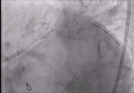
How do you rule out CAD ?
A middle aged man or women with diabetes with a T wave inversion and non specific ST segment is being planned for ca-prostate or breast surgery .Both of them couldn’t do stress test due to associated OA knee.
If coronary atherosclerosis is defined as CAD , there is no way you can rule out CAD.In fact near 100% of elderly population will have evidence for CAD ( at least some degree) in the walls of the coronary .All that is required is just few minutes of heightened adrenergic drive or prolonged fall in blood pressure to trigger acute coronary syndrome in any person who may have shown even a normal coronary angiogram. How does it happen ? We have sufficient technological jargons to use in such situations endothelial dysfunction, plaque erosion erosion ,micro or macro vascular spasm coronary auto circulation failure etc ..
Is exercise stress test , Doubtamine stress , or CAG must for all persons suspected to harbor CAD ?
This could be the key question that makes most cardiologist tentative in their office .suspicion is relative and subjective term .So we have the guidelines .Guidelines are simply guidelines. It may give you comfort if you follow that either academically or legally .
Iam not convinced .Iam new gen cardiologist. Iam unable to rule out CAD without CAG , my cardiology training over a decade has never taught me to r/o CAD clinically
I will go ahead with a screening coronary angiogram in all persons in whom I suspect CAD strongly .If the patient is not willing for CAG I will do a doubtamine stress echo.
What if you detect a positive Doubtamine test or a significant multi-vessel CAD in an other asymptomatic person ?
Now you are stuck again !
- Are you going to postpone the surgery pending further evaluation possible revascularization
- Are you going to clear the patient with added risk frightening every one from surgeon to anesthetist, pateitn and their family.
How guilty are we ? If we fail to predict a cardiac event during non cardiac surgery ?
We need not feel guilty at all as long as you have done the basic tests and given your learnt opinion.I would think no court of law can plead guilty for that. (But your local reputation may be at stake !)
Final message
It is very important to realise , pre-op screening should not be a “hunting ground for CAD”.What we refer to as cardiac fitness is actually is a logical guess considering all risk factors and comorbid conditions and make a learnt decision depending upon the aim of surgery and the urgency of surgery .(Read at least once the meticulously prepared ACC guidelines of 2014)
Forbidden thoughts
In real world , it appears the task of risk stratification and pre-op evaluation is mainly driven by the fear of litigation rather than true concern about the impact of surgery on the ultimate outcome.In this gentle world of noble professionals one can’t question the true Indication of a surgery however dubious it may appear as it considered serious violation of Hippocrates oath* (Not respecting or suspecting your colleagues’s credentials !) But , I earnestly believe a genuine review of decision about surgery or procedure is to me made.
In my humble opinion , if surgery can be postponed or( if could be altogether avoided in few !) till complete cardiac stability is achieved is the most desirable option in high risk patients .
* Am I misquoting Hippocrates ?
Consider this true story . Recently a elderly women came for cardiac clearance for laproscopic Cholecystectomy . As I was doing echocardiogram the patient asked me with real concern , “Is the surgery really necessary doctor ? my doctor says its urgent “ . I casually told her since its a incidentally detected small gall stone , if its not troubling you surgery may be not be urgent , rather may be avoided. Few days went past. I don’t know whatever happened to that women , she opted out of the scheduled surgery .
Next week, , there was huge uproar from the surgeon , who called my associate and censured my behavior.He went on to add , as a cardiologist I have no business to comment about the gall bladder condition. Even if what I uttered could be truth , as a doctor you don’t have a right to breach other doctors opinion.
After few days of self deliberation , I agreed with him and realised in harsh way , freedom of expression can never be taken as granted especially in dealing with others patients !
My colleagues ridiculed my Ignorance , aren’t you aware every patient is a registered property of some doctor ? An Anesthetist friend of mine working in a corporate hospital, said If I start scrutinising the indication of surgeries I assist, I cant win my bread for my family beyond few days !
I simply couldn’t comprehend .” A patient is a patient “ What is the demarcation between my and your patient. She asked me a question I answered it to my conscience , that’s it. Should I behave like a deaf mute ?after 30 years in to medical practice !
I was still restless over the week .Finally, I decided to get the answer from Horse’s mouth and mailed the query direct to father of medicine,
To query@hippocrates.heaven
Dear Mr Hippocrates ,
Here is a story of “Fit gall bladder and an unfit surgeon” . Did I really err on that day or was my behavior unprofessional in any way as others thought ?
Yours greatly
S.Venkatesan.Physician,Chennai .India
I got a surprisingly shocking reply ,
From query@hippocrates.heaven to drvenkatesans@yahoo.co.in
Dear Dr.Venkatesan
I could feel your inner fight about the things happen in medical profession . However genuine your thoughts are, I am sorry , I say this with pain , you are largely unfit to practice medicine in the planet earth. Please try to change yourself or try changing the planet !
Learn to take things easy in life !
With regards.
Hippocrates. (Digitally signed )


 ’
’


