Feeds:
Posts
Comments


asd-closure-device-www-drsvenkatesan-com

The following  factors are critical determinants of the success of ASD device closure

  1. Location of the defect ( Only ostium secundum )
  2. Size of the defect (<35mm .Never forget  simple truth , larger the defect shorter would be the  rim )
  3. Shape  of the defect (Please note ,none are strictly circular but most devices are ! )
  4. Eccentricity of the defect (RA aspect of ASD need not match LA aspect)
  5. Length of the rim ( 5mm said to be adequate)
  6. Thickness of the rim ( Least respected parameter .Thin filament like rims are notorious  in sagging the device into RA)
  7. Pre and Per- operative TEE (As Vital as the procedure)
  8. Technical expertise . (This includes extreme patience  of the primary operator .Most sub optimal results and complications are related to this.
  9. Good team ( Not every  interventional  cardiologist should  attempt this !)
  10. Courage to abandon the procedure
  11. Device brand (Probably less important )

One of my otherwise  well behaving  patient ,  suddenly asked me this question ,  before leaving my  clinic  after a 15 minute consult .

Doctor  . . . I am taking the clopidgrel and aspirin  for 5 years like a vitamin tablet  . . . is that all right  doctor ?

I just got curious, I checked  the prescription again . Yeh ,  he was right !

I have  been mechanically writing  Tablet Clopitab  A  since 2008 !

Clopidogrel abuse long term

For what ?

Some sort of CAD !   Was it for ACS ?   No , it was for chronic stable angina . No PCI,  . . . no DES !

Why the hell  he is taking dual anti-platelet  therapy for 5 years ?

Some  body  , some  where  , has  prescribed it . This man  is taking it  for years together with absolute sanctity.

I was amused  . . . it is also  my mistake . Why it never struck me  to scrutinize the prescription ?

I thanked him . I  removed clopidogrel  from the list , and asked him to continue tablet Ecosprin 150mg  for some time .

( And  now I  had a genuine doubt  ! Does he have CAD at all ! I browsed his file , I couldn’t  find a true documentation for CAD  as I feared  !)

I asked him to get back with an  exercise test ,  . . .  if it comes negative i  can even stop the aspirin as well  I  explained  him  ! (Now he got amused !)

Final message

Who wants Knowledge ?

It is  dumped every  where , free of cost  . . .  both in real and cyber world .Applying it requires more sense  .  and my patient  taught me that  !

Patients  not only  help us  earn  our  bread  and butter , they  do  enrich  our brain  as well ! Never get humiliated when a patient teaches  you a lesson in medicine !

Hypertension is  probably the most   important clinical entity for physicians
for decades .With the advent of modern interventional cardiology management of HT with  drugs have become a  less glamarous job for us. Still , the quantum of the problem and it’s impact on the  risk of CAD and progression   remain a major issue.
There many  different bodies periodically coughing  up guidelines  to manage HT.
  1. JNC from USA
  2. British Hypertension society from UK
  3. European society of cardiology
  4. World hypertension league
  5. Finally WHO guidelines* ( It is not a regular exercise ,WHO releases it  as and when it feels like !)

The stakes are high for the drug industry .Anti hypertensive drugs are the  major source of revenue  to them . Any dip in per capita consumption will have direct impact on their health ! ( WHO bothers about public health ? )

The so called scientific  guidelines,  are generally made balancing patients health vis a vis drug companies health .I have found more often than not it was tilted towards the industry .

The fact that there are multiple guideline with varying impact factors makes sure the confusion among the global physician intact . This is one of the aims of the pharma companies as they influence heavily  when to initiate the treatment ,  and what we are  supposed to prescribe.
Some of the guideline are notorious for insinuations . One example was about the definition of pre hypertension  few years ago .It has since been removed  from the literature after a critical debate .

* One may wonder why I’m focusing always  on non scientific  issues more than academics .(I some how feel non scientific factors are going to impact our health more than any other factor in the coming  generations  )

Now is the beginning of a balance .

European society of cardiology 2013 guidelines for hypertension
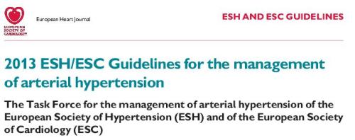
Among these guidelines  I would  think  ESC is close to reality and fairness.
Even    it was carrying dubious advices till recently .Now they have come out with new one in 2013.Most changes are  welcome.
  1. It is essentially about cleansing the contaminated guidelines
  2. Removing unnecessary medications
  3. Unified definition.
  4. More efforts to identify true secondary HT
The salient  points
There are  18 point update in the ESC 2013 . All of them are great . Essentially they are about the basics we have been  taught as we learnt in our final year MBBS. (The rest of our life we have to unlearn  the junk we have accrued over the years  from various CMEs )
I can modify it and  short list
  1. Do not start too early .Have universal definition (Now 140mmhg)
  2. Respect non drug treatment ,( However attractive the  gold tipped pen the  representative leaves  in your consulting suit !)
  3. Avoid using multiple drugs
  4. Never miss a secondary HT .( If  diastolic BP> 110mmh almost always a renal component would be there .Remember Conn syndrome (Primary aldosteronism )  is 10 times more common than much hyped pheochromocytoma ! Just do K+ levels to detect this )
  5. In CAD patients never treat HT in isolation .( Measure blood pressure with sugar and  lipid 120 /70 mg of LDL )
ESC 2013 is a commendable Initiative . It has  tried to remove most errors of the past .obviously  the pharma industry will be unhappy as it will definitely bring down  total drug consumption the  population.
Final message
HT  is an important target  for prevention and management of CAD
Thanks to the much maligned pharma industry  .
We have good drugs.Use it judiciously . Try to reduce the number of drugs .
If possible make them drug free.
If a patients taking   beta  blocker for associated  cardiac condition do not add another anti HT drug . (Recall  from your distant memory , beta blocker is a anti HT drug too !)
Simply follow common sense . (* If you think you  lack  it  ,  get  it from your learnt patients .Many  of them have in plenty . I often do that . One  question they keep asking  “Should I take this drug  life long doctor ?”  is a definite common sense booster!  )

A Cardiologist will never accept  the diagnosis ,  if a technician reports   a  ECG as normal in a  patient with chest  pain   . . .

While , the same cardiologist  gleefully  accepts  an  echocardiogram   done by a technician  and  treats  the patient without  verifying the veracity of the finding !

Why ?

Some where along the cardiology training  , we have been made to believe Interpreting  Echo Images does not require serious medical knowledge . . . but we strongly believe  ECG  cannot be read by technicians however well they are trained .(In-spite of the fact , Echo images are highly dependent on the person who does it , while ECG wave forms are  totally independent of the person who record it ! )

ECG, still has a  prestigious place  in cardiologist’s mind ,  while Echo is often considered  an inferior  Investigation.  Many of us consider ECG interpretation  as a  brainy work while Echo image acquisition and  interpretation is perceived a dumb job* !

Lastly , probably most importantly ,  performing  Echo  is   a time intensive process for the  present day cardiologist  who’s hands are tied with  catheters and  guide-wires  .He has little time for  the meanly echo . .  . hence  ready to compromise on the quality .

* With due respects to all non invasive cardiologists (That includes the author !)

Final  message

I would think it is  fundamentally inappropriate  for technician  to  report Echocardiogram (Of-course they may record it  ) . Unfortunately , for some reason this practice is continued  in many  parts of world .

The other day my fellow got a call  from surgical ward for emergency ECG opinion for a  suspected Inferior MI .It later turned out to be an acute cholecystitis.

One of the important  anatomical mis-perception  among physicians ,  is to consider  inferior, posterior  and diaphragmatic surface  of heart  as separate entities .They are all  closely linked.In fact, they  more often  mean  the same  anatomical zones !

Heart is a dynamic suspended organ within the middle mediastinum .It  can assume a vertical or horizontal position due to number of surrounding anatomical  and physiological factors. (Diaphragm, Lung , being  important ).The ratio of intra thoracic vs Intra abdominal  volume  &  pressure determine whether the posterior surface of the heart is going to face the back of chest  or simply sit and  rest on the diaphragm .We know a horizontal heart is likely to inscribe q waves  in inferior leads .

acute abdomen diaphragm inferior wall mi cholecystitis pacreatitis

Courtesy : Basic image source from digitallab3d

The  diaphragm can be termed as an  anatomical causeway , that isolates   thorax  from the  abdominal  cavity .Close encounters between the organs separated by this delicate biological  membrane is  always possible .This is especially true for electrical signals  which show little  respect for anatomical barriers .

This is the reason there are too  many abdominal conditions that mimic  inferior MI during a painful  emergency (and vice versa  when inferior  MI mimics  acute abdomen .) In  our  department , we   have witnessed  the following conditions mimicking Infero-posterior ACS.

  1. Acute ascites with polyserositis
  2. Gross obesity with APD
  3. Posterior fat pad ( Necrosis ?)
  4. Thickened pericardium
  5. Minimal posterior pericardial effusion
  6. Diaphragmatic pleurits
  7. Esophageal spasm
  8. Fundal air  trapping and ballooning after a heavy meal !
  9. Acute duodenal ulcer perforation ( With gas under diapharam causing q waves)
  10. Acute cholecystits
  11. Diphragmatic hernia
  12. Achalasia cardia
  13. Pancreatitis

Final message

Do not rush to make a diagnosis of inferior wall MI when  you encounter inferior q waves  with  or without ST /T changes , especially  when the symptoms are atypical .

cardiac auscultation murmurWe know clinical auscultation is an art . It is more of a  special sens rather ! It is a combination of natural and acquired ability of your brain to phase out a sound or series of sounds . Sound perception also has  two point discrimination  like touch .(Auditory cortex -Temporal lobe maturity)

It involves selective blanking  and noise cancellation techniques. Ambient noise  contamination is more in youngsters . Elderly men  often have otosclerois so they are benefited by  this handicap .Your chief maybe one of them .So simply do not bother.

Finally ,  clinical acoustics   require lot of imagination . Seniors professors  know what they are expected to hear in a given patient . They look for it rather than  they hear  it  .This is the secret of their  magic ears .

The famous quote  “What the mind do not  know  . . . the eyes do not see”   is very much  applicable  for the ears as well ! 

What your  temporal lobe  do not expect  . . . the ears do not hear!

Beware , even experienced cardiologists  mistake   systolic events   with that of  diastolic and vice versa !

Final message

With due courtesies  to great men like  Potain ,  Leatham , Austin flint,   and other pioneers of cardiac auscultation , I would modify  the  title  of this article .

The science of  cardiac auscultation  may appear more of an  auditory illusion to many  youngsters today  . Still , dedicated  auscultation , with a sound clinical knowledge in a quiet  room  with  a good stethoscope  would  make this illusion  into a  reality !

Dual LAD is an interesting coronary artery anomaly proposed  originaly  by Spindola in 1983 .He classified it into 4 types. In recent years the  dual LAD has increased from 4 to 6 types.

The essential criteria to diagnose  could be summarised.

  1. Two LADs should be identified.
  2. One would be  large and another small
  3. Both should give a  diagonal  branch .

* Ramus is virtually unknown  if there is dual LAD .

The origin  of second LAD can be from

  • LMCA
  • RCA
  • Direct from Right coronary sinus

Course

  • Can be epicardial
  • Intra -myocardial
  • Or both

Branches

  • Diagonal
  • Septal
  • or Both

Drainage area

  • Highly variable

Implication for intervention

  1. Apart  from  the surprise element , the second LAD   has  little impact on the interventional protocol.
  2. However , it may confer a  ischemic protection  as the critical anterior wall has a twin blood supply.
  3. Whether  they are protected from primary  VT or VF is to be studied  because of better electrical stability
  4. Second LAD may act as an additional collateral channel.

Spindola’s  classification of Dual LAD (Types 1 to 4  was called sometimes A,B,C,D )

classification of dual LAD

Source : Lee et al. BMC Cardiovascular Disorders 2012, 12 :101

Spindola type 1 to 4 classification of dual LAD

An illustration of  first 4 types  of Dual LAD .Note  the type 4 originates from RCA. Image courtesy : Prachi P. Agarwal Ella A. Kazeroon . AJR:191, December 2008

Surgical issues ( This is  excellent data  from India . I convey  my   greetings to one  the authors Dr D.B Baruah,  my friend  from  CMC Vellore !)

dual lad classification Spindola-Franco H, Grose R, Solomon

Reference

Spindola-Franco H, Grose R, Solomon N. Dual left anterior descending coronary artery: angiographic description of important variants and surgical implications. Am Heart J 1983:105;445-55

Dual Left AnteriorDescending CoronaryArterySurgical Revascularization in 4 Patients Tex Heart Inst J 2000;27:292-6

http://www.ncbi.nlm.nih.gov/pmc/articles/PMC3509398/pdf/1471-2261-12-101.pdf

Dual LAD  CT  Angiogram : http://www.ajronline.org/doi/pdf/10.2214/AJR.08.1193

culotte technique for bifurcation senting
Original article
Chevalier B, Glatt B, Royer T, Guyon P. Placement of coronary stents in bifur-

cation lesions by the ‘culotte’ technique.Am J Cardiol 1998;82:943 – 949

The humble looking strip of ECG  recorded  in millions ,  every day across the globe  has a complex definition.

And this definition  is  the most apt I have found.

An electrocardiogram (ECG) is a curve showing the potential variations against time in the whole body stemming from the heart, which is an electrochemical generator suspended in a conductive medium.

Einthoven(Dutch)  is the the man who invented the ECG  ,  got a Nobel price for not only inventing the string galvanmeter to record ECG,  but also  making us understand   the rules  of the  electrical wave front  that emanates from the heart.( Not to forget the  original concept  of who demonstrated electrical activity from heart by by capillary electrometer.  by Waller.(British). In my opinion waller should have shared the Nobel prize. I am  sure ,even Einthoven would agree to it.

Of course , do not ask  which  comes first  ” Ionic  flow” or  “the current”  that  comes with it !

Credit goes to  the creators of this  wonderful book  on medical physiology and put that free on the net . Three cheers to  them .

The book is from  University of Copenhagen  , Pannum Institute of Medical Physiology.

physiology text book paulev Zubieta sweden

Reference
British physiologist Augustus D. Waller of St Mary’s Medical School, London publishes the first human electrocardiogram. It is recorded with a capilliary electrometer from Thomas Goswell, a technician in the laboratory. Waller AD. A demonstration on man of  electromotive changes accompanying the heart’s beat. J Physiol (London) 1887;8:229-234
Willem Einthoven introduces the term ‘electrocardiogram’ at a meeting of the Dutch Medical Association. (Later he claims that Waller was first to use the term). Einthoven W: Nieuwe methoden voor clinisch onderzoek [New methods for clinical investigation]. Ned T Geneesk 29 II: 263-286, 1893
Even though multiple mechanisms operate  the major mechanism is due to augmentation and  diversion of blood flow towards sub endocardial region* which is  main area of ischemia in most patients with Angina .
subendocardial-blood-flow
*Beta blockers  does this by smoothing   the  myocardial contractility there by  reducing  trans-myocardial gradient. The coronary arterial perforators which traverse from epicardium to endocardium gets less squeezed and promotes sub endocardial perfusion. 
Link to  a related article from this site