Feeds:
Posts
Comments

Archive for the ‘cardiology -ECG’ Category

We know q waves are not synonymous with Infarct . It just represents electrical activity going away from the electrode.This is why it can occur  even in physiologically in many leads.

Non  infarct Q wave can be recorded with

  • LVH
  • Fibrosis
  • Fluid/Air in beneath  the recording lead
  • Thick chest wall/pericardium (More often Poor  R wave )

rv cavity potential in inferior leads mimicking inferior mi q in

When a chamber enlarges (Any chamber )  it is  brought near the chest wall the electrode may pick up the intra cavity potential that is recorded as q waves .

(The q wave in V5-V6 in severe volume overload of LV may represent LV cavity potential )

Similarly qR complex in severe RV  enlargement  in V1 represent RA cavity potential.Right ventricle is anatomically a difficult chamber to understand. It is located anterior below the sternum  the inferior and posterior aspect of the RV  is facing the diapharagmatic  surface

copd ra rv enlargement mimic inferior mi q waves in 2 3 avf differential diagnosis

In huge RV enlargement , RV cavity potential or( even RA )  can be picked up by limb leads . While cavity potential is well picked up by unipolar pre-cadial leads , it is uncommon for limb lead  record  intracavitory  potential. However  this patient , who was diagnosed  as inferior MI by a  resident ,  turned out to be a clear case of severe  pulmonary hypertension due to  COPD .

Final  message

One  more differential diagnosis for  inferior MI in ECG  exists. A grossly dilated RA, RV due to COPD  with  severe  pulmonary hypertension.

Read Full Post »

Surprises are hall-marks of medical science . The cardiologists do  get  it ,   in enough doses   from  echo  labs  on a regular basis !   . One such thing is  the total ECG-ECHO myocardial  territorial  mismatch following  a STEMI .  Human myocardial segments are divided by cardiologists  by 17 segments by echocardiogram . Long before  echo came into vogue ,  electro-cardiologists  divided the  heart electrically into three zones to  localise MI . (Anterior , inferior and  the  poorly defined entity  lateral walls* ) .Inferior and posterior  segments are  almost used interchangeably. So , when we have 17  echo  segments to be fit into these three electrical category !   were  bound to have  some overlap . The issues of fitting in septal segments is really complex as septum  is a three dimensionally engulfs all three electrical surface of the heart .

* By the way , anatomists  never agreed about existence of walls in heart.They simply said  , heart has smooth  surfaces that blends with one another.  We cardiologist have  built imaginary walls and struggling to come out it !

We will   try to answer the question that’s been asked here .  “Inferior MI”  by ECG   . . . “Anterior MI”  by  echocardiography . How common is that ?

Possible causes for this wrong call

Technical errors  in  acquiring echo  imaging plane  or  it’s interpretation is the commonest . Many  times  ,  obliquely obtained long axis view  wrongly and strongly  suggests  a septal  MI  instead of   inferior posterior MI. This is  because  in  apical 4  chamber view  bulk of   septum  (Basal and mid third )  lies   in the  infero-posterior region .

wall motion defect

Perhaps ,  misunderstanding this  septal  geography is  the  commonest cause for  erroneously  calling inferior MI as anterior  in echocardiography . (A simple clue is the presence of MR . (It  fixes the infarct in infero-posterior zone with 90% accuracy )

Rotation  and  posture of heart

Alignment of the septum to the rest of the chambers  can influence  , how three inferior leads is going to look  at the septum (There can be  considerable errors  -Electrical myopia ? as these leads are located distantly )  . The plane of the septum is such that  in horizontal hearts  septal electrical activity  will be directed infero posteriorly inscribing a q waves in inferior leads rather than anterior leads . One can expect such ECG /Echo discrepancy in the following subset as well

  • Post CABG patients (Any pericardiotomy will make the septal motion  erratic )
  • Obese persons
  • COPD

There are three  more  situations  ,  which   mystified me   with  definite  ECG/ECHO  mismatch

  1. LVH and STEMI  is always an engima . Counter clockwise rotation when accopany  LVH  that masks anterior MI  electrically . It  however inscribes a   q wave in inferior leads.
  2.  In dominant LCX lesions  ( with at-least  one  major OM    )  and  left main bifurcation  STEMIs  ,  combination of  anterior and inferior  wall motion defects are  quiet common . When a such  a  MI evolves ( with or without  revascularization )   regeneration of R wave can be  time shifted . Septal R wave may appear  much earlier and inferior R may follow or vice versa . .Further,  anterior MI  may  evolve as  Non q MI  making it  ECG blind ,   still  echo may pick up the WMA . So there can be important  ECG-ECHO mismatch in myocardial segmental geography .
  3. Further , WMA  need not  always be an  infarct  .Any new episode of ischemia  can result in WMA . Hence a patient  with inferior Q waves  in ECG may experience anterior wall motion defect meagerly  due to fresh episode of   ischemia (This we should not attribute  to  old anterior  MI. It is also possible intra-myocardial conduction delays can elicit remote wall motion defects.

Final message

By general rule  , ECG  correlates  well  with  ECHO  for localising myocardial segments   . At times ,  it  can  really be tricky , and we  get into above situation  in echo labs.

While ,  it is common to observe  ECGs  to mimic  inferior MI  at the first look  and  subsequently echo  revealing  anterior  infarct ,  the reverse is also very much possible .

The  mechanisms are varied and technical  issues are for more frequent than true clinical discrepancy .The issue has important management implications.

Of course ,  coronary angiogram will pin point the   anatomy , still  it also has  strong limitations in localizing myocardial segments (to which it supplies ) especially with multi-vessel  CAD and  collateral dependent circulation .

Read Full Post »

A 60 year old man with chest discomfort and severe breathlessness  and  blood pressure of 160/110 was  wheeled into CCU. A diagnosis of  acute anterior  STEMI was made and he  was about to be  thrombolysed . Since  his blood pressure was high they were waiting for it to come down with IV  Nitrglycerin

I was called to see this  patient  .Here is his ECG .

Q-LVH INCOMPLET LBBB STEMI DIFFERENTIAL DIAGNOSIS 2

Though   ECG  suggested anterior  STEMI  , I  was  fairly  convinced  it  was  in fact  LVH and  incomplete  LBBB.

I confirmed with the  patient  about the onset of symptoms . It was primarily  breathlessness and only a  vague discomfort .Meanwhile , the troponin came as positive and CPK MB    was  normal. The combined troponin  positivity  and ST elevation  almost confirmed the STEMI ,  and  the  urgency for  thrombolysis was  intensified . One resident suggested  an  emergency PCI.

My self ,  in spite of  being a cardiologist was isolated among the physician team .  I  had to  urgently  prove to them it is indeed  not STEMI !  I did a bed side echo and showed  the  physician colleagues   a vigorously contracting  hypertrophied  left ventricle  with a EF of 68 % . There  was  negligible wall motion defect  . . .  if at all any !

They were still far from convinced ?  They  were  sort of  amused .There is   ST elevation ,  there is  troponin  positivity. . . what else you want  . . . they seemed to ask  ?

I asked them  . . . How can an  acute  extensive anterior   MI contract so well ,  without a trace of   wall motion defect ?

It took me considerable time and effort  to  convince them  that the whole thing was not a STEMI.  Finally they agreed .It was  a simple LVH with secondary ST elevation  due to incomplete  LBBB .  Troponin elevation  simply  represent minor myocardial  injury associated  with hypertensive  LVF . This  patient was discharged within 24 hours  in perfectly stable  condition . Since he had mild elevation of creatinine and was  sent for  nephrology  work up.

Final message

LVH with secondary  ST elevation in V1-V4 is a common situation that mimics  acute STEMI . Cardiac failure can result in non ischemic troponin  release .  Acute medicine is  an unique art . Some times it demands all your senses to be on alert mode . Realise ,  in the above case ,   in spite of the   the classical   triad of  chest pain ,    ST elevation , troponin positivity  it  almost led to a wrong diagnosis of  acute myocardial Infarction .

After thought : What  if they had thrombolysed this patient or taken for a PCI ?

When  the clinical suspicion is high and  circumstantial evidence  point to an ACS   ,   this error can be  justified . After all ,  5 % of   famous ISIS  study population were not suffering from STEMI  but got thrombolysis !

* One real possibility in this ECG is  old AWMI with re-infarction  or a dyskinetic septum lifting the ST segment .But both were excluded by the rapid bed side echo.


Read Full Post »

As I expected ,  my earlier algorithm “An Idiot’s approach to tachy-arrhythmias” has  elicited  mixed reactions  .Some  EPs calling it a dud while few  physicians termed it awesome . Here is  a follow up .

Heart rate of a tachycardia is the most neglected parameter by physicians .  They are often seen spending  hours together for decoding  arrhythmia , splitting the brains   for P wave  location , VA conduction, Fusion beats etc .Finally they end up  either administering  Amiodarone a broad spectrum anti arrhythmic agent or DC shock.

Here is an unusual algorithm  for arriving at a diagnosis in all tachy-arrhythmias  based only on heart rate and the width of he qrs complex.

(Click over the table for high resolution image )

approach to cardiac arrhythmias narrow qrs vs wide qrs brugada wellens criteria

General principles in diagnosis of tachycardia

Narrow  qrs tachycardias.

90 % rule : If regular It is sinus tachy if irregular it is A-FIB . Take some efforts to r/o sinus  tachycardia . (In children and young adult it can be extremely difficult at times )* Please note : Sinus tachycardia can show some irregularity due to sinus arrhythmia and  frequent  APDs and JPDS . Further at  fast rates P may fuse with T it should not be confused with  A-fib .

Wide qrs tachycardia

Common things  are common , if  you sight a large animal with a huge trunk  in a Kenyan safari ,  it is most likely to be an  elephant and not a Dinosaur !  Please diagnose VT  when you encounter wide qrs tachycardia by default especially when the BP drops  !

  Management issues

It  would be  foolish to split our heads for decoding an arrhythmia when a patient is unstable .Any hemo-dyanmic unstable tachycardia needs DC shock . (Synchronized will be better unless it is dire emergency )There are very few arrhythmia where DC shock is contraindicated   ( MAT/Dig toxicity/Underlying sinus node dysfunction )

Only if the patient is hemo-dynamically  very much stable   the  physicians  have enough time to  confuse themselves  and the real  ordeal begins .Please remember  the 5 arrhythmias  constitute 98  % of all known tachy-arrhytmia . So where ever  you practice ,  whether  in remote Nigerian village  or  sophisticated  Cleveland  university hospital , when you are  confronted with a tachycardia  the diagnosis  should be one among the  following  five  !)

  1. Sinus tachycardia .
  2. AF/A-fib
  3. Atrial tachycardia  with  or without blocks
  4. ventricular tachycardia /VF
  5. AVNR/AVRT with or without aberrancy

All  other tachy-arrhythmiaa  are  largely  academic !

Regarding  drugs

Knowing the mechanism of  arrhythmia genesis  is less important  at bed side . They are  triggered , sustained, and maintained by either functional or structural component .Ionic basis operates in every arrhythmia  , but it is the anatomical  substrate that maintains it .This happens in only diseased heart.The only point worth remembering regarding mechanism of arrhythmia  genesis  is ,  automatic and focal tachycardias  will not respond to DC shock . All other can be termed some form of re-entry . Micro reentry  for all practical purposes behave like  triggered  activity. Ischemic and electrolytic VTs are primarily ionic based and often polymorphic.Structural VT are commonly mono-morphic. Any VT just prior to degeneration to VF become polymorphic

Every patient with cardiac arrhythmia should be checked for hypoxia,acidois , electrolyte defect or exposure to any  pro arrhythmic drugs. (The commonest  cause of tachycardia in any  IMCU , is inotropic induced (dopamine /doubtamine ) tachycardia .

We  have  5  pharmacological options

  1. Blocking  adrenergic  receptors(IV Esmolol, Metoprolol)
  2. Blocking calcium channel (Dilitazem,Verapamil)
  3. Blocking potassium channel  (Amiodarone  ,Sotolol Adenosine  to a cetian extent )
  4. Blocking sodium channel . ( Procainamide , Lignocaine (Wonder drug almost forgotten now ! ) Flecanide Mexilitene etc)
  5. Digoxin ,Adenosine  magnesium are special  anti-arrhythmic  agent which  has very useful role in certain specific situations (Magnesium -Torsades/Polymorphic VT / Adenosine in LVOT/RVOT VT etc)

General principle is ventricular arrhythmias  are blocked successfully  by sodium or potassium blockade  Atrial and functional tachycardia are blocked by calcium or adrenegic blockade  .Of course,  there would be  some degree of overlap  when the arrhythmia  origin  hovers  around the junction  on either side of the AV  ring . This is basis of verapamil sensitive VT .Clusters of  calcium  channels are scattered  in the junctional  region

Refractory tachycardia

  1. Consider ablation  in AVNRT/AVRT
  2. ICD +Drugs  in VT
  3. Ablate and  Pace(Some A-fibs)
  4. Ablate and ICD (Some  incessant VTs)
  5. Surgery in minority

In AVNRT/AVRT 90 % success can be achieved  in most EP centers .VT ablation  is still a complex process  with  success rate around 60 % ICDs  are indicated in all recurrent VTs except incessant forms .(Where the battery will deplete within a month !) Surgical cure (Maze etc  ) is possible in selected few while undergoing mitral valve surgery.Contrary  to the modern scientific  mood ,  I can ay with conviction most A-fibs can be managed medically except a fraction will require pulmonary vein ablation / isolation .

Final message

Mastering the field of of  cardiac  arrhythmias ,  though  appear a daunting task ,  it does not  require   immense  sense  to understand real world problems are  only a  few and can be tackled in a simplistic manner !

Read Full Post »

Heart rate of a tachycardia is the simplest of all  . . . but   neglected parameter by physicians.  They are often seen spending  hours together for decoding  arrhythmia , splitting their brain for locating P waves ,  VA conduction, Fusion beats etc Finally , most end up  either administering  Amiodarone a broad spectrum anti arrhythmic agent or a DC shock  without arriving at a correct diagnosis.

Here is an unusual algorithm  for arriving at a diagnosis in all tachy-arrhythmais  based only on heart rate and the width of  the qrs complex with acceptable accuracy.

(Click over the table for high resolution image )

approach to cardiac arrhythmias narrow qrs vs wide qrs brugada wellens criteria

Caution :

The above table is  an extremely simplified approach for tachy arrhythmias. Not applicable for scientifically inclined . But in my personal opinion ,  in an emergency room  pure science matters less !

Read Full Post »

When I posed the above question  to few  cardiologists including electro physiologists , the answer I got was surprising .  In the process ,  I could understand why cardiology is such fascinating subject !   Each one gave a different answer and all the 5 responses were forth coming .

The following post in my blog which  I wrote years ago tries to decode the reason for such wide variation in our understanding of AVRT of WPW.

By the way ,  is there a  real risk   for an  ortho-dromic AVRT into anti-dromic AVRT by a definite block in AV node  ?

Read Full Post »

This paper is to  be presented in the the Forth coming   Annual CSI meet New Delhi  December 2012

Beware of Primary PCI : Is there a Low risk STEMI where  pPCI is potentially contraindicated ?

Venkatesan Sangareddi  . Department of cardiology  . Madras Medical college

Primary PCI has proven to be the best  option for management of STEMI . But it need to be done early,  by an experienced team , in a good facility . It is not the individual expertise that matters !  Any treatment , which has great therapeutic potential  also  carries a hazard . So , these treatments  must be used with caution.  Not every STEMI patient , carry a high risk for death.  In fact , the mortality  in some of the subsets of STEMI  can be as low as 1%. If , a  STEMI patient , with a likely 1% mortality is going to get a procedure with  3 – 4 % ,risk it should (And Must !) raise a validity question  But,this issue is rarely addressed in the interventional summits.

In a case pool of 56  randomly collected primary PCIs from various institutes , the outcome  of pPCI  was analysed .It is a retrospective , observational study .STEMI was graded as high risk when one of the following features  were present and it was “low risk” when none of the feature  was  present ( Second STEMI , Extensive  anterior MI , Class 3 /4 killip, An episode of VT/VF, Complete heart block, Diabetic individuals )  High risk STEMI  constituted 22 patients .The overall in hospital  mortality  was (5/56) 9 % In high risk STEMI it was (2/22 )9.5 % in low risk  STEMI it was 3/34 6.4 % .In the corresponding period 40 patients with STEMI who were treated by only thrombolysis or heparin (If beyond time window ) was used a control . 15 patients  were in high risk In the this group the  mortality in high  risk STEMI  was (3/15 )19% and low risk STEMI  there was nil mortality (0/25) 0% .

There was an unacceptable moratlity  with  pPCI  in the low risk STEMI which fared worse than even simple administration of heparin.These data reveal a dangerous fact , that is , primary PCI does not differentiate in the procedural  risk with reference to the patient profile it deals with .While , it dramatically reduce the risk in high risk STEMI It confers a astonishing risk to low risk STEMI .The exact cause for this risk is not known . Common sense would tell , pPCI is  expertise driven driven while thrombolysis is not .Our analysis also suggest bulk of early hazard of pPCI is also logistics related.

Primary PCI could be  cautiously and consciously avoided  in  patients with  low risk STEMI even if it is technically and academically indicated. This can have a great impact in the overall outcome of STEMI management.It is suggested every STEMI patient need to be risk stratified on arrival.(It is still a mystery , why we do this for NSTEMI and not in STEMI ) . A change in the current PCI guidelines to this effect is to be considered.

Read Full Post »

Some general rules are available

RBBB -Morphology -LV origin

LBBB morphology -RV origin

Exceptions : Interventricular  septum  is electrically  RV or LV ?

Electrically it is more of  a  LV .  Septal  focus often have RBBB morpholgy . Exist points  do  matter

Three lead  approach

Rapidly looking at lead  V1 , V6 and AVR  can give us a clue

AVR +ve  will immediately tell us the VPDs are  firing  towards right shoulder .

RBBB morphology points to  a  LV focus .

Negative VPD in V5 will further confirm  LV apex is in the trailing  end  of VPD

Common  sites  for  post MI VPD

  1. LV apex and Apical septum
  2. Infero posterio MI
  3. RV origin more common

Which VPD  morphology  has better localising value  RBBB or LBBB ?

It is  the LBBB  that has more localising value . LBBB invariably fixes the right ventricle

RBBB can either be  right ventricle or left ventricle .

To be continued .

Read Full Post »

Top 5 conditions that closely mimic and often mistaken for STEMI !

  1. Early repolarisation syndrome
  2. Left bundle branch block(LBBB)/ Left ventricular hypertrophy(LVH)
  3. Hyperkalemia
  4. Pericarditis
  5. Brugada syndrome

ERS

The repolarisation is due to  K + efflux . The  K channel porosity  is subjected to high degree of genetic  variations .If the repolarisation starts even by 10 milli- second earlier,  it would have early take off from descending  limb of R wave  and  the J point  ST segment appear elevated.

  • Common  in young  males . Especially in vago-tonic persons with relative baseline bradycardia
  • The ST elevation in ERS is often global .
  • Concavity is upwards .
  • ST elevation can be dynamic ( Further  confusing the picture ! )
  • On EST it  is expected to the  touch the baseline .
  • Benign entity in most . ( False alarm of STEMI is the major risk !)
  • There is some evidence ERS may confer a risk  of  primary VF ,  if they  experience a true STEMI  (Michel Haïssaguerre 2008  NEJM )

* STEMI in ERS :  The issue becomes too delicate ,  if  a  patient with ERS  develops  a true ACS .   ERS being a common ECG pattern in general population , it is not wise to label  every  chest pain in  ERS patient as benign . Suspicious  ones demand observation in step down units , at least !

LBBB

 “Any patient with  LBBB & chest pain . . . suspect  MI”  .

Unfortunately,  this rule is  too reverently followed by  physician community.  In fact ,  ACC/AHA guidelines  reinforced this behavior ,  as it  added a key word  in  their STEMI guidelines   “New onset”  or   “presumably new onset ”  LBBB is  an  indication for PCI/Thrombolysis    .( Physician presumption is a too delicate thread  to hang  our concepts !   )

               Every LBBB is new onset unless you have  a  documented proof otherwise  . . .   it seems to suggest !

Probably , this  is the reason many of the LBBBs are thrombolysed when they present to ER in an acute fashion . Of course , we can apply criteria of  Sgarbossa  to differentiate !  however flimsy it may appear . It  help us to exclude few benign LBBBs. Still ,  Sgarbossa will  struggle to  differentiate  an acute STEMI  in Chronic LBBB  from an  acute LBBB in  old AWMI .

Simply put . . . even old MIs  are at risk of  acute intervention if they have LBBB  and vague chest pain !

How to overcome this ?  Always rely on clinical  features  . If  STEMI is causing the LBBB ,  it  should be a large extensive one and you can not  expect the patient to be  comfortable .(Logic  would suggest necrosis of  large  parts of IVS is necessary to cause LBBB ) Chronic  LBBBs  are relatively comfortable  .

Of course , there  is one another  issue to comprehend  ie  transient ischemic LBBB .We do not know the true incidence  and long-term significance of this entity . Here , LBBB is  not due to necrosis of  the bundle but due to ischemia . (Almost impossible to differentiate it from  rate dependent LBBB  with  aberrancy  )

Role of enzymes and Echocardiogram in LBBB  and suspected STEMI .

You can always ask  for   Troponin  T / CPK MB .(They are helpful only  if 3 hours have elapsed , can we afford to wait ? ) . LBBB  due to STEMI  will  purge  a large quantum of cardiac enzymes from the infarcted zone . (So a marginal elevation is not going to help!)

Unfortunately,  LBBB  can induce wall motion defect in septum that may awkwardly simulate an ischemic wall motion. Even experts have erred in this . One clue  is,  the motion defects  can  not  extend   into anterior wall . It  is confined to septum ,the second clue  is a little delayed  post QRS  thickening of IVS (Septal beaking sign will vouch  for benign LBBB with fair degree of success  )

LVH

  • LVH can mimic a STEMI due to secondary ST/T changes . (Secondary to tall R wave )
  • LVH with incomplete LBBB  – A very common association that can further elevate ST segment in v1 to v3 .
  • Left ventricular hypertrophy  mimics old MI as poor R wave progression in V1 to  V3.
  • Contrary to our belief even Inferior  leads can  show q waves due to  inferior  septal hypertrophy.

Hyperkalemia.

With aging population and rampant  acute and chronic renal disorders it is becoming  a daily affair to get calls from medical units for ECG changes .We know  the rapidity of  efflux  potassium is responsible for ventricular re-polarisation .Phase 2, and 3 are K + exit zones. This is the same phase ST segment and T wave are inscribed.In hyperkalemia  K + accumulates inside the cell and keep  ST/T  segment  elevated .T wave also  becomes tall . It can mimic  both as hyper acute  STEMI .

Read a related article (Dialyisable current of Injury )

Pericarditis

  • ST elevation is not confined to an arterial territory
  • Can be global .(Regional ST elevation  does not exclude pericarditis)
  • ST elevation is concave upwards as in ERS

Link to Read regional pericarditis
Brugada syndrome

Brugada syndrome  is  an ECG -Clinical complex in which ST elevation in pre-cardial leads is associated with  ventricular arrhythmia. The defect lies in sodium channel . It reflects  a mis -match between RV and LV epicardial repolarisation forces .It keeps the RV epi-cardial current afloat and  the pre-cardial leads  facing the RV records ST elevation that  mimics  STEMI. It often  shows  a RBBB pattern and varying patterns of ST morphology  . The  ST segment is  also  subjected to dynamism  , due to change in autonomic tone and myocardial temperature  .(Febrile VTs)

After thoughts

Other close contenders for the top 5 slots

Myocarditis

Acute pulmonary embolism

Dissection of aorta

More

  • Acute stroke (Neurogenic ST elevation )
  • Stress cardiomyopathy (Takot Subo )
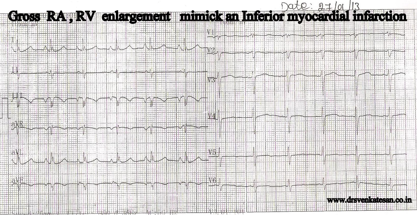
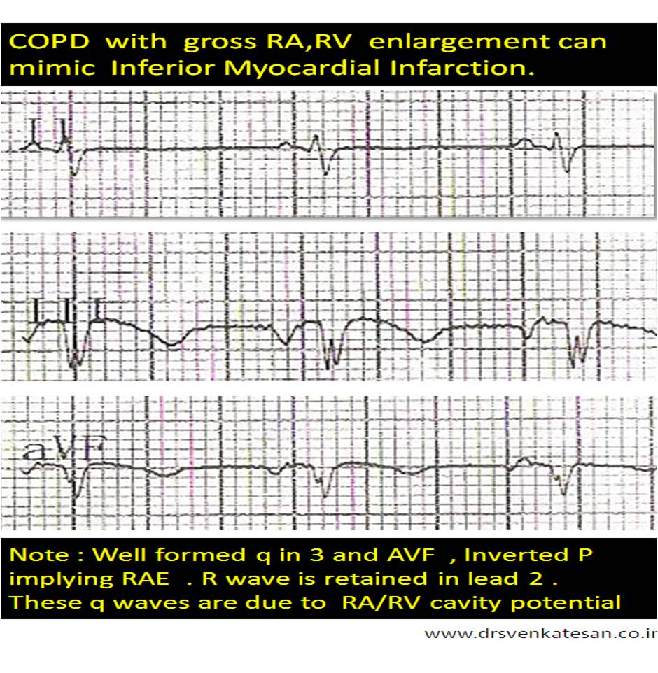
  • Acute abdominal conditions mimicking inferior STEMI.
  • Panic attacks /Anxiety states / chronic anti psychotic  medications which are known to elevate ST segments.
  • Contusion chest

(Cocaine hearts / Coronary arterial spasm / LV dyskinetic segments  and  LV aneurysms  were not nominees ! )

Read Full Post »

A young man with   VT  (LBBB morphology ) and  apparently normal heart by   echocardiogram  is  a  real  diagnostic challenge .
Here is a comparison  of  the two  closely mimicking  entities. RVOT VT and ARVD .
Please note -Micro reentry and triggered activity  are very closely related cellular evens. For all clinical reasons there is generally no purpose in differentiating the two.

*Please note -Micro reentry and triggered activity  mimic each other at the cellular level . For all clinical reasons there is generally no purpose in differentiating the two.

*RVOT- Right ventricular outflow tract. ARVD/ARVC -Arrhythmogenic  right ventricular dysplasia /cardiomyopathy

(Caution : RVOT vs ARVD  -In  the traditional medical teaching system , we are often taught to differentiate  two closely related  entities.Our brain also loves to look things in either black or white . Realise , medical science always brings  surprises . There can be significant overlaps between the very entities we want to differentiate.Bear that in mind)

Reference

1. Hoffmayer KS, Machado ON, Marcus GM, Electrocardiographic comparison of ventricular arrhythmias in patients with arrhythmogenic right ventricular cardiomyopathy and right ventricular outflow tract tachycardia. J Am Coll Cardiol. 2011 Aug 16;58(8):831-8.

2 .Ainsworth CD, Skanes AC, Klein GJ Differentiating arrhythmogenic right ventricular cardiomyopathy from right ventricular outflow tract ventricular tachycardia using multilead QRS duration and axis. Heart Rhythm. 2006 Apr;3(4):416-23.

T wave inversion in V1 TO V3 for diagnosing  RVOT VT .

3.Daniel P. Morin,  Andreas C. Mauer, Kathleen Gear, Usefulness of Precordial T-Wave Inversion to Distinguish Arrhythmogenic Right Ventricular Cardiomyopathy from Idiopathic Ventricular Tachycardia Arising from the Right Ventricular Outflow Tract .Am J Cardiol. 2010 June 15; 105(12): 1821–1824

Read Full Post »

« Newer Posts - Older Posts »