Feeds:
Posts
Comments

Archive for the ‘cardiology -ECG’ Category

Often general practitioners refer  ECGs  with abnormal resting  ST/T wave patterns  to cardiologists .

Following are few of them

  • ST elevation
  • ST depression
  • T wave inversion
  • Tall T waves
  • A relatively uncommon  finding is  a flat ST segment  , which  is discussed here.

The commonest( benign) abnormality   is  T wave inversion  in women and tall ST /T waves   reflecting  early repolarisation  pattern in men. A flat ST segment is an occasional finding in general population.

ST segment is inscribed  during the most important  time  of  cardiac cycle.This is the period the ventricle is doing its prime function , namely ejecting the blood in systole .Hence it is subjected to maximum stress . During times of ischemia  ST segment  gets elevated or depressed depending upon the severity of ischemia. For the same reason , even  subtle changes in this segment is  frowned upon by cardiologists. Most of them would receive a EST.

It is ironical to note  , few normal people  show almost silent electrical activity during this  crucial  phase of   their  ECG .ST segment is often  a flat line  in them . This is a ECG of a women referred as CAD. She was asymptomatic . Echocardiogram  was normal . She was asked to do  a EST.

This asymptomatic women was refered for ECG opinion

The peculiar thing about T waves  are ,   a 10 mm upright  as well as  5 mm inverted T wave ,  both can be normal. So .  there is no element of surprise  to note absent  T waves  or a flat  T wave  to be called as normal .

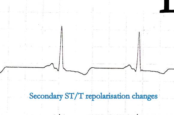
The curious case of lost ST segment !

* T waves are recorded when K+ efflux occur rapidly out of cells . Hypokalemia  can be an important cause of flat T waves.

It is still a  mystery to me  why some people inscribe a tall T when  potassium comes out  of cell and  an equal number (Esp women)  record a down ward T wave  for the same event !  I wish  I get an answer  to this  lingering  question from  any of the readers !

Is a flat T wave represent  a T wave in  transition  to become inverted T wave  later ?

Possible .But we  are not sure ! A static T wave is safer than a dynamic T wave .

Final message

Flat ST segment and absent T waves  represent a same spectrum of ECG  findings  which  are  referred to as  non specific ST segment changes in  clinical practice .Generally , they have  little clinical significance.* In our experience we have found , female patients, Anemia  hypothyroidism  are  often associated with flat ST segments  . If CAD is suspected exercise stress test  should be done. Some believe a flat ST segment  is more likely to  result in EST positivity (Not necessarily true positive !)

* Non specific ST/T changes by itself is a  huge topic.  Ideally the term non specific ST /T changes should be avoided , as it  primarily came into vogue  to denote non ischemic ST segment (Still , other pathologies are very much  possible) It is estimated there are about  50 causes for non specific ST/T changes , right from a  benign situation  like deep   respirations , to significant  myocardial disorders. However , it still makes   good clinical sense for a  general practitioner  , to refer to a cardiologist , whenever ST  segment deviates  without any reason .

Read Full Post »

Can you diagnose inferior MI with poor R waves ?

No , you need  a “Q ” that’s  for sure !   Do not diagnose inferior MI without a  q wave  . ( The luxury of diagnosing MI without q waves  is available  only for LAD region )

Any axis deviation ( even 30 degrees) from  base line  can alter the inferior lead qrs morphology to a great extent. R wave amplitude is  primarily determined by the  initial septal depolarisation .  So if the  inferior septum is intact  it will never allow to inscribe a q wave  . Further ,  limb leads are bi polar leads and they are   sum-mated  potential  reflected along the entire  bottom half of the  torso . Hence it is not  reliable to attribute  significance  to presence or absence of  r wave (Unlike  chest leads).

The lung and diaphragm  exert  not only electrical insulation but   also mechanical  alteration of septal profile with phases  of respiration.

Counter point

Not really  . . .  you do not need a  Q   waves  to diagnose inferior MI  ,  electrically  diminutive R  is same as  “Q”

There is  an alternate way of  reasoning  too  . R wave is muscle , We diagnose LVH with tall  R waves so muscle loss should be equivalent to R wave loss .We have innumerable examples where  low voltage R waves are  recorded in inferior leads after a well documented inferior MI.

How do you diagnose old inferior MI by ECG ?

  1. Near normal ECG with degeneration of q waves and regeneration* of  R waves
  2. Residual T wave inversion
  3. Simple low voltage inferior leads
  4. Slurred or notched qrs  complex in 2 3 AVF
  5. Rarely with atrial abnormalities and AV nodal prolongations

The concept of regenerated R is well established . And it brings to the age-old debate of R with live muscle Q is dead muscle

Regeneration is salvaged muscle (Natural salvage , awakening from hibernation etc)

How good is Echocardiogram in diagnosing old Inferior MIs ?

Surprisingly , echocardiography do not help much either .Technically inferior transmural MI  is expected to  leave  a residual wall motion defect.  But many times it do not. Many non q inferior MI (Is there such an entity ?)  do look perfectly normal by echo .

The primary reason  for this is ,  infero-posterior surface is anatomically remote and it makes  wall motion analysis difficult .Newer tissue motion analysis (Velocity vector imaging)  could aid us better.

Some times a trivial or mild  mitral regurgitation is the only sign of   old inferior MI  as  the pap  muscle  lags behind in it’s  functional recovery  while  free posterior wall is  fully salvaged and contracting well .

Final message

It needs  that extra bit of   of  knowledge to  expose  our ignorance.

Even in this  maddening   scientific  era  we have valid  reasons to  go back to fundamentals  of  R wave and Q  wave genesis in MI ,  where clarity  is lacking .

Read Full Post »

We presume  ECG  fails miserably against echocardiography for assessing hemodynamics , while  echocardiogram  has  little value  when it comes to studying   electrophysiology .  Ironically ,  we often  ignore  the fact  ,   ECG can  provide  important long-term   hemodynamic  data . The pattern of  chamber enlargement  give us  vital clues to the prevailing hemodynamic  stress and loading conditions. While echo  can be termed as an  anatomical and  physiologic   modality  , ECG  apart from  its unique capacity to record cardiac  electrical finger prints ,  it  provides  useful ,  anatomical ,  hemodynamic information too !

While Doppler is a  fascinating modality to measure hemodynamic data in a moment to moment fashion it can never ever tell us  , what has been going around in the preceding months or years. This  is were chamber size helps which  give us chronic physiological information (Chronic  Doppler ?)

A simple E:A reversal  in  mitral inflow doppler can be a  innocuous  finding in isolation  . If it is associated with even   minimal grades of  LAE  it gains huge importance. That is why left atrial size is  funnily referred to as HB A1C of diastolic dysfunction ( A marker of chronicity  of  diastolic dysfunction)

If LAE is so important to diagnose diastolic dysfunction , why  we are so  obsessed  with doppler filling profiles  of mitral valve ,pulmonary veins, mitral annular tissue Doppler and what not ! .Many of these sophisticated doppler methods are extremely operator dependent  and are  subjected  to technical and mathematical errors. Especially , with  tissue doppler where we  magnify the errors as we  filter  extremely  slow tissue motion .

For  many  decades  we  have failed  to impress ourselves  , about the importance of subtle P wave abnormalities in the  ECGs   of  hypertensive patients.

In fact those  innocuous looking  slurs and notches   in P waves ,  suggest the left atrial  stress and a definite marker of underlying LV diastolic dysfunction .

P wave is the only electrical wave that occur in diastole .Hence there is no surprise  ,i  gives us enormous information about this phase of cardiac cycle .

If only we look  at them carefully, zoom it (Now it is made easy with so many softwares)  analyse critically we can find a wealth of information about the atrial behavior in hypertension.

Experience from our hypertension clinic  with periodic echocardiograms suggest ,  the following  ECG  findings   can be   good markers  of significant  diastolic dysfunction .

  1. Notched P wave
  2. Wide  P waves
  3. Slurred  P wave
  4. Bi-phasic P waves

* Surprisingly  , these abnormalities correlated with at least grade 1 diastolic dysfunction even in the absence of  for LAE or LVH by echocardiogram.

** In an  occasional patient  P waves  can widen due to inter atrial block or conduction delay. This a rare exception for wide P waves without LAE.

Final message

A well recorded and   analysed   ECG can  predict diastolic dysfunction  with fair  degree of accuracy .This fact need to be emphasized  by every one  .  Next to ECG ,  LA size and volume  by 2d echo are excellent parameters  to assess diastolic function in a long term fashion. Sophisticated  but  error prone ,  momentary doppler parameters are getting too much attention  at the cost of simple ,  shrewd ECG and 2D echo  !

Read Full Post »

LVH can be diagnosed with fair degree of  accuracy  by surface ECG . We have a set criteria .The Estes  scoring is  the most popular. Very rarely we have all  the classical features of LVH in a given ECG .

With the advent of echocardiography ECG diagnosis of LVH has become redundant . Still , it is essential to  build the  foundations  in cardiology  for the current generation cardiologists.

The following are the  magnified views from the above   ECG

High Voltage

High voltage QRS is a hall mark of LVH .It increases in both chest  and  limb leads .In chest leads , both R and S wave gets amplified , while in limb leads only the R wave  is taller . We have to sum up R  from lead  V 5 and S from V2  (Practically any deep S and tall R can be added . LVH is diagnosed  if  sum qrs voltage  is  >35 mm . Voltage criterias in limb leads do not require these  addition business . An  R wave amplitude > 11mm  in limb leads by itself  would indicate an LVH (In the absence of bundle blocks )

Pit falls in voltage  criteria

It is our belief    qRS voltage  would faithfully   reflect the   quantum of cardiac muscle mass ,  but in general  to equate qRS voltage  to myocardial  mass  is   a  huge error we make ! (Of course  It  may be true in  some cases  following MI )  .

The qRS  voltage is determined by   numerous  factors (Important ones are :  chest wall thickness , age , LV cavity size ,  amount of blood inside LV cavity,  heart rate , conduction delays  etc ) This is the reason a 10-year-old boy’s   ECG will  satisfy the criteria of LVH  by 100 % .Do not ever report a ECG without knowing the age of the patient .

At high heart rates R wave amplitude increases(Broddy effect) due to high conductance of blood

Chest lead always balances RV and LV forces .One can mask the other .So be ready for surprises when you find a perfectly normal ECH in bi-ventricular  hypertrophies ) A balancing act !

Mini summary : Never diagnose LVH with high voltage alone

Left axis deviation

The axis deviation is again non specific  . The LV mass shifts the mean axis to left (Beyond -15 degrees) .The axis shift would also be contributed by mild forms of LAFB . This  fascicle  which criss crosses the LVOT  easily gets injured to hemodynamic stress ( or rather insulted ) and  lose its function . So its job is  transferred to  the posterior fascicle  which  shoots  towards  anterior and superior and left , hence the  left axis deviation) .The LAFB is generally a benign defect unless it occurs in an acute fashion as a response to ischemia.

Mini summary : Never diagnose LVH on the basis of left axis alone

Left Atrial  abnormality

This need not be present in every one with LVH . It happens only  if  LVH  is associated with relaxation defect , when   it calls for  LA’s  assistance .(In other words , presence  of LAE in hypertensive  patients is  a  sure and simple way to confirm diastolic dysfunction ) . Similarly absence of  LAE (  with a   significant LVH )  is a good sign as the LV is able to tackle the hypertensive stress in solo fashion in all likely hood free from significant diastolic dysfunction.

Apart from LAE , note also the p wave encroaches good part of PR interval .

Mini summary : LAE can be very useful parameter to diagnose LVH . (Is it not ironical  to note   LAE is more reliable to diagnose LVH ! . This is because qrs morphology is unreliable as it influenced by many factors  while p wave  changes are  not subjected to such influence )

Secondary repolarization changes

We know ventricular depolarization and repolarization are interlinked phenomenon .Both  occur in  opposite directions still  , able to  record   ECG deflection  in same direction  (positive QRS/positive T)  . This is due to the fact  the epicardium and endocardium has  action potential with different velocities . At times of   LVH this epicardial  , endocardial heterogeneity in repolarization becomes void. (Note : This is a simplified statement of a complex repolarization process)

Because of this the repolarization is recorded opposite to that of depolarization .Hence we get all sorts of secondary ST /T changes. (The  term secondary is used to denote secondary to alteration  in depolarisation ).

Many times  all of the following  could   mean the same  in the bed side clinical parlance !

  • Secondary ST/T changes
  • Non specific ST/T ,
  • LV strain
  • LV systolic over load etc .

Note : Primary ST depression occurs in true ischemia without any alteration in LV Mass or conduction defect.

*** For advanced readers  only : Some of the ST depression that occur in ischemia could again be secondary changes. This  needs further reading.

Definitions

Echo is the gold standard for diagnosing LVH .There are two definitions .

  1. Based on septal thickness
  2. Based on LV mass*

LV mass > 200mg in men and 175mg in women is considered LVH . LVH based on LV mass is  ideal . But can be misleading in a dilated heart where the mass may be increased with a  relatively   thinned  out IVS .

Final message

There are numerous  ways to miss    LVH in ECG,  But the definite way  for not missing  is by echocardiogram !


Read Full Post »

Coronary collateral circulation can be termed as one of the  mysterious  circulation in our body.Cardiologists generally do not  give much importance to it and some interventionists even ridicule it !  . But  ,  God has given it ,  with  a purpose. He adds a riddle though !  .Collaterals  grow  in  almost  every  individual  when   obstruction occurs gradually ( chronic coronary syndrome ) but only in  a few ,  it  will open up  during a real emergency like ACS !

How and why , only  few of us can  recruit  coronary collaterals   during   acute occlusion ?

God  blesses acute coronary collaterals only in selected few  , who  are on the right side of his good books .This can be  the other name for our  destiny !

Role of coronary collateral circulation  in acute coronary syndrome.

  • Limits  infarct size and volume
  • Promotes salvage
  • Converts q  MI to non q  MI
  • Prevents Unstable angina from becoming MI
  • Prevent primary VT and VF*

All  of the above can be vital  in saving a life  . Even as  we realise 30 % of STEMI do not even reach hospital  , it seems certain men and women with early collateral recruitment  will never  fail to reach the hospital alive

Is there a simple  method to identify  people who are blessed with acutely recruitable   collaterlas ?

I am afraid it is  almost equal to  asking   for a glimpse of GOD   !

Wait . . .  when we were on cath lab  few  days ago a  stunning  phenomenon happened  that could pave way for identifying  potential acute  collaterals  in any human being.  Follow this site  . . .the details will be posted !

Read Full Post »

Left bundle branch block (LBBB)   has a curious but important relationship with  STEMI . LBBB inflicts a dramatic change in qrs morphology   with  a diagonally  opposite   polarity of ventricular activation . This masks    the initial qrs vector  and  makes it a difficult task  to diagnose acute MI in this setting. The ST segment which is of primary importance  in STEMI is   lifted  up due to altered repolarization .

LBBB can be associated  with  STEMI in the following ways

  • Acute necrotic LBBB  with massive myocardial damage – Impending shock
  • Chronic LBBB with acute STEMI
  • Transient ischemic LBBB during STEMI
  • Rate dependent  LBBB (Usually tachycardic  ,  rarely bradycardic  )
  • STEMI in pacemaker rhythms

While every one of the above can be experienced ,  the most common diagnostic conundrum  occurs ,  when a patient   comes with acute  chest pain and LBBB . There has been many criterias  suggested to diagnose STEMI in the presence  of LBBB.

The criteria  proposed  by Sgarbosa  (A  GUSTO   off shoot )  in 1996  caught our imagination .One prime reason for this is ,  it came from the prestigious NEJM and Duke university combine. Suddenly this became the de- facto standard to diagnose  STEMI 

In the  past 15  years  ,  our experience in one of largest coronary  care units in India , we have   found this criteria   to have  little utility value  in STEMI and LBBB  . Most of the time  a correct diagnosis was made  by   simple clinical guessing .

Next to  clinical assessment, we found cardiac enzymes (Troponin and CPK ) were reliable in diagnosing  STEMI with LBBB.

Surprisingly ,echocardiography  was as unreliable as ECG .( The paradoxical  septal motion invariably confuses the already  confused  cardiology fellow who usually does the emergency echo  !) 

Even as our  CCU documentation was  far from satisfactory  , now this article from Mayo exactly reflect  our observation.

Sorry   Sgarbosa . . . the criteria was  based  on  sound observation and a  good  electrical principle  . . . still LBBB is able to beat   it convincingly ! ( Very low sensitivity !)

Read Full Post »

WPW syndrome remains as  a   fascinating ECG entity ,  ever since it was described by Wolf , Parkinson and White in the year 1930.It is  primarily a  disorder of cardiac embryology . Heart is an organ made up of  tissues from mesoderm and neuro ectoderm.The muscle which comes from mesenchyme has to be incorporated with specialized conducting system. This is a complex  process .It is determined by the bio-genetic forces. When errors happen in the embryonal  tissue  flow  congenital anomalies occur.

In  WPW this  error   happens  exclusively in the conduction  tissue movement  . Normally the specialized conducting system    pierces  the  entire  AV ring and connect atrium  and ventricle  .Later ,   it regresses in  all areas  except in the AV nodal zone  . When  It  fails  to regress ,  these  remnants of  conductive  tissue act as AV accessory tissue  and create electrical  short circuiting .This is the reason , all these pathways are located in the close vicinity of AV ring.

Accessory pathway shows   varying conduction velocity , but generally devoid of  decremental conduction properties .  The presence of such pathways make these individuals prone for variety of cardiac arrhythmias .It can range from  simple AVRT  to  malignant antidromic  AVRTs  that can end up in  VT /VF.

Resecting  these  pathways surgically was once popular.  Effective blocking  of  the pathways with  drugs  is a good option. Currently ,  it is possible to  locate and  ablate  most of these  pathways   successfully.

Even though there are many protocols to locate accessory pathway the one that is very popular is  simple   Type A and type B  WPW , which locates the pathway either in the  left  or  right  ventricle  respectively.

Huge data base  has been accumulated over the past 80 years  regarding WPW syndrome,  still   many questions are unanswered.  One of the important clinical issue is  multiple  accessory pathways , scattered  at  random  across the  tissue planes of atrium and ventricle  .

The other issue is intermittent pre-excitation and shuffling  of path ways during tachycardia  .

It is very rare to see a patient who manifests both Type A and type B pattern during sinus rhythm .Here is an  article from  unexpected  quarters  , Colombo Sri-Lanka in the year 1972  candidly  describes a patient with classical  combination  of  Type A and  B  WPW . It is great to see such an interesting  observation in the pre  EP/Echo era from a remote island nation.

Now , let us ponder over  these questions

    1. Can a pre-excitation  happen simultaneously in both right and left free wall pathway ?
    2. How will the ECG look like  when impulse travels over multiple pathway ?
    3. When dual pre-excitation combines   with  normal AV  conduction  ,     will  it not make  a  triple AV pathway ?
    4. How does a supra-ventricular impulse decide ,  which pathway it is going to travel  when confronted with a choice of  three or  four pathways ?
    5. How do you plan ablation for such a patient  ?

Read Full Post »

What do you advice a patient with single vessel  CAD  with milder forms of angina or no angina ?

Medical management ?  May be you are right . But most of us do not  follow this  correct advice.  Why ?  We have a inherent bias against  medical management  . Cath labs exude  unmatched glamor and  attraction in various forms  to both cardiologists and their patients.   Now , here is a   surprise  finding  , unpalatable though , for many of  us !  Simple jogging or bicycling can have equal if not  more relief  than even a angioplasty . This study which came in 2004  was made sure , not to  gain a prominent place in cardiology literature.

http://circ.ahajournals.org/cgi/content/full/109/11/1371?linkType=FULL&resid=109/11/1371&journalCode=circulationaha

Let me pedal faster . . . cardiologists are after me !

The circulation article

How does exercise help in reversing CAD  ?

We know the prerequisite for plaque formation is the endothelial  injury along with lipid accumulation. Further ,  high local adrenergic(vasomotor) tone ,   growth factors and inflammatory activity would accelerate the plaque formation.

Regular exercise  has been  convincingly  shown to improve  the endothelial function. It  restores  the optimal adrenegric tone in the coronary micro circulation so the blood flow is brisk and pro-coagulant  activity is reduced .

It is easy to accept  the fact ,   exercise  can  prevent   progression of plaque   . . .A question that lingers in many including  many  cardiologists is this   . . .How  is it possible   for exercise to  regress well  established plaques ?

When   exercise  can   dissolve  huge  fat loaded  abdominal  tummy  in matter of weeks ,   there need not be any doubt  about the efficacy  of   exercise  in regressing   minute  lipid laden  coronary tummies (also called as plaques) .

(Of course , the  above statement  is supported by  documented  angiographic evidence  as well !)  Read below and  also the AVERT study .

Final message

Attention  all CAD patients ,  empower yourself , you  can become your own  cardiologist . You can perform angioplasties with bicycle  at zero  cost ,  of course  you have to pay for the bicycle !

This article “hypes up” the importance of physical activity in the management of CAD. But , it has to be  combined with optimal anti anginal drugs, good lipid control , blood pressure and diabetes  control  if present  , stress reduction  and good  sleep  to keep the CAD and cardiologists  at bay !

Read Full Post »

Sudden cardiac death is the  most common cause of instant death.  We know , heart is  also  under massive neurological control . Still , heart can run for days even after brain dies ,   if respiration is supported.This implies ,  heart is independent  neurologically  !  what a paradox !  But this  independence of   brain  function after death makes the  human heart transplantation  possible.

Even  as we “wow” about this cardiac independence , we   witness  widespread deaths due to sudden neuro- cardiogenic  deaths .This makes medicine . . .  a wonderful puzzle  and  compels  us  to pursuit  the eternal    journey  of  knowledge !

When an area of brain fibrillates what will the heart do ?

When  the brain suddenly discharges  huge amount of electricity (Load shedding )  as in epilepsy or some other neurological  injury  , it  may  travel down  and make sure the heart also  shares  the electrical insult .Sudden deaths have been reported in many epileptic individuals and in  some forms of stroke .To distinguish   sudden brain deaths  from sudden cardiac death in such patients  is a very difficult task.

The message is ,  hypo-functioning  brain does not generally harm the heart (Men in coma live for years !) but , over -active brain can inflict major electrical damage to heart. This may  indirectly explain ,  how an  episode of  severe mental stress  could act  as a  trigger  for acute coronary  syndromes.

* We do not know , whether brain stem which has the cardiac high command  can fibrillate independent of cerebral  cortex .

Reference

Ictal asystole

Asystole during epilepsy could occur in significant numbers. As this  review   from France  reveals


Read Full Post »

SA node is  the ultimate   power  center  of heart located in the junction of SVC and right atrium .In normal physiology  it fires  at a rate of  60 -90 /minute   that  dictates  the  ventricular rate  .

SA node is a linear  spindle shaped structure with a length of  1.5cm . The P cells with unique mitochodria  are  responsible for pace making activity  . The ion responsible for pacemaker current is mainly  calcium  with the initial 25 % push given  by  sodium current as well .  These cells are predominately under vagal control.Even though  pace making activity  is normally restricted to the SA node  , the vagal innervation is such that  the pacemaker  has a  potential to shifts it’s activity  both functionally  geographically.

In fact , there is constant flux of pacemaker activity  with  the entire length of SA node.The  cranial   aspect  SA node has more fire  power than its caudal tip . It is possible Sinus tachycardia  and sinus  bradycardia could represent  minor changes in the firing focus in its cranio-caudal axis.Further the P cells of  sinus node can spill all over the atria and even up to AV node.

What is wandering  atrial pacemaker ?

This entity is poorly defined  in literature.  With pace making cells scattered all around  there is no surprise to note dynamic pacemaker  shifts  even in healthy people. This is  especially common in young athletes.

Wandering can occur

  • Within SA node ( Shift of focus of p cell firing .No visible changes in ECG )
  • Within SA node and atria
  • Between SA node and AV node. (Sino-Junctional rhythm )

Effect on ECG

  • Baseline bradycardia.
  • Changing P wave morphology
  • Change in PR intervals
  • Intermittent absent (Rather concealed  )  P wave if  is also possible
  • RR interval can also show minor variation.

Image Modifed from http://www.eheart.org

Clinical significance of  Wandering pacemaker(WAP )

  • A Benign condition generally has no clinical significance.
  • It is often an expression of  high vagal tone.
  • Usually transient.
  • Can be unmasked by beta or calcium blockers.
  • Severe forms of wandering  pace maker can be a marker of sinus node dysfunction  and  would need  further evaluation
  • In  the coronary care units it is  associated  with infero-posterior MI when the vagal fibers are  insulted.

Differential diagnosis .

  • Some times it  need to  be differentiated form ectopic atrial rhythm /Low atrial/Coronary sinus rhythm etc .
  • Sinus  slowing  followed by a  functional escape and  reemergence of sinus beat   can be a termed as a form of wandering  pacemaker

Final message

WAP : This attractive and  descriptive ECG entity  is   largely insignificant in clinical cardiology .

It should not be confused with more dangerous cardiac arrhythmia  like sinus pauses and arrest .

Read Full Post »

« Newer Posts - Older Posts »