Feeds:
Posts
Comments

Human body is  now  approached by many of the physicians as  collection of  multiple  organs . This is  the price we pay for modernity in medical science. The era  of great physicians  in general medicine has gone . Now, a  super specialist  of one organ  is  rarely concerned about what is happening to the patient’s  other organ ,  it is  considered    foreign to him  ! While ,  this is the dominant thinking pattern of   modern-day specialist

Let us  travel intime  and  go to the year 1954 . . .

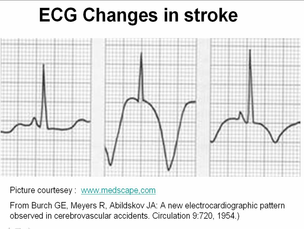
Three  physicians from Michigan ,USA  published  one of greatest observation in clinical sciences , namely the ECG changes in various forms of stroke .

Now , a shrewd physician  , will  suspect a subarachnoid hemorrhage (SAH) by looking at the ECG when the clinical situation demands . But , what we need is every one should develop that skill . We have seen errors happening  even in big institutions (or is it because it is big ?)  when  an elderly person comes with deep T  inversions with or without  altered sensorium being rushed into  CCUs  & cath labs instead of  neurology units.

We  need to teach  our junior  colleagues  . . .  That ,  ECGs of patients with  acute neurological syndromes  (ANS)  can mimic as acute coronary syndromes (ACS) ( especially in elderly ) .

The following ECG changes * are observed during stroke

  • Deep  T wave inversion –   Sub arachnoid hemorrhage
  • Cerebral thrombosis   –      Prolonged QT interval, U WAVES
  • Cerebral hemorrhage –      ST segment  shifts /T inversion


The ECG changes tend to occur very early after CNS injury.May last up to 1 week. They are not useful to identify the type of stroke. But , deep T wave inversions strongly suggest SAH rather than ICH or thrombotic stroke.

What is the mechanism of these ECG changes ? 

It is a clear proof that heart and brain are interconnected by neural network. All the noted changes occur during myocardial repolarisation . (ie ST segment )  The current thinking is  (Ofcourse , it is same as our thinking  in 1950s !)  it is mediated by adreneergic surge  initiated by CNS insult  transmitted to  myocardium by the sympathetic system.

Why should SAH produce more  ECG changes than others ?

It is possible the net adrenegic drive from the brainstem and spinal cord will be greater in SAH as it  spreads the entire CNS  through the cerbro spinal fluid. While localised ICH and infarct is  likely to generate less adrenergic impulse. 

Reference

Read the link to circulation 1964 .With courtesey to circualtionaha.com

http://circ.ahajournals.org/cgi/reprint/9/5/719.pdf

This came 50  years  ago , we still quote their work and no one has improved their work . 

Final message

If  only  we make the  clinical bed side teaching as a  regualr habit ,  we  do  justice to   our  great  physicians of the past ,   who enriched  our  life  with their  clinical  skills  and  passion for knowledge  sharing .

As the name suggests   dilated cardiomyopathy  would imply  cardiac chambers will dilate , at least some time in the course of the disease .It can be minimal, mild or massive. A new entity called  non dilated cardiomyopathy is also gaining wider acceptance . (That will be dealt seperately )

Logic would suggest , the first chamber to dilate in DCM  should be the left ventricle because it is  facing the direct load of systemic blood. But we also know , whenever  LV is stressed , left atrium comes to it’s assistance .

Left atrium does this    by total self sacrifice ( by all  means!)  increases  it’s  force of contraction, elevating it’s  mean pressure or even increasing it’s rate (AF) .

Like most  other critical questions in cardiology  ,  the factors that determine LV dilatation in DCM ,  is  also poorly understood !

  1. Is it the after load ?
  2. Is it the  muscle mass ? or it’s turgid  or flabbiness ?
  3. Is it the interstitial integrity?
  4. Is it the blood volume ?(LVEDV ,  LV residual volume )

When the issue is complex , it is  usual  to  make the   the unknown  genetic defects  ,  the scapegoat !

As of now the most important determinant of LV dilatation  could be  the behavior of the desmins, the gap junctions and myosins the titins etc

If  the LV of a DCM patient  refuses  or  resists  dilatation what  might happen ? Is it good or bad for the patient ?

Here is a catch .  A  LV  that does not dilate  obviously should be  be good for the patient  is in’t ? Medicine is not that simple.

When   LV  fails to  dilate  it means it has become  too  stiff and rigid    and pass on the  burden to  to LA which  faces the music. And in the process it dilates.This is the reason , we  observe  diastolic dysfunction in vast number of DCM patients.( Currently it is estimated > 75% DCM will have significant diastolic dysfunction )

So , now we can imagine how complex the sequence of hemodynamic stress in DCM that determine the chamber enlargement.( RA, RV  dimension in DCM is a separate issue !)

So now answer this question :  Which chamber dilates first in DCM ?

  1. Left ventricle
  2. Left Atrium
  3. Any of the above
  4. Both of the above dilate simultaneously

The answer must be 3 .

Why  recognising this sequence of  chamber enlargement  in DCM   is important ?

  • It gives us an opportunity to assess the dominant mechanism of LV dysfunction.There are reports , where some  DCMs  have more diastolic dysfunction than systolic dysfunction  .This will have important therapeutic implication.Further , many of the infiltrative   disorders of LV can have features of both DCM & RCM .
  • When a RCM begins to dilate it is usually  a harbinger of terminal heart failure. But,  it need not be always true .  A small restrictive LV  , when  dilates ,   may acquire a  slightly improved diastolic properties , as the  LV becomes more placid . And ,  if it happens the LA size may regress.
  • The role of LV restriction devices like, Acron mesh, Dor procedure, plication  in refractory  DCM is not well defined. All these   modalities actually  adds  a small dose of diastolic dysfunction in these patients who have grossly dilated ventricles. This fact is  very important  , as presence of any preexisting  significant diastolic dysfunction in DCM makes  the role of LV restrictive devices and surgery a big question mark !

Imaging  coroanry artery is  generally  in the   domain of interventional cardiologists. MDCT has helped us to change that.

The  humble echocardiography can   identify the origin* of   coronary arteries   in  most   persons. The resolution power of modern day echocardiography is  2mm and the left main  ostium is >3.5mm in 99%  of population . If some body says one can’t  visualise the coronary artery by echo ,   it can only reflect their ignorance or lack of patience to get an optimal image. Of course technological limitations are there.

*  To be emphasised again , only the origin can be identified.

Can we identify ostial leftmain or proximal  left main disease  by echocardiography ?

It should be possible in  few .

Can we place  a doppler sample volume  within  the left main and measure coronary flow velocity ?

When obsterticians are able to  assess the  uterine artery flow  in a bulky uterus ,  it should be possible to do the same in  a coronary artery . Motion artifacts is the issue in the heart.  Micro sample voulme (<1mm) are expected in the future  that will make a non invasive coronary flow assesment a distinct possibility.

Hypotension is one of the dreaded complication of acute STEMI.

  • It can be due to either a  mechanical complication or hypovolemia.
  • The hypotension in inferoposterior MI is  often related to enhanced vagal tone and easily correctable with atropine  and fluid  administration.
  • RVMI is the classical example of hypotension that may improve with fluid resuscitation
  • Hypotension,  if  not reversible within 12  hours  ,  is more likely to  represent a more sinister mechanism like pump failure, MR or ventricular  septal tear etc .

A new mechanism for persistent  hypotension is increasingly recognised.

This is due to the

1.Loss of LVOT dynamic activity.

2.Excessive  dynamism of LVOT.

LVOT contractile and ejectile falure

Even though LV  outflow tract  contain  less  contractile myocytes  , it has an important mechanical  job to do. We know , it’s  primary job is that of a  conduit  but  it also  has to  eject the blood into aorta with sufficient force.  In fact, it is thought much of the acceleration of blood velocity occur in LVOT . So, LVOT  plays a key role in maintaining the cardiac index.  An excessively dynamic LVOT will impede the forward blood flow as in HCOM.  Similarly  less dynamic contraction  of LVOT  results in  low velocity propulsion , that interferes with   proper delivery of blood from LV cavity into the aorta .

These factors get amplified in  acute MI , as it is a compromised situation with fluctuating HR and contractility. So a properly functioning  LVOT conduit is  absolutely mandatory.

STEMI due to a proximal LAD obstruction   located can involve the septal .If the first septal branch  happens to be a major one,  there will be  definite impact on the LVOT function.

Excessive dynamism  , LVOT   desynchrony  LVOT collapse .

LVOT has a medial border formed  by IVS , an  anterior surface and  a posterior surface .The lateral border is relatively boundary less , except it is guarded by  the anterior mitral leaflet.

But one should recall , the AML comes towards the LVOT only in diastole . When it comes in systole it becomes a pathological event  called  SAM  (Systolic anterior motion )

The LVOT wall desynchrony can occur in both anterior and posterior MI.In a mulivessel CAD  this can happen when there is disproportionate inferior to anterior wall motion defect.

Management.

  • There is no specific management strategies aimed at restoring LVOT function.
  • Emergency revascularisation will attenuate the mechanical dysfunction
  • Dosage of powerful inotropic agents should be moderated in dynamic LVOT obstruction.
  • Spontaneous recovery  may occur in few

http://circ.ahajournals.org/cgi/reprint/116/5/e110.pdf

Haley et all Mayoclinciproceedings 1999

  • It is only rarely a journal of International caliber is published from India . IJEP is one such journal.
  • Cutting edge articles on Electrophysiological science  break here !
  • This is an online journal . No print issues . Enjoy, it is free !

Here is the  Link

Just sample an article  : A great review about cardiac arrhythmias in congenital heart diseases , Must read by  all cardiologists    http://www.ipej.org/0906/khairy.htm

  • It is going to be the era of non invasive imaging  in  cardiovascular  diseases .Future looks very exciting
  • We have now ability to slice the heart 356 times a second !
  • Image resolutions are getting sharper .
  • The only worry ( Of course a major one !)  would be the radiation , that has to be addressed .

Now we have a dedicated journal for cardiovascular CT scan .

Does it surprise you  ?   For me  . . . It  is  !

Link to the current journal page . Get updated  !

http://www.journalofcardiovascularct.com/current

There are millions of  articles in cardiology . Some  simply  occupy   valuable spaces without any purpose  . Some give us knowledge . Some enlighten  us. While few are  so vital , it is almost a crime  if we do not read such articles and apply  it in day to day  practice .

This an article  written by Henri Justino that has a immense importance for the patients as well as the physicians .

Do not think  the article which came in pediatric radiology  is not applicable in adults !

  • Here is a site which has dedicated  resources for learning echocardiography .
  • The site has collection of various work shop and conference highlights
  • The basic echocardiography with classical line diagrams  would be very much useful for the beginners,

Cheers to duke university for sharing ! www.echoincontext.com

STEMI is the major cardiac emergency .The acute mortality is about 20% (Both prehospital and in CCU )  STEMI occurs whenever a coronary artery is occluded  suddenly in toto .We traditionally believe that STEMI occurs only in the major  epicardial vessels. (LAD/LCX/RCA .)

The total length of   coronary tree is much  longer than the length of the three vessels put together.The diagonals , the ramus, the OMs ,the septals  run for varied  distances. The caliber of these vessels can be quite large.It is estimated the diameter  of   first diagonal ,   the first OM  or the   ramus can be as big as LAD proper in 30% of CAD population . Law of statistics tells us sudden occlusion can occur any where in the coronary tree in  ACS prone patients.

What is the real incidence of side branch STEMI ?

The  dogmatic answer is   ” We do not know”

Will we ever know ?

How will a  Diagonal / OM /Ramus   or PDA   STEMI  behave ?

It is surprising this question is not addressed by us  for  so long . Some may even question  the existence of  such an entity(Side branch STEMI ). This is most likely ,  reflect our ignorance on the issue . We know  bifurcation lesions at  the   side branch  origin is very common . Further , thrombus can migrate from a main stem to a side  branch  immediately after formation .

Clinical presentation of side branch STEMI

  • Acute presentation is identical  to  that of a major main branch STEMI . The  pain  can be severe , the primary arrhythmic threat is real . Ischemic VF , once initiated does not  modify it’s  character  according to the  quantum of insult .
  • ECG is the major variable.  You ,  don’t expect gross , ST elevation in many leads as one would see in LAD MI /RCA MI.
  • The  age old teaching that  an ECG can be entirely normal in acute MI ,  could actually imply the side branch STEMIs . When a small D2 or D3  gets occluded the ECG may not pick up the ST shifts .
  • The commonest site of atherosclerosis apart from proximal LAD is the bifurcation of PDA in RCA.  STEMI due to PDA occlusion is  the most  difficult thing to recognise. Many of them have very subtle ECG and clinical findings.
  • There has been reports of acute complete heart blocks with isolated AV nodal infarcts. Here sudden cardiac deaths are reported

It is very much possible ,  many of the  side branch  MIs   may be wrongly diagnosed as unstable angina by us , for the simple reason the myocardial necrosis is not large enough to produce ST elevation .They may actually respond to thrombolysis ,  as there is total occlusion in the coronary artery.  Since, they do not manifest ST elevation there is a lost opportunity here  . This ,   probably  is the population in TIMI 3B  trial that showed some ( statistically   insignificant ) benefit for   thrombolysis in NSTEMI.

Is primary PCI justified in side branch STEMI ?

May not be . The chances of side branch STEMI   to result in LV dysfunction and progressive adverse remodeling is considerably less . The hazards of primary PCI for exceeds the risks of  MI  due to a   septal  or diagonal branch lesion .

Final message

  • STEMI due to   branch coronary artery  occlusion is a less recognised entity among ACS.
  • Cardiologists ,  need to  look into this  issue with little more seriousness as it could represent a new  intermediate risk  category   among the much  flaunted  classification  of  acute coronary syndrome. Triaging and risk stratification of  ACS  needs  a revamp.
  • It is possible  many of the UA  patients  ,  may in fact represent total occlusion of side branches.
  • There is a  definite  case  for showing less aggression in these patient  subsets  ,  provided we are sure  about  the location of lesion.

Counter point

* Identifying  a side branch STEMI with confidence  may be very difficult at bed side in an emergency . Implication of wrongly  calling a STEMI  as benign  can be  dangerous . So it will be argued ,  one need not do this exercise of traiging STEMI into main branch or side branch .

Image courtesey

Coronary tree : http://www.southcharlestoncardiology.com/64cta.html

Modern medicine promises   healthy new lives  to millions. We all , enjoy  the   fruits of  great scientific discoveries.  But do we  realise , medical science is nothing but experiments done on live  human beings . All  treatment modalities   are under constant scrutiny.  A great drug becomes the most harmful drug over a short time.

A drug or a device which is banned in one country is freely used in other country , marketed by the same parent company . How on earth this can happen ?  A device  ( Eg : A stent )  which is found  inferior  can  still can  be used legally elsewhere ,  hiding the information .

  • Do we divulge all vital information to our patients ?
  • Do we reveal all our conflicts of interest to our faithful patients ?

Informed consent is  the  “greatest  invention” in medicine . That is democracy in medicine . Doctor  patient conversation is   supposed to be  most noble of all communication !

But . . . this is under  genuine threat .  In this ” New medical AVATAR ” truths  remain only as thoughts , they  rarely  come out as words or action !

One such  situation a  physician  often  faces  in his office ,   as he is compelled  to act against his  conscience

If only we  have an ability to  read  his silence it will go something like this  . . .

For the sake of  Science &  Commerce , I have to implant this device  in your heart , and  my gut feeling says  ,  you will do much better without this device as well !   But ,  I am sorry . . .  I can’t avoid it .

Doctors are not be blamed  . . . rather  , we can’t blame  any body

Patients believe in doctors , and doctors believe in science  And the irony is ,  doctors have no other option as they are  coerced to  believe ,  in whatever is published  as science even if it is  half baked , unproved,  unapproved or  even dangerous science .

Let us prey for the genuine science to prevail  at least in human health  and the mankind  reap the maximum  benefits !

Let us  recall  Mahatma Gandhi’s    “Seven Social sins”   That included one  advice for the scientific world  nearly a century ago when it  was at it’s infancy !

Seven Social Sins By Mahatma Gandhi


  • Politics without Principle
  • Wealth Without Work
  • Pleasure Without Conscience
  • Knowledge without Character
  • Commerce without Morality
  • Science without Humanity
  • Worship without Sacrifice

– Young India, 22-10-1925