Feeds:
Posts
Comments

Left main bifurcates into two , that’s  the classical anatomical behavior of LCA. (Or it trifurcates) When left main divides , it tends to share its diameter between its two siblings LAD and LCX with considerable  whims and fancies.(Though Finet* et all thought it has a working rule !) * From  Biomedical Engineering, Cardiovascular Hospital and Claude Bernard University France

Now , have a look at this , its a rare example of  how a left main might Ignore the rule of bifurcation just like that !

Left main simply continues as left main* after giving off a casual side branch from mid left main shaft .Yes , Its a innocuous looking LCX which would be non dominant as expected

LCX arises exactly mid way in left main , (Technically LAD begins at this point ) but , can you find any difference in the left main after giving off  LCX branch.

Can we say left main continues as LAD without a bifurcation ?

Or shall we say  left main gives off a premature early side branch ( true LCX)  non bifurcating  branch ?

It is an unusual anatomy and  as expected , this patient had a dominant  RCA .

What could be the clinical implication for such a premature  LCX ?

We can only guess . May be nothing !  Obviously ,these patients are immune to develop true bifurcation lesion. Does it in any way mean they have anatomically blessed coronaries !


Reference 

1.Finet G1, Gilard M, Perrenot B Fractal geometry of arterial coronary bifurcations: a quantitative coronary angiography and intravascular ultrasound analysis. , EuroIntervention. 2008 Jan;3(4):490-8.

Heart is not like a rigid structure built with  bricks . . . . so , its too architectural mindset to describe cardiac chambers to be made up of walls. Rather , Its a four chambered muscle mass moulded together in a complex 3D interface with distinct surfaces rather than walls. It’s also important to realise, since the heart is positioned (rather hanging )delicately in the middle mediastinum resting on the diaphragm , its subjected to one more dynamism due to respiratory motion blurring the definition of surfaces as well. (Vertical vs Horizontal)

4415_21_26-heart-human-posteriorly

Posterior surface is now referred to as infero-posterior

The posterior aspect of heart contains essentially the venous channels and the atrium (LA in particular)pulmonary veins and coronary sinus.  This happens right from 8 week heart open stage when venous end of lower straight heart tube folds up and posteriorly .

cardaic-looping-posterior-wall

It should be recalled only a small portion of lower aspect of posterior wall is alloted to left ventrilce.Instead the Infero diaphragmatic surface is formed by two-thirds  the LV and one-third Right ventricle.

anatomy-of-heart-posterior-wall-mi-lcx-rca-grays-grants-anatomy-netter

Image courtesy : From the great Netter

Nomenclature  issue 

The term posterior wall is now abandoned in most Echocardiography texts its replaced by inferior .The implication is more for Electrophysiologists with reference to accessory pathway localization

What is true posterior wall MI ?

As discussed before ,posterior surface of heart is different from posterior aspect of left and and right ventricle.

What does leads V7 V8 V9 record ?

It actually records electrical signals arising from posterior  aspect  of heart.  Left atrium,  pulmonary vein along with insulatory  effect of lungs dampens the potential . This makes the sensitivity of ST elevation  in posterior MI is low.

Blood supply of posterior surface

It’s highly variable.Both RCA and LCX arteries contribute with its  posterior left ventricular branches (PLV)

It can be inferred , LCX has more territorial rights than RCA in this unique zone of heart as the artery covers more posterior areas.

Read a related article

Back pain from anterior MI : Is it possible ?


Hippocrates is bestowed with the Immortal  tag “Father of Medicine”, not because he invented  any miraculous gene therapy or a modern virtual imaging of human organs, nor did he found any magic drugs .He didn’t receive a single award  even from his local village, forget about any Nobel prize to him !

hippocrates

Mind you , he lived before Christ ,2ooo years ago , there was nothing in the hands of noble professionals, not even a piece of paper and pencil to note down patient’s symptom .They didn’t even know what organs human body contained no basic medical tests . They just had a pair of hand, working brain with six senses , strong will to work hard and most importantly a caring mind and a constant search for answers to lingering scientific queries .

good medical practice hippocrates medical oath ethics drsvenkatesan dr s venkatesan

Yes, Hippocrates is still holding the post of father of medicine because he was the first human  to propagate  thought that human diseases are not evil forces beyond our control, it may have scientific basis , every disease has a specific cause that arise from derangement of body function.

More important than this , he formulated a way to practice this profession in a dignified manner. He also predicted common sense may prevail over science  in innumerable instances. Going through his quotes , one could wonder , he probably predicted technology might hijack human Intellect as well !

hippocrates quotesQuotation-Hippocrates-disease-Meetville-Quotes-149262Now,we have every thing. Students read medicine  in animated 3D class rooms , physicians  get a deluge of body system data &  images beamed straight into their ipad . One can perform  complex interventions with ease in almost every  organ or  even replace it , if it doesn’t work .

Still , as on 2017  ,there is something huge , that is missing  in the Noble profession when compared to ancient days  (2 millenniums before!) when people thronged Hippocrates  clinic in the remote Koss Island of Greece, where he used to sit  with almost  nothing , but was able to offer definite cure for many .

What is that missing link  ? Without realizing  what it is, we enter the Noble profession and fervently  take the  customary Hipocratic oath . For many (or most ?)  of us it is amusing to read and practice that. Life has moved in  fast lane since then. It is a tragic truth , Hippocratic oath  have become  redundant , obsolete .or outright humiliating for few !

Final message 

Whatever you say, still Iam compelled to feel sorry for that “Good old man” who miscalculated the Integrity modern day Noble professional , (I would say, Mr H failed to  realise doctors are also made from ordinary human beings ! )

It’s ok . . . here is a “Doctor’s life maintenance” manual : Keep reading it periodically !

British General medial council , has done a wonderful job . It has published a practical life maintenance and behavioral guide for  doctors  which I feel is most important text to be read periodically and of course  followed  !

Link to Good medical practice


A 50-year-old man was referred for dizziness, bradycardia and dysphagia .He was very clear in describing his symptoms and  landed up in Gastro- enterology  OPD , from there was referred to my clinic for cardiac work up . His ECG showed a sinus bradycardia HR of 48 /mt.

Screenshot_2017-07-05-19-09-12

Echocardiogram revealed a structurally normal heart as we expected , but was surprised to spot suspicious shadow in para-sternal long axis view , beneath left atrium.

A well demarcated large mass compressing left atrium.  Trans Thoracic Echocardiography  may not be looking at the heart alone ,(Its technically Thoracic Ultrasound though we may refer it as Echocardiogram   )

  • Aortic aneurysm ?
  • Mediastinal teratoma?
  • Bronchial adenoma ?
  • Esophageal mass ?

The Answer is none of the above

As I was wondering what it was, the staff nurse in charge threw a heavy folder with well worked up gastro Investigations.

That moment , diagnosis became obvious , without a need for further scrutiny to my medical acumen.

Note: The barium swallow of the Esophagus reveals the Intimate relationship between the food tube and the heart as it descends vertically downwards posteriorly  . Realise , how the proximity of these two structures could  confuse a physician when symptoms spill over on either way. (I would have expected a lateral view to show the compressive effect of Esophagus on the left atrium the radiologists felt its not important !)

Yes , it is Achalasia of the cardia , dilating the lower end of esophagus with fluid /mass effect  , compressing the posterior surface of Left atrium.He underwent a myomectomy surgery.

Why bradycardia  ?

There is well described esophago-vagal reflex reproducible by stressful swallow or balloon inflation in the lower end of esophagus at D7 level.(Ki Hoon Kang,Korean J Intern Med. 2005 Mar; 20(1): 68–71.)

Achalasia cardia is known to be associated with symptomatic bradycardia, dizziness, and rarely swallow syncope,though this patient didn’t have a classical syncope.The bradycardia is probably due to high vagotonia, (Hugging effect on posterior surface of heart known for rich innervation of vagus.) . Complete reversal  of bradycardia after esophago -gastric surgery is expected.

Implication for cardiologists

There has been instances of patients with esophageal syncope and reflex bradycardia getting permanent pacemaker therapy. I think , clinical or sub clinical esophageal disorders should be included in the work bradycardia before labelling them as intrinsic sinus node dysfunction .(Ref 1,4)

Final message 

The field of Cardiology  is often referred to as a super specialty atleast in India . I disagree with it strongly. Cardiologists are neither super(eme) nor special .We need to be reminded  its afterall a sub-specialty of Internal medicine and each specialist should undergo retro-training in medicine periodically .This patient is a typical example of a gastric problem entering the domain of cardiac Imaging.Strong foundations in symptom analysis and some degree of medical  curiosity will enable an occasional cardiologist to make a correct diagnosis belonging to a remote foreign specialty.

Reference 

1. Palmer ED. The abnormal upper gastrointestinal vagovagal reflexes that affect the heart. Am J Gastroenterol. 1976;66:513–522. [PubMed]

2.Armstrong PW, McMillan DG, Simon JB. Swallow syncope. Can Med Assoc J. 1985;132:1281–1284. [PMC free article] [PubMed]

3.Turan I, Ersoz GBor S..Swallow-induced syncope in a patient with achalasia
4.Dysphagia. 2005 Summer;20(3):238-40  4.Basker MR, Cooper DK. Oesophageal syncope. Ann R Coll Surg Engl. 2000;82:249–253.

Some of the noise bites from a busy cath lab after a  mid noon angioplasty

         Oh’  that  looks bad , whats that projecting !

There is some haziness too ,

            Make sure its not a flap,

  Better to do IVUS or should I OCT ?

           Shall I  post dilate with NCB ?

Should we cover with  another stent ?

           I think we can manage with Tirofiban or Reopro 

Call the chief ! suggested a first year resident,that seemed to be the most reasonable noise bite among all .Yes, the final command came from the chief cardiologist after a 10 second glance over the workstation ,”Guys,  forget it , . . its acceptable  pinching, DAPT will take care of it , just ensure adequate ACT till night , put the next case . . .on table” !

That’s fairly common chat session in any high volume cath centres (Which ended abruptly  in this case with the chief’s uttering)

Does any body know  what the chief meant by  the term pinching ?

  • Is it the  pinch of Intimal fold ?
  • Is it pinch of plaque ?
  • Is it a flap ?
  • Is it a plaque prolapse within the strut ?
  • Or just a evaginated thrombus
  • A subintimal calcium speck ?
  • A longitudinal stent deformation?

Any one knows the histology ? Is there any natural history  study of such pinching ?

Iam afraid no one knows . But common sense tell us it can be anything  between a totally benign entity to  Imminent nidus  for an acute stent thrombosis , depending upon the patient’s destiny and physician’s luck !

How does one make a decision in such an uncertain situation ?

The decision to leave that pinching is taken by any  cardiologist  based on his past experience or  Inexperience or both. Some do IVUS/OCT , many don’t . Whatever the decision  ( empirical or scientific ) its  going to be tentative  and  outcome is any body’s guess.

Final message

Coronary arterial pinching is a dangerous cath lab slang used exclusively by expert Interventional cardiologists , often after a hurried PCI ! It may sound  innocuous .To label a protruding plaque as a “safe pinch”demands heavy courage that is an essential requirement  for a successful Interventional  cardiologist , which most of them are blessed with !

Reference 

1.No Reference as such :There is no specific study about histology of coronary pinching  .Though , IVUS and OCT data are available for various post PCI shadows , it never addresses the issue of pinching specifically as no one is clear about what they mean by it.  Hence ,we are planning to decode this long pending mystery with our own  PINCH-iVUS  study.

2.This article from Circulation Imaging  new generation IVUS could reveal  histology of pinching

f5-large


Professional competence is defined as doing things, always in the Interest of patients. It’s generally believed small hospitals are not competent enough to treat cardiac emergencies . . .Do you agree with that ? No, Its largely a myth . Do you know there is a absolute lack of proficiency threatening to plague our country’s coronary care system. ? It’s the professional Incompetence by the space age, star hospitals (mis)managed by masters of the noble business. None (am I right ?) of this hospitals either monitor or publish the outcome of their treatment.

Backed by pseudo scientific data , amplified by unrealistic expectations of ill Informed patients , some hospitals are avoiding Initial emergency treatment of acute MI , instead they waste time ( load DAPT ofcourse !) in securing the finance for the costly Invasive procedures or refer them out of their premises if they can’t afford for it.In the ensuing emotional and financial melee many of the ill-fated patients lose vital time window of thrombolysis as well ! and carry risk of fatality or damaged myocardium.

Every stake holder in the current coronary care system simply assume the enforced modality must be far superior because they administer the most modern and costly treatment suggested by few high intensity cared clinical trials originating from west. The wisemen who run the corporate hospitals never realise medical competence and outcome is not entirely defined by science. Their primitive cognition wouldn’t allow to think beyond business equations either.

Please believe me, time and again, I have witnessed patients reaching Government hospitals after being shunned away by big (Some times even medium sized ) hospitals who boast themself only as PCI enabled care. Even if they want to lyse they stock only the Tenekteplace .

I think tragedy is a lesser word to describe the scenario , where a distressed family is trying to arrange for a Rs30,000 shot of Tenekteplace when thirty times cheaper still equally efficacious (Rs 1000 Streptokinase) is concealed from their visibility .The Govt should urgently look into instances of large private hospitals avoiding Govt insurance scheme patients even in cardiac emergencies ! To label our poor patients as unaffordable ones is a outright misnomer, rather its the rich hospitals that are “not affordable” to lose profit and treat our countrymen , in a cost effective manner is the reality !

Who is Poor ? You decide.

Two forbidden things in coronary care

1.Cajoling and manoeuvring a distressed family for a primary PCI as a routine treatment hyping its beneficial effect and underplaying the true advantages of thrombolysis in largely technical jargons is the current norm in most coronary care units.

2.Another issue is , after confused confabulations with the duty medical officer, if a rare patient family choose the option of thrombolysis , comes the next googly*. Many noble minded hospitals do not stock the low-cost and equally efficacious thrombolytic agent and offering only the costly option to the anxious families when the myocardium is on fire.

Hospitals that practice these two coronary protocols need to be shamed and labeled as “Coronary Incompetent ” In spite of having 24/7 cath labs. (Realise , they are just like any remote rural hospitals , at least the later can’t be faulted as they don’t withhold a reperfusion strategy !)

Final message

I think , mindless proliferation of cath lab based cardiac care , which follow this theme , ie “Thrombolysis incapable but PCI capable “ are biggest threat to coronary care in our country ! For the best coronary care for any country ,what we need is efficient prehospital thrombolysis team .We have conveniently forgotten the great study of CAPTIM wherein the ambulance drivers replicated the same effect of primary PCI performed by highly trained cardiologists in modern labs.

In India, primary health centers which is within few km reach of entire population can be designated as static ambulance equivalents with basic resuscitation facility . If a multipurpose health worker can be trained to lyse, with remote supervision that will accomplish 90 % of what the cathlab guys can achieve ! Selective shifting is suffice.

Postamble : Ofcourse, not doing pPCI for high risk or complicated STEMI is unscientific and we need to have proper consenting and referring frame-work for such patients.

Counter point : One of my colleagues asked me ? Why do I enjoy attacking the established scientific practices ? May be I have a problem , yes, but I think in a true medical democracy we have right to debate anything , absolute truth is a ongoing journey !

*Googly: An unplayable ball delivered to a batsman in the game of cricket.

A 45 year old man came with  recent onset breathlessness.His left ventricle was dilated along with left atrial enlargement.The LV EF was 42% (By current definition mid range preserved systolic function( Circ Heart Fail. 2016 Apr;9(4))

But, he was severely symptomatic because of combined  systolic and  diastolic dysfunction.Diagnosing and grading diastolic dysfunction has been extensively done in last decade.Now , we realise without significant diastolic dysfunction symptoms of pulmonary congestion can never occur in patients with DCM.

We don’t require complex tissue Doppler parameters to diagnose high-grade LV diastolic function.Just have a look at LA dimension,  concentrate the E to A ratio. A tall E that humbles the A by more than 2 to 3 times is clear evidence for  LA mean Pressure exceed  18 to 20 mmhg or so.

This , in combination with dilated LA is a marker of chronic severe diastolic dysfunction.The fact that A is diminutive in no way takes the Importance of Atrial contribution to LV filing at this critically compromised LV status.

Note E:A ratio is 3:1 .This simply means the early (and mid to a certain extent ) diastolic pressure in LA is high and most of the filling takes place before Atrial contraction .There is one more reason for diminutive A . Atrial contractility fails to prevail over E in late diastole as LV end diastolic pressure is significantly high in these patients with diastolic dysfunction.

A dilated left atrium is an Independent marker of significant LV diastolic dysfunction (In the absence of MR) .When does LA begin to enlarge in diastolic dysfunction ? There is uniform rule.Generally LA size more than 4.5cm indicate grade 3 or 4 LV diastolic dysfunction.

LA size and Pulmonary congestion 

It’s a paradox , a roomy  LA dampens the LA pressure curve and A reversal into lungs may not happen.

*AF irony on A reversal

Logic might suggest , loss of atrial contraction might attenuate A reversal and less blood flooding into pulmonary veins.No, It doesn’t happen that way.If  AF is precipitated for any reason its going to be “switch on”  for acute pulmonary edema.

What is the relation between systolic and diastolic dysfunction in DCM ?

We find about 30 % of DCM has documented resting diastolic dysfunction.This is actually a underestimation of true diastolic dysfunction as it can very well manifest only during exertion.

Though generally , there is good correlation of grade of diastolic and systolic dysfunction in terms of severity , some of the patients show severe diastolic dysfunction out of proportion with systolic dysfunction.

Note : In the above patient it’s actually a fairly preserved systolic function but still has advanced diastolic dysfunction.

Grading of diastolic dysfunction .Image courtesy MM Redfield et al: JAMA 289:194, 2003. Note E:A >1.5 is

Final message

Relying on E:A ratio to diagnose diastolic dysfunction  may appear  amateurish for some of us .The rampant reporting of E>A for grade 1 diastolic dysfunction has made this parameter a “Doppler cliché”. But , the fact of the matter is,  it does help us confirm severe (Grade 4) diastolic dysfunction when E stands  tall and towering over an almost dwarfed A.

Clinical Implication

Please realise ,In patients with DCM  when you find an  A that is too diminutive in combination with  a menacingly tall E , it may be prudent to raise diuretic dosage. It’s a sure signal for impending pulmonary edema.

Queued queries 

Can DT and IVRT normalise with progressive diastolic dysfunction ?

Continue Reading »

Brugada syndrome is  as an  Inherited sodium Ion channel defect leading to loss of /or reduced sodium channel function.This specifically causes RV epicardial Imbalance of In-flowing(depolarising)  and out-flowing (repolarising)current , potentially triggering ventricular arrhythmia. This happens either spontaneously or during electrical stress times which include, fever, various drugs , adverse autonomic fluxes etc. So far,  we have been thinking it as primary electrical disorder with no macroscopic/ histopathologic  defects.

Newer Insights are emerging

But, how is this primary electrical disease , harbor a well demarcated  RV epicardial phenotypic substrate ? .  . . ablation of which eliminates the VT.

Zone of probable structural defect over RV epicardium (Pink zone) amplified by infusion of Ajmaline. Note the ECG showing typical ST elvation lead V1 to V3 .(Image courtesy Carlo Pappone et all  )

A recent study from Italy from the original founders  (Brugada team Ref 1 ) has confirmed RF ablation of RV epicardial tissue  is indeed feasible in many and should be considered in high risk Brugada syndrome. (Then should we suggest , ICD is no longer a choice in Brugada ?)

MRI findings in Brugada has shown some structural defects .(Ref 3,5) .It seems  Brugada is an Inherited electrical cardiomyopathy with a structural defect. (The overlap between ARVD and Brugada syndrome appear more real than we thought before ! (Ref 7 )

Final message

Still , Brugada is more of a electrical disorder,  but soon we may refer it as structural heart disease.

Reference

4.Catalano O,Antonaci S,Moro G,Magnetic resonance investigations in Brugada syndrome reveal unexpectedly high rate of structural abnormalities. Eur Heart J. 2009;30:22412248Abstract/FREE Full Text/Google Scholar
A best review comparing Brugada vs ARVC

An event that happened recently  that shook my country’s  collective conscience .It was, loss of  hugely popular and beloved  President of India , Dr Abdul Kalam on 27-07-2015. He was 84.Death came in a most dramatic way when he fell down midway  during his lecture to students of Indian institute of management ,Shillong in the state of Megalaya.

image

Indian President Kalam addressing the students snapped moments before he dropped down dead due to cardiac arrest

What is the implication of this VVIP’s death for  cardiac Academic ?

I believe , there is lot .The presumed cause of death was cardiac arrest . As we know , it must have been an instant electrical death as the local medical personnel  couldn’t  revive him after an Initial  CPR and later shifting him to state of the art facility . The ex-president was known to have a good health record and the heart should have been normal until prior to the cardiac arrest.

Now coming to the key question what is the chances of survival of cardiac  arrest ?

While there have been many survivors  of cardiac arrest within hospital premises  and coronary care units . . . still ,  life cannot be  guaranteed even if prompt CPR is initiated .

It’s the height of  Heisenberg  irony,  some lives can be saved  even when cardiac arrest happens out of hospital , while it’s also a fact deaths due to cardiac arrest happen right inside the cath lab where all emergency strategies are in place .

How much delay is permissible in resuscitating cardiac arrest ?

Cardiac arrest is nothing but activation of  the switch of death . Evey second is important . Experience suggest if reversed within first 2 minutes maximum survival is expected .Beyond 5 minutes and within 10-15 minutes most deaths will ensue.Up to 20 minutes survival is possible though with a risk of brain permanent brain damage.

A  recent study  from Sweden which addressed this  issueand  confirmed a  dismal fact  that even in the presence of best emergency care system,  survival is meager  4 -8% . However , the positive outcome was , If the bystander does some form of CPR till the arrival of  emergency service reaches the spot it can go up to 10.5 %.

cpr cardiac arrest

It is very clear , surviving an unexpected cardiac arrest is in the domain  of  God , still  we must encourage lay persons  to know and learn the techniques of CPR. The protocols has been simplified now , to include chest compression alone as the Initial measure.

Coming back to the question of death of our president,  as some one asked me direct question if the death could have been prevented ? , I said , yes if God willing !

Post-ample

Note : Cardiac arrest in patients who are at high risk with underlying heart disease (structural or genetic ) will require implantable cardiovertor  defibrillator ( ICD ) that can save thousands of life every day across the globe .


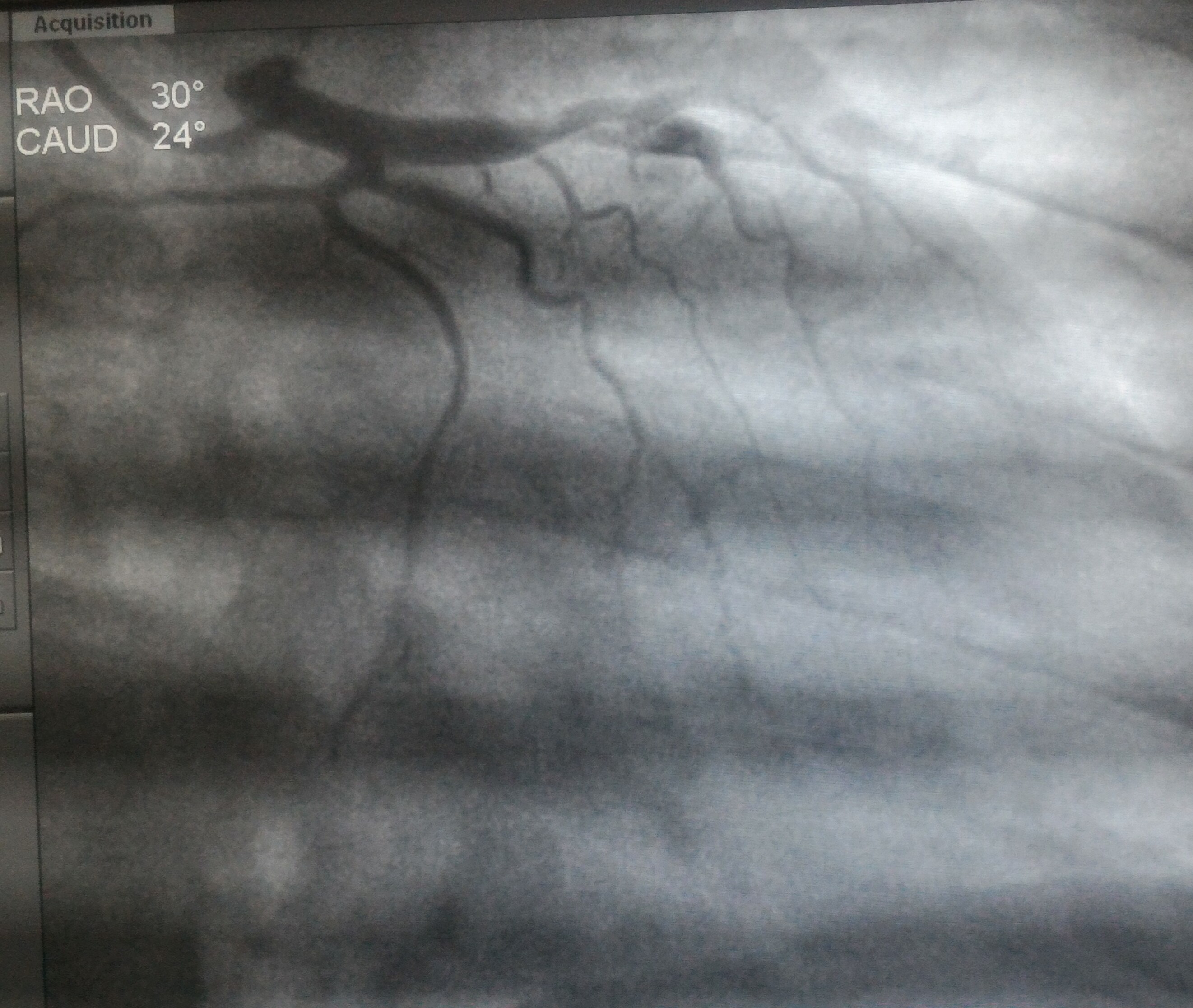
BMS, the original stent technology with meticulous metallurgy and design has been silently replaced by the drug eluting stent (DES) for over a decade. DES was introduced to bail out BMS from perceived high rates of  restenosis . It was a fundamental flaw, we failed to give due  weightage to the multiple variables like  operator expertise, lesion morphology, patient factors that determined the restenosis  rate .

There was never a single study done in large scale that compared a well deployed BMS with a poorly deployed BMS/ DES in terms of restenosis rate.This would have clearly quantified the technical component in the  restenosis rate that brought  pseduo -bad name for BMS in early days.

Without applying mind, wrong questions were asked and tested. No body could refute a “novel concept” , when some big names in industry  suggested  we must involve an anti cancer drug to prevent cell growth and neo-vascularisation  and hence restenosis. But , in reality  the technology of DES essentially complicated the metal behavior by adding a drug and drug adhesive agent(Polymer)  to the otherwise inert metal. Further , the , metallurgy  engineers had to restart / reduplicate from the scratch since we had  already well developed stent technology for BMS . The manipulation was  to add a drug to the metal.

The  irony of DES lies in the fact it Intentionally allowed to interfere /damage the endothelial healing and make the extended anti-platelet mandatory.  Still , DES was able to rule the world backed up by hyped data  with   bloated  reduction in restenosis rate. (Now we realise  the true benefits of DES  are nil  or at best marginal or even harmful in certain subsets of ACS .Read NORSTENT Trial linked below  )

Yes DES has a concern , but its not the drug you know !, 

Off late , since the polymer was  assumed as  culprit, variety of new generation stents with disposable /Non durable /Zero poylmer were developed. Still, polymer could not be proven as true culprit , some have started blaming  the drug again. Recently, It led to one famed DES based on Paclitaxel (which has a pride of place in the Land mark SYNTAX  study ) exited the human domain  with disgrace . (I wonder can  we conclude then SYNTAX study is also become invalid !)

This study done with over 9000 patients  concluded  like this  . . .

The DES industry was (is) so powerful it could easily shrug  the challenge of truth that came out briefly  in early 2000s when DES got hit with increased  acute complications.

Now, in 2016 NORSTENT study again showed us BMS is as good as DES in all walks of CAD.  Let us see what happens , still  its very unlikely mature cardiologists do not trust BMS.

*I have a belief  (Paranoid or not time will tell !) one of the reasons  DES are strongly promoted  is to sustain DAPT market alive and kicking for a long haul !

Scenerio  in India is frightening.

While the developed countries have DES usage rate around 65 % , India leads the world with DES constituting 95% (NIC registry 2017) of all deployed stents.What a way for a poor country  to  tackle CAD , which doesn’t even have prompt prehospital Aspirin for  bulk of their ACS patients, ready to waste  its resources in DES.

India , a country Infested with an unregulated health industry  became the perfect battle  ground for abusing the stents. With direct collusion with the large hospital managements the issue got exploded recently  .The Govt was compelled to come out with urgent restrictions and price control  in the use of stents.

Funny world this. World’s richest economies  are worried about the cost and want to phase out inappropriate therapy whenever possible, its absolute arrogance most of us feel shamed to keep BMS in their cathlab.

Final message

A  good metal based flexible ,trackable , thin struted  BMS should be the default choice for coronary stenting .( We used have one , now it vanished !)It avoids unnecessary prolonged DAPT .Most importantly one BMS costs 25 % of the cost of DES   . . . think of 4 critical proximal LAD lesions of a poor man can be fixed at the cost of one DES , that’s  definite way forward. Govt of India can pass another regulation in this regard. If you think  NORSTENT is NONSENSE  let us atleaset  insist for a large scale Indian  study for BMS /DES and  Cardiological society of India has much work to do !

Future for BMS  . . . looks bright !

While the  superiority  of DES is being increasingly questioned , the concept of surface modified BMS is being tested .This I believe is a face saving way to bring back the BMS in lieu of DES. There is a distinct  possibility of many of the new generation  DES going the BVS way in the near future.

Reference 

1.Hassan AK1, Bergheanu SC, Stijnen T, van der Hoeven .J Late stent malapposition risk is higher after drug-eluting stent compared with bare-metal stent implantation and associates with late stent thrombosis.Eur Heart 2010 May;31(10):1172-80. 

2.Zhang K1, Liu T, Li JA, Chen JY, Wang J,   Surface modification of implanted cardiovascular metal stents: from antithrombosis and antirestenosis to endothelialization.J Biomed Mater Res A. 2014 Feb;102(2):588-609.

3. https://www.pcronline.com/eurointervention/114th_issue/volume-12/number-17/350/ultra-hydrophilic-stent-platforms-promote-early-vascular-healing-and-minimise-late-tissue-response-a-potential-alternative-to-second-generation-drug-eluting-stents.

4.Drug-Eluting or Bare-Metal Stents for Coronary Artery Disease NORSTENT Investigators N Engl J Med 2016; 375:1242-1252

Post-ample : Only For non believers  ( who think this article is near rubbish ) 

I am  very much  convinced DES should be superior  for the simple reason it elutes a drug and the whole world believes it works !

Do you know, what these drugs do, and what they are expected to do ! In this elegant study  by Hassan AKEur Heart J. 2010 May;31(10):1172-80.  Its proven with IVUS , DES is many fold likely to cause late stent apposition than BMS.( Thus carrying the risk long term )  Reason is simple , patchy and incomplete endothelisation on the luminal side and pathological metal vessel wall interface  in abluminal promoting late mal-apposition.