Feeds:
Posts
Comments

Left main ostial lesion remains a  challenging task .A new stent design is  proposed here.

The lesion

Left main  ostial  stenting lesion003

The hardware 

left main ostial coronary stent drsvenkatesan

The technique

Left main  ostial  stenting lesion002

Final message

This thought came  when I  recently encountered a patient with a left main ostial  stent which was projecting well into aortic root .It is an open access patency ,whoever is capable of converting this idea  to a clinically applicable technique is welcome to proceed !

Epicardial fat is increasingly recognised to be a coronary risk factor.
Methods to measure and quantify epicardial fat has not been standardised.
Nomenclature issues
There is some confusion in the literature regarding  the definition epicardial, pericardial , paracardiac fat etc.
Epicardial fat is the true visceral fat located in proximity with myocardium .Epicardial fat shares the same blood supply as adjacent myocardium and also show  para-crine functions.This is the risky fat that is metabolically active.
Paracardiac fat  is  the fat deposited outside the parietal  pericardium .It is also referred to as  thoracic fat .They carry less risk and behave more like somatic non visceral fat.
Pericardial fat is a loose terminology .Many  use epicardial and pericardial fat interchangibly .It is to be realised , fat either deposit inside the visceral pericardium or outside the parietal pericardium. It almost never deposits  within the pericardial space.

* Currently it is better to use  only two terms .Epicardial fat and  thoracic fat .Other terminology should be avoided.

How to measure and quantify epicardial fat ?
Distribution of fat over heart is not uniform .Though it appears a random distribution there app eras to be a pattern to it .It is largely loaded anterior surface of the heart, over the AV grooves, invariably hugging the coronary arteries and great vessel origins.Fat deposition posterior to heart occurs in gross obesity.
There is no accurate way to quantify it . A rough estimate can be arrived . The following investigations are useful.
  • Echo
  • Multi-slice CT  Scan
  • MRI
Epicardial fat pad thickness by M-Mode /2D  is simplest,  but considered crudest modality to quantify.  .Still , it is a useful screening test. In our experience , if there is distinct anterior fat pad > 5mm it is likely to be significant.
However, epicardial fat volume is ideal method to quantify it.(Read below)

Which part of cardiac cycle it is measured ?

Since epicardial  fat is a compressible structure , in diastole the myocardial mass encroaches the epicardium  any imaging modality the distinction between epicardial and para-cardiac fat  tends to attenuated.

Even MRI images obtained with  diastolic  gating ,  may  not be reliable .End systolic frames are now considered ideal.

(J Am Coll Cardiol. 2011 Oct 4;58(15):1640;Distinction of “fat around the heart”.Kaushik M, Reddy YM.)

What is the normal  epi-cardial  fat volume ?

There is no standard normal.In one large study from  Boston in people  with a BMI between 25-30 the  mean epi-cardial fat volume was 125 cubic cms

normal epicardial fat volume

(*Intrathoraic fat  same as para-cardiac fat located outside parietal pericardium )

How to differentiate fat from pericardial fluid ?

It can be a very difficult issue especially  when you expect both as in hypothyroidism .It needs proper  gain settings and interpretation.

Here is case report of how epicardial fat was mistken for PE even in TEE

Can we measure  the  net mass of  cardiac  fat , does it compress the heart ?

When we say average heart weighs 300 grams it includes fat embedded in it. I don’t think it is possible to measure the fat mass ,but clinically it may be important.Read this .Can-epicardial-fat-constrict-the-heart-an-authentic-yes-from-ulsan-korea ?

Final message

Estimation of epicardial fat volume is largely a research tool.In grossly obese patients it may  have some clinical utility.Increased anterior fat pad in routine 2D echo is an useful  screening test. However, it requires MRI/CT imaging for accurate quantification.

References

how to measure epicardial fat framingham study
2. Marwan J M, Achenbach .S  Cardiovasc Comput Tomogr. .Quantification of epicardial fat by computed tomography: why, when and how?2013 Jan-Feb;7(1):3-10
3. Sarin S, Wenger C, Marwaha A, Clinical significance of epicardial fat measured using cardiac multislice computed tomography. Am J Cardiol. 2008 Sep 15;102(6):767-71.

CHB with CAD is a  common combination especially in the elderly.

Which will you Intervene first ?  Is the AV block related to CAD  ?

How to differentiate Ischemic from degenerative AV block ?

Differentiating is often difficult.Even coronary angiogram may not answer the query unless it is totally normal . For AV block to occur usually  LCX / RCA lesion is required.  LAD lesion in isolation are rare to cause CHB .

How often re-vascularisation  reverses  ischemic CHB ?

Logically  you expect more reversals.In real world it rarely happens.

Therapeutic options in combined CAD and CHB

  1. PCI and pace maker in the same sitting .
  2. PCI first followed by pace-maker at a later date.
  3. Pace maker first followed by PCI at a later date if required.
  4. CABG  and epicardial pacemaker ( best option In all critical TVD and CHB)
  5. Pace maker followed by CABG later
  6. Pacemaker followed by medical management (CHB with Insignificant CAD)

Can worsening of ischemia  occur after pacemaker  ?

Very much possible . Since the patient  has been benefited by low heart rate in terms of MVO2 consumption .(Inserting a pacemaker  is  like sudden withdrawal of beta blocker !)

Rate adoptive pacing can confer chronotropic competence which  may bring back the angina.So,what was a insignificant lesion  can become hemodynamicaly relevant  and  may require  angioplasty  later.

*The above clinical issue is applicable  for sinus node dysfunction and CAD as well.

Final message

There is no  fixed rule in the management strategy in combined  CHB and CAD .

Generally , electrical  therapy  should be given preference .Symptom guided approach  may be practical.

In this scientific era , one may argue to deal both issues  together by simultaneous  PCI and pacemaker ,  still  option 3 and 6  remain clear  favorites !

If angina  occurs  even in  baseline bradycardia  it is obvious the obstructive CAD  is  significant and needs immediate fixing .

Finally , though it looks an attractive  concept , It is very rare for CHB  to get reverted by PCI or CABG.


All left to right shunts are  acyanotic heart disease to begin with. Cyanosis appears if there is progressive PHT and reversal of shunt .We know this happens late in ASD.(third decade)

It is important to remember some of the  patients  with large ASD  can show significant desaturation without severe pulmonary HT.  This should not be mistaken for Eisenmenger reaction.

How ?

In  any large ASD ,

  • IVC blood can stream into LA by hitting preferentially the lower part of IAS.( It is the old fetal route that heart does not forget  and indulges whenever the  local hemo-dynamics permits !)
  • During straining , (Valsalva and equivalents)  right atrial pressure can exceed LA and small amount of shunts occur across RA.
  • ASD is often (15%) associated with systemic venous anomaly. The common one is persistent LSVC.  LSVC  is usually connected to coronary sinus . If it has a communication with LA (Un-roofed CS) , there can be significant cyanosis .
  • Further , a large ASD can act as a single atrium and considerable mixing happens and cyanosis results.

Finally ,two conditions should always be considered

  • ASD if associated with VPS auguments R-L shunt .
  • TAPVC can be mistaken for Eisenmengerisation of  ASD in bedside which presents as clinical signs of ASD + Cyanosis

* It is useful to recall ,even PFOs can shunt right to left at times of extreme RA pressures like during PEEP ventilation and orthostatic  deoxia in sick ICU patients are reported (If PFO can shunt R-L , why not huge ASD ?)

Final message

Cyanosis  in ASD is not always  an ominous sign .There are few important causes other than Eisenmenger. Though it  occurs intermittently , persistent mild desaturation is also possible.

Any new ST depression occurring during  EST is sine qua non for inducible ischemia.But,this rule does not uni-formally apply  in all 12 leads .ST depression occurring is certain leads is more important. While severe global ischemia can depress  ST segment  in most leads ,factually  only the leads V 5 and V6  predict true Ischemia.This because , bulk of LV muscle mass faces these two leads.

Isolated ST depression in inferior leads  during exercise

  • Is a frequent issue occurring at the peak exercise.
  • Is least predictive of significant CAD.
  • The exact mechanism is not clear.
  • Some continue to  believe it is indeed significant .
  • We have  observed  isolated  ST  depression > 2mm in inferior leads with significant CAD.
  • What really matters is the quantum of ST depression , symptoms, and exercise time and preexisting CAD .

Probable mechanism

  • Apart from true ischemia ,ST depression may indicate relative sub endocardial strain rather than ischemia.(By the way can simple stretch can cause ST depression ?)
  • The Infero posterior surface of heart represent  right ventricle .RV volume overlooked peaks exercise.Some think it represents acute raise in RV load during peak exercise.

How to report such EST ?

You can report it as such,  what you have observed.

  • ST depression noted in Inferior leads at peak exercise.
  • Mention whether it was angina free,
  • At what METS,
  • Total exercise time .

If you are statistically inclined  you can also mention the likely hood of CAD by positive predictive value (PPV) of the test (Low with isolated Inferior ST depression )

If you are really confused , and do not want to scratch your brain we have the most convenient terminology  invented by cardiac physicians ie Borderline EST, or Mildly positive EST “

Should we do Angiogram for such patients ?

In this era of catching normal people  who attend master health check ups  for a day care CAG  . . . it is not all  a crime to do angiogram in a  patient who shows suspicious  ST depression in three of his leads (2,3,AVF) especially if he also complains of vague chest pain.

Alternate investigation

Of course , we  always have the luxury of using  MDCT  that can stunningly  photograph the coronary arteries.

It is a mystery investigation, if it comes entirely normal every one is happy.Even slightest  defects in the photography  has a potential to confuse both physician and the patient .

What I do ?

I hesitate to  do routine CAG  if ST depression occur exclusively at  peak exercise beyond 10-12  METS , which disappear fast.(Many times we can apply this rule  to classical ischemic ST depression of lead V4 as well !)

ST  depression  in any leads (with any degree) following an episode of  ACS seems to be important.

Related topic

The WordPress.com  prepared a  official 2013 annual report for this blog.

Here’s an excerpt:

The Louvre Museum has 8.5 million visitors per year. This blog was viewed about 540,000 times in 2013. If it were an exhibit at the Louvre Museum, it would take about 23 days for that many people to see it.

Click here to see the complete report.


Dear friends,

It all started in 2008. This is  6th year of my attempt to share knowledge in cardiology.The followers of my blog is the only  strength that sustain my writing .

Wishing you all a Happy , wonderful  and  a prosperous , New year 2014

But . . . please be reminded we don’t require a New year to bring a bout of happiness , it is sitting right in our minds every day  !

On this day let me quote my most revered quote of Hemmingway.

Ernest hemmingway quotes

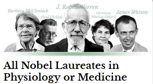
Ernest Hemmingway the Nobel laureate  who was born in USA, Lived in Paris , fought in world war 2 , lived in the deep forests of Africa with wild animals during the fag end of his life .He had a Intimate relationship with Cuba,  made a passionate appeal to end the man made disaster called wars in this planet , before his life ended in 1961.

To connect with  this noble (Nobel ) soul  reach through Wikipedia Link

Auscultatory  findings of mitral stenosis is the most common question asked in clinical cardiology right from first clinical year to higher specialty examinations . It should be answered in a systematic way.
What happens to  the murmur and sounds in mitral stenosis after the onset of AF ? When I asked this question  in my class  , many struggled . Hence this post .
 Mitral stenosis auscultation
* Correction
The A2-OS interval in critical MS  is relatively short and only is minimal variation is expected in spite of AF .This is because, A2-OS interval is primarily determined by instantaneous  LA pressure  at the beginning of MV opening which is always high and does not vary much with RR interval .
Reference
Criely mitral stenosis pressytolic accentuation ausultation atrial fibrillation

Following are revered  facts  . . .  among the  “Guardians of   Cardiology” !

myths-truths-300x300

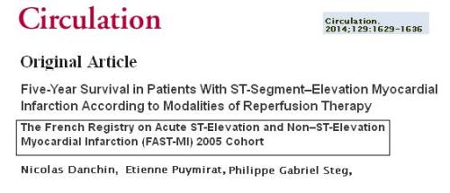
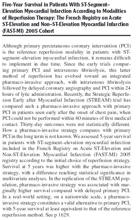
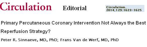
When false truths are synthesized to conceal a true myth . . . where will the poor myth complain ?Following are revered  facts  . . .  among the  “Guardians of   Cardiology” !

  • Primary PCI  is a greatest innovation  in modern day cardiology .Without this modality  most  STEMI patients will buy Instant  tickets to grave yard !
  • A cardiologist who intends to  thrombolyse  a STEMI is considered as a low quality cardiologist .
  • Streptokinase should have  no place in the crash carts of modern coronary care units.
  • There is nothing called “Time window” for rescue angioplasty.
  • VVI pacemaker  will convert an electrical problem of heart block into a mechanical one by depressing LV function .
  • Digoxin is an obsolete  drug even in well established cardiac failure with dilated heart.
  • Beta blockers not only fail to control  blood pressure smoothly , it often converts  a hypertensive individual into a unhealthy one  by it’s prohibitive side effects !


CokeMini_Happiness_03

  It costs just one Dollor !

Harvard school of Public health has  recently  published some  hard data about  the adverse effects of bad diet habits especially the impact of  soft drinks on long term cardio vascular health.

soft drinks and impact on healthAnother prestigious journal confirms it . . .

risk of soft drinks and coronary artery diseaseThe ultimate journal for the cardiologists Circulation reaffirms it .

soft drinks carbonated and coronary artery diseaseWe  have overwhelming evidence , prolonged use of soft drinks
increase CAD (Like tobacco ). Still , we haven’t added the customary caution on the bottles of these soft drinks ?

Why  ?

My guess would be, there are powerful forces in  modern society that would love to keep  the  threat of diseases high in the  human domain  so that all  stake holders  make a profit .

The poor , World health instigation(WHO)  and various public health forums  simply watch it .If they protest , they are at risk of sucked into the loop of deceit !

Failure to reverse public health hazards  may be a  lesser crime  in modern days  . . . But , here comes  a series of  advertisements in Indian media , that is a strong indicator that human  intellect and the sixth sense  is going nuts !

It suggests , Do you want  ever lasting happiness . . . Drink  it  . . . earnestly believe  in it  . . . and drink again  !

Coca-Cola-12

I was also told  a shocking fact that , The beverage war in India has  started and the soft drink giants have heavily Invested to take it into the rural markets .Their aim is to  provide  “health  for all Indians”  by 2020 !

Concluding question