Feeds:
Posts
Comments

Archive for 2008

Pulse deficit is a clinical sign wherein , one is able to find a difference in count between heart beat (Apical beat or Heart sounds ) and  peripheral pulse .This occurs even as the heart is  contracting , the pulse is not reaching the periphery.This can occur in few clinical situations .

1 . Atrial fibrillation.

2. Very early diastolic  ventricular  ectopic beats

3. Some patients with Pacemaker.

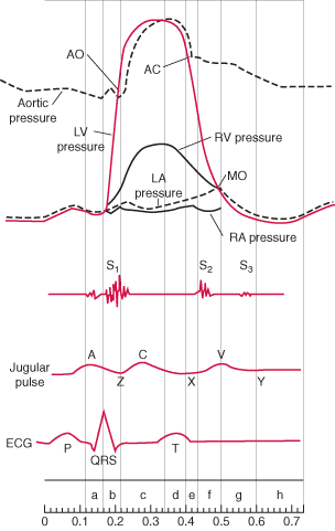
The mechanism is  , the ventricular contractions are  too weak and unable to open the aortic valve  (Or opens feebly*)  , but at the same time they are good enough to close the mitral valve. To open the aortic valve it has to generate atleast 60-80 mmhg pressure , while mitral valve closes even  as LV generates  8-14mmhg  .(LV/LA pressure cross over). So intermitently the  second heart sound  is missed while S1 is retained,  producing more heart sounds and less pulse count in the periphery. The S1 is either felt or heard at the apex but the corresponding pulse is missing . Further , this intermittent absence of  S2  results in totally irregular S1 /S 2 relation.


Why some of the contractions of LV is too weak to open the aortic valve ?

Because the RR interval varies , the ventricular filling also varies , diastole duration is constantly changing some of the diastole are too short  and LV hardly gets filled , as the LV force of contraction is directly decided by the LVEDV and LV  fibre length these contractions are too weak.

Other published studies

There has been some doppler observations ,where there is a midventricular LV blood flow reversal in atrial fibrillation that could  explain the pulse deficit . Mechanism of production of pulse deficit in atrial fibrillation: assessment by blood flow dynamics

Second Department of Internal Medicine, Kagoshima University School of Medicine.

 http://www.ncbi.nlm.nih.gov/pubmed/3221309

What is the clinical significance of pulse deficit ?

Currently there is no great clinical significance of this phenomenon. But an astute clinician will pick up this sign and it may indicate underlying LV dysfunction. In patients with PPM,  pulse deficit  suggests  pacemaker malfunction .Some patients with cardiac tamponade &  pulsus paradoxus  systolic  blood pressure falls too low to make the pulse feeble or not palpable in the periphery .This situation may mimic a pulse deficit if not recognised.

Dr.S.Venkatesan ,Madras Medical College , Chennai, India

* What is the evidence for intermittent absence or feeble Aortic valve opening in Atrial fibrillation ? I could find this from the book written by Harvey Feigenbaum. whom we consider Father of Echocardiography


Read Full Post »

The long standing controversy about diastolic heart failure is settled !

The perception that diastolic heart failure ( Now renamed as heart failure with preserved EF ) is less dangerous than systolic HF has been exposed by this land mark study by Owan TE, in 2006 (nejm) But unfortunately this information is not yet fully disseminated among the physician community. Hence this post, with due acknowledgment to NEJM & Owan et all.

Experts from the article

“The nosology of heart failure has been the
subject of much current debate, and some extreme
positions have been taken. The observation
that 22 to 29 percent of patients with diastolic
heart failure die within one year of hospital
discharge, and 65 percent die within five years,
is a reminder that we are facing a lethal condition,
regardless of its name. Owan et al. also
show that, in recent years, there has been little
improvement in survival rate among patients with
diastolic heart failure, in contrast to the improvement
in survival rate over time among patients
with systolic heart failure”

Have a look at the survival curve below, almost similar , surprise surprise ! DHF survival is not only worse ( in many ), than systolic CHF and further they respond poorly to treatment, compared to conventional systolic CHF .

Click below for the link to full text article

Short abstract :

Trends in prevalence and outcome of heart failure with preserved ejection fraction.

Cardiorenal Research Laboratory, Mayo Clinic College of Medicine, Rochester, Minn 55905, USA.

BACKGROUND: The prevalence of heart failure with preserved ejection fraction may be changing as a result of changes in population demographics and in the prevalence and treatment of risk factors for heart failure. Changes in the prevalence of heart failure with preserved ejection fraction may contribute to changes in the natural history of heart failure. We performed a study to define secular trends in the prevalence of heart failure with preserved ejection fraction among patients at a single institution over a 15-year period. METHODS: We studied all consecutive patients hospitalized with decompensated heart failure at Mayo Clinic Hospitals in Olmsted County, Minnesota, from 1987 through 2001. We classified patients as having either preserved or reduced ejection fraction. The patients were also classified as community patients (Olmsted County residents) or referral patients. Secular trends in the type of heart failure, associated cardiovascular disease, and survival were defined. RESULTS: A total of 6076 patients with heart failure were discharged over the 15-year period; data on ejection fraction were available for 4596 of these patients (76 percent). Of these, 53 percent had a reduced ejection fraction and 47 percent had a preserved ejection fraction. The proportion of patients with the diagnosis of heart failure with preserved ejection fraction increased over time and was significantly higher among community patients than among referral patients (55 percent vs. 45 percent). The prevalence rates of hypertension, atrial fibrillation, and diabetes among patients with heart failure increased significantly over time. Survival was slightly better among patients with preserved ejection fraction (adjusted hazard ratio for death, 0.96; P=0.01). Survival improved over time for those with reduced ejection fraction but not for those with preserved ejection fraction. CONCLUSIONS: The prevalence of heart failure with preserved ejection fraction increased over a 15-year period, while the rate of death from this disorder remained unchanged. These trends underscore the importance of this growing public health problem. Copyright 2006 Massachusetts Medical Society.

Other interesting article

Heart failure with preserved ejection fraction: dangerous, elusive, and difficult.

Eur Heart J. 2008 Feb;29(3):339-47. Nielsen OW, Køber L, Torp-Pedersen C.

BMJ editorail 2009

http://www.bmj.com/cgi/content/full/338/jan27_1/b52?ijkey=c7a29d35dc9d9dddf7d0e75c5b8d05014315c564

Read Full Post »

Is reciprocal ST  segment changes  occur  only in STEMI ? Can it occur in UA/NSTEMI ?   


                      Even   after   100 years of electro cardiology   the electrophysiological mechanism of ST elevation in STEMI  and ST depression in Unstable  angina is   still in the hypothetical stages. One popular theory   says that the   current of injury   as we see   as  ST  segment elevation  in surface ECG  is  actually   an illusion. It’s   apparently due to   constant negative current    pushing down the   rest of ECG segments. Ironically   the concept   of  reciprocal ST depression in patients who have  ST elevation is well debated  for over 3 decades and  is considered  a  settled issue. It   probably   represents , a  purely electrical phenomenon where the  tail end  of the  lead   picks up the opposite vector. Even   as   conflicts   continue to confront the basic electro physiological   concepts management    strategies   of   acute coronary syndromes is witnessing   great strides.
Aim
                                     We   hypothesized   if   ST depression occurs as response to ST elevation it’s logic to expect strong ST depressive forces should  possibly elevate The ST segments in the reciprocal leads .
In fact  we have seen this phenomenon in three distinct  clinical situations. 
1) ST   elevation   in posterior leads: Patients who present   with isolated   ST depression in V1,  V2 , V3  and  ST elevation in posterior chest leads V7, V8 .These patients were initially thought to have isolated posterior MI. But later the cardiac enzymes were found to be normal   indicating no myocardial necrosis   echo evaluation revealed wall motion defects in anterior segments rather than in posterior segments. CAG revealed critical   LAD disease .  This we believe a pure reciprocal ST elevation in the posterior leads to  a  ST depressive forces in anterior leads.
 2) Inferior  ST   elevation   with ST depression   in   V4- V6 : Few  patients who present with  infero    lateral STEMI   later  do not  evolve into  Q  MI but as a NSTEMI .The initial ST elevation  was found  be transient and  disappeared  much earlier,  while the  ST depression  lateral leads persisted.
 3) ST elevation in AVR   in high risk unstable angina :As   already reported in the literature,  we have seen  ST  elevation in AVR  in patients with  high risk unstable angina. This was   more often observed when there is > 3mm ST depression in V4-V6. The AVR  ST elevation  possibly   represents   the  reciprocal  vector.
    Conclusion
                                            ST elevation in certain   specific leads in   some of the patients with ACS,   could   be   a pure reciprocal   electrical phenomenon   to   dominant ST depressive forces in Opposite leads .  And hence   ST elevation in the surface ECG during early hours of ACS   should be interpreted more cautiously. The   sanctity   assocociated with ST segment   elevation   could  be  opened   for debate. 
 
                                     To down load full PPT  click on  the slide

//

Read Full Post »

                    Lateral myocardial infarction is not a common site when compared to anterior and inferior MI. But the lateral MI has some unique features, since it involves free wall of the ventricle.The laplace law mediated wall stress is more as the dyskinetic segments bulge with a long radius .Due to this,  lateral MI has a distinctly high risk for free wall rupture .Further pericardial rub is more common in thse patients.Ischemic mitral regurgitation and vulnerability to LVF is also more prevalent if the lateral wall is involved.  Generally lateral MI pateints have a turbulent and complicated course than a simple inferior or anteroseptal MI.

The angiographic correlation of  lateral MI is rarely reported in literature.

The following leisons are commonly observed.

1. Proximal  LAD with large D1  involvement

2. Isolated large D1/D2 disese

3.A left dominat LCX  with large OM1 disease

4.Large ramus disease

5.LAD total and RCA to LAD/D1 collateral pattern

Final message

If we encounter a lateral MI either alone or in combination with inferior/  posterior MI , it is better to manage these patients  aggresively with early triaging for CAG and revascularisation.

Read Full Post »

To download complete presentation click on the slide

“ WAVE RED FLAG” FOR ANTICOAGULATION, WHEN YOU ENCOUNTER MOBILE LV CLOT !

Venkatesan Sangareddi , G. Gnanavelu ,M.A Rajasekar, V.Jaganathan

Department of cadiology , Madras medical college , Chennai.

Formation of LV mural thrombus is one of the important sequel of STEMI. The natural history of LV clot is variable. Spontaneous dissolution often occur . Stroke and peripheral embolism, are other natural events by which left ventricle get rid of the clot. The morphology and the behavior of LV clot is determined by endogenous procoagulant and fibrinolytic mechanisms. Drugs administered in the peri infarct phase also play an important role. In current thrombolytic era ,the incidence of LV clot has come down. Once the clot begins to form over the raw area adjoining a dyskinetic segment, it follows the local hemodynamic factors , that determine the shape , size of the clot which varies from linear , layered , projectile or pedunculated.

Administration of oral anticoagulants remain the standard practice in patients with LV clot. It is prescribed , in the hope that it will prevent the progression of clot and prevent thrombo embolism . Whether, long term warfarin dissolve , regress or dislodge the thrombus is not known. We have observed the incidence of CVA is high in the first few weeks following introduction of oral anticoagulants . We report our experience in 8 patients, with LV clot in Acute MI . All patients were male . Age range 22-58 .All had anterior MI. The mean EF was 38%(28-43%) the mean size of LV clot was 1.4cm (7mm -24mm) mobility was graded with reference to independent movement parallel or perpendicular to the LV. 3 had highly mobile clot. 5 had relatively fixed clot. All were put on titrated warfarin. Two patients who had large LV clot with a stalk got dislodged after starting anticoagulation. The CVA occurred on 12 th and 14 th day after starting warfarin .The pedicle is probably the vulnerable point and is exposed to greatest risk for dissolution . On the other hand the 5 patients who showed relatively stable clots are attending to our cardiology OPD without any events . One patient who had a mobile clot , which got organized at 4 weeks , incidentally this patient had discontinued anticoagulants.

We conclude, oral anticoagulation has a potential to destabilise and dislodge a mobile LV clot in the early days following STEMI .Existing anticoagulation protocol recommends, oral anticoagulation for all patients who have LV clot. This need to be redefined. If surgery is not an option , temporary withdrawal of anticoagulation may be indicated in selected patients with LV clot, to facilitate organization of clot.


!

Read Full Post »

Coronary artery disese  predominantly  occur in the proximal segments of coronary artery.The fact that CAD is mainly a proximal disese , implies  that  clincal impact is likely to be more . But we now recognise distal coronary artery system is equally affected .But isolated distal CAD  is a not a common finding .We describe our analysis on the topic .

distal-cad-csi-2005

Click on the slide to download

Read Full Post »

Transient ischemic attacks are not exclusive to cerebral circulation.

Many such episodes can occur in coronary circulation also .

TIA of heart PPT presentation

Click here to download  tia-of-heart

Read Full Post »


Is it a crime to do a plain balloon angioplasty in 2008 ?

Plain balloon angioplasty,   the greatest  innovation in   cardiology  when it was introduced in 1977 in a Zurich cath lab , has now become an  ugly  word for most of the cardiologist !

Why this turn around ?  Has technology ,  really overtaken a great procedure and made it obsolete now ?

The answer is a definite ” No”

The restenosis which was the villian in the plain old angioplasty has never been overcome even today. Stents initally used as a bail out procedure during  abrupt closure , later it was used conditionally, followed by provisional stenting and now in 2008  we are made to believe  it is mandatory.

When we realised , bare metal stents are equally  bad (If not slightly better ) in arresting the restenosis drug eluting stents came into vogue with a big bang in 2002. It was projected as the ultimate breakthrough in interventional cardiology and  in 5 years the truth was exposed and it not only failed to prevent the restenois but also had a dreaded complication of acute stent thrombosis.

Now we know , metals  inside a coronary artery  carry  a life long  risk of sudden occulusion , and we talk about biodegradable stents (With poly lactic acid ).

 Common sense ( Unscientific truths)  would suggest

Plain balloon angioplasty still has a major role in our global  cardiovascualr population.

Since restenosis is the  only issue here, ( about 30% )  we can choose patients in whom even if restenosis is likely to happen  no major harm is done . A vast majority of chronic stable angina patients  fall in this category.

Aggressive lipid lowering with plain  balloon angioplasty has never been tested properly . In future also it is unlikely,  such trials will be done as it would be considered unethical . But that would be a premature conclusion.

The other major issue is the cost of stenting , the procedure of PCI/PTCA  has become unaffordable for most of the population in developing countries .The primary reason being the PCI without stenting is considered  ” A untouchable” . If only we remove this stigma from the cardiology community   a signiificant population will be benefited.

A patient with chronic stable angina treated with POBA ,if develop further angina after few years , he  is likely to get a recurrence of  relatively safe  stable angina.  While in a post PCI patient  any angina after the procedure becomes a unstable angina ( Braunwald classification)  and requires emergency care . Angina in a  stented patient is can not be taken lightly as  the the course of angina is unpredictable .

POBA in primary PCI ?

Many may think it is a foolish idea . It has been found many times,  when we rush the pateint to   cath lab after a STEMI  we are in for a surprise !. About 30% of times it is a very complex lesion profile  like diffuse disese,  tight bifurcation lesions , loaded with thrombus or a left main disese.

We fail to realise a basic  fact  , the  initial aim of primary PCI is to salvage the myocardium ,and the next comes the prevention of restenosis . It may even , be argued salvaging  myocardium is the only aim ! Myocardial salvage sould be done urgently . And even  removing the thrombus and opening a IRA can be suffice in a patient who is crashing on table.  Of course stenting can be done whenever possible. But for IRAs which has complex anatomy attempting a perfect stent PCI   (Some may require more than few stents)  as an emergency procedure invariably affects the outcome. One should spend  shortest possible time  inside the  illfated coronary artery. Prolonged manipulations within the coronary artery in an unstable patient  aiming at  longterm patency of an IRA  is to be avoided .The pending procedures can always planned in a next stage. 

Final message

So it is not a crime to think about plain balloon  angioplasty  in some of  our  patients  with acute or chronic coronary syndromes .  Hope Gruentzig  is listening from the heaven and hopefully agree with me !

Dr.S.Venkatesan, madras medical college, chennai, India .

Read Full Post »

LV clot formation is one of the important complications of acute myocardial infarction. Preventing this is difficult and managing this problem is still more difficult.Some of these clots are linear and laminar along the shape of LV apex and carry less risk of dislodging.

 While mobile LV clots , even if it is small can cause a embolic episode. Most of these patients have a significant LV dysfunction and they are candidates for early CAG and revascularisation. Even If the coronary anatomy is very ideal for a PCI these patients are often sent for CABG and physical removal of LV clot . If  only ,we have an option to remove these LV clots by a catheter based modality, we can offer them a totally non surgical cure.

This is not impossible,  considering  we are in the era of percutaneous implantation of prosthetic valve in Aorta ! The only issue is potential embolism into carotids and periphery .A temporary distal protection at the level of aortic root will prevent that .

Device companies shall produce one such exclusive catheter system to remove LV clot.

Dr .S.Venkatesan, Madras medical college, Chennai,India

Read Full Post »

Orthopnea is a classical sign of established CHF.

While paroxysmal nocturnal dyspnea is an early sign of cardiac failure,orthopnea is a late manifestation of cardiac failure .This symptom was mainly attributed to volume displacement from systemic venous to pulmonary circulation when the patient goes to recumbent posture.The exact mechanism of this has been speculative. Now with liberal usage of bedside echocardiography, we have found out there is postural variation in the diastolic function of the failing left venticle.

Many patients develop a restrictive ventricular filling pattern in recumbent posture (Grade 3 diastolic dysfunction). While sitting up some of them revert to normal or downgrade to grade 1 diastolic dysfunctionThis observation proves another fact that every patient with severe systolic dysfunction also has significant diastolic dysfunction at some point in their course of illness.

Read Full Post »

« Newer Posts - Older Posts »