Feeds:
Posts
Comments

In the early  20th century , Waller invented the ECG machine. Wilson  created  leads and  methods to record it. Eienthoven  formulated the concepts the electrical theory behind ECG.

Between 1940-197os one man ruled supreme in the world of electro cardiography .   He is Dr Sodi pallares from Mexico. His deep insights revolutionised  and helped  us  understand how the cardiac electricity is generated  and propagated  in various pathological states that is the beginning new age electrocardiography! It adds much to his  credit , as in those days scientists  from non American and European countries   were hard to come  in the  global limelight.

Some of his thought processes and Inventions

  • He laid the foundation  for deductive electrocardiography.
  • Applied  vectorocardiographic  principles to scalar  ECG and helped  understand mechanism of ventricular chamber enlargements
  • He was instrumental  to analyse  the genesis of current from the myocardium . He tried to reason out  the contribution  of  cell metabolism, hypoxia, electrolytes to the current genesis.
  • Polarising myocardium with GIK infusion STEMI was proposed by him
  • Finally he tried to incorporate the laws of thermodynamics into electrocardiography .

Please remember , Sodi pallare’s  conscience  is still largely unexplored .There are lots of hidden truths .We now know fever can influence qrs voltage and febrile illness can trigger  ventricular tachycardia as in  of Brugada syndromes.

Today’s youngsters can take a  few cues from this great man and enlighten the field of electro-cardiology

DOCTOR DEMETRIO SODI PALLARES

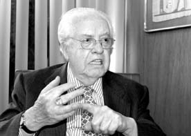
Demetrio Sodi Pallares (1913-2003 )

If  modern-day cardiologist is able to  interpret the  ECG by a cursory 3 second scan of  strip of waves  , we are greatly  indebted to the knowledge imbibed by this great man from Mexico !
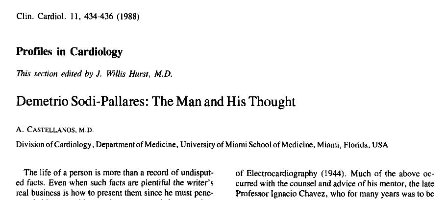
Sodi pallares

Reference

http://onlinelibrary.wiley.com/doi/10.1002/clc.4960110616/pdf

Sodi-Pallares D,Medrano GA, Bisteni A, Ponce de Leon J:
Deductive and Poly-paramerric Elecrocardiography.Instituton acionalde
Cardiologia de Mexico, Mexico,DF1970,VII,VIII
Sodi-Pallares D, Testelli MR, Fishleder BL, Bisteni A, Medrano GA, Friendland C, De Micheli A. Effects of an intravenous infusion of a potassium-glucose-insulin solution on the electrocardiographic signs of myocardial infarction: a preliminary clinical report. Am J Cardiol. 1962;9:166–181


mid diastolic murmur

mid diastolic murmur in  sinus rhythm

With the onset of Atrial fibrillation

  1. The  first heart sound becomes variable in Intensity (Soft to loud in pliable valve / Soft to softer in calcified valve )
  2. Opening snap continue to occur as long as the  valves are  pliable and noncalcified  .The timing may vary but Intensity remain same .
  3. A 2-OS interval is usually less influenced by AF  as it is primarily determined by mean LA pressure at the onset of diastole which has little variation beat to beat .
  4. Length of the diastolic murmur varies . In Short cycles  MDM  can be  very brief  or even in audible.
  5. In long cycles MDM will be distinct.
  6. A late diastolic murmur in long cycle indicates severe MS.
  7. Even pre-systolic accentuation may be appreciable in some of the long cycles .

Link  to  Echo Image of  Mitral stenosis .

https://www.youtube.com/watch?v=3qvOMwOshg4

How to assess the severity of  MS in the presence of AF ?

  1. Presence of AF by itself indicate presence of severe MS  in most .
  2. A2-OS interval  may be useful
  3. Concentrate on long cycle .Look for ( rather listen !)  for late diastolic component of  MDM .If  you hear it ,  MS is usually severe

Other signs  are often useful

Low volume pulse

Inconspicuous LV impulse

Presence  of  any significant Pulmonary hypertension

Squatting is a simple physical  maneuver that can  be done in bed side.

  • Squatting  increases  systemic venous return.
  • Raises aortic after load and SVR

It is  ideal to do  “Stand -Squat -Stand” sequence to appreciate the attenuation during squatting and augmentation during standing

squatting dynamic auscultation mvps mitral valve prolapse

Hemodynamics

MVPS-MR is  a pre load (LV volume )  dependent phenomenon.The degree of prolapse is inversely proportional to the LV size.

Squatting increases the venous return and after load both tend to  increase the LV volume. More blood in the LV  , means mitral leaflet floats much closer to the  mitral annulus . Hence the  force of LV contractility is not only  less on the prolapsing  leaflet , it  reaches late ,  hence the click is delayed  and murmur is short (It may be less intense as well * )

(* Squatting increases aortic after load hence the murmur of MR  may  get amplified .)

Reference

We know  new onset LBBB  creates considerable anxiety . We  experienced a  reverse situation recently . A 72 year old  man who is known to have chronic LBBB  for over  5 years came  to CCU with vague  chest  discomfort .

His   ECG  was  perfectly normal . . . every one  was  curious !

My ECG always looked like this doctor  !  Now you say it has normalised and you say it concerns you  ! I am really worried  doctor  !

What does it mean doctor ?

Cardiologist : I do not know . Any sudden change in rhythm even if it is from abnormal to normal is to be given importance .

Patient : Is  the  going bad ?

Cardiologist :  I do not know

Patient : Should I  get admitted ?

Cardiologist : I think so  but you need to undergo few blood tests and repeat an ECG .

Patient : Oh  what ?  you  are not sure either !  Are you not an expert in heart  disease doctor ?

Cardiologist : I think I am . I wish I have an answer to  your question .

Follow up

This patient was admitted in intermediate care ward and observed for 12 hours .

His enzymes and Troponin were negative . Echo showed normal LV function .

He was discharged later and adviced  a stress test .

What is the the mechanism of normal ECG  here ?

Intermittent LBBB due to rate dependency is common .But this  man  had persistent chronic LBBB for > 5 years which got normalized .That mystified us !

Can transient ischemia of left bundle  accelerate  the conduction ?

Can there be a small r wave in V1 and V2 in LBBB ?

Yes .

Though we expect the  reversal of septal depolarization that will   extinguish  the initial r in v1 to v3 . It is  preserved in  many. Hence the  presence of small r in v1 to   v3 does not rule out LBBB.

  1. The commonest explanation given is un-masking of RV free wall forces which is   normally  masked by early LV forces .
  2. Another possibility is the   orientation of septum  in pathological states.
  3. Third possibility is  “r” may  actually represent  the  septal q waves as in LVH or old AWMI  .(Counterpart of small  q in lateral leads )

Normal sequence of  S2 split is A2 followed by P2. This split is heard only in  inspiration . The split is due to pulmonary low vascular impedance and is represented by the hang  out interval (40-80ms) . Inspiration transmits the low intra-pleural  pressure into pulmonary vasculature  and split widens. In expiration the reverse happens and split narrows.Normal human ear does not recognize the normal expiratory split.

If the split S2  is well  heard in expiration especially in sitting posture it is abnormal .

What is paradoxical split of S 2?

If split occurs and is well heard only in expiration and  becomes single in inspiration , It is classical of paradoxical split.

What is the paradox ?

In physiological conditions  S2  splitting occur during inspiration and become  narrow or single during expiration.

In paradoxical split ,  the physiological  behavior with reference to respiratory phases is lost and it can even become single in  inspiration .(It is also reversed  because A2 follows P2 )

s2 splitting second heart sound split a2 p2 paradoxical split reversed split

Conditions causing paradoxical split of S2 .

  • LBBB
  • RV apical pacing .
  • Aortic stenosis
  • Chronic sever AR

Japanese are the pioneers in CTO reopening .(I understand they do less   CABG surgeries  for  religious reasons ) CTO is the ultimate test for cardiologist patience .  it may  take  hours to open up a CTO (or even to abandon it .)  Here is a  success prediction tool from Japan .

cto score success in chronic total occlusion

j cto score  sheet

Source courtesy  : JACC: Cardiovascular Interventions Volume 4, Issue 2, February 2011

Reference

http://www.sciencedirect.com/science/article/pii/S193687981000912X

When I asked this seemingly simple  question to my cardiology fellows , I found they struggled  to come out with a proper  answer .I hope this will  make the  issue simpler .

Why the onset of PAH in VSD is early and late in ASD ?

Though number of factors are involved in the genesis of PAH , the single important reason  is  behavior of pulmonary circulation  especially the pulmonary arteriolar muscle .

Normal pulmonary vasculature losses it’s muscle rapidly after  birth and the pulmonary vascular resistance (PVR) falls to the adult level by 6 months .(Bulk of the fall occur in first 60 days) This is the same time the RV dominance is lost and RVH regresses . This also coincides with peaking of  left to right  shunting peaks and may result in cardiac failure .

Though  both ASD and VSD shunts are  highly dependent on PVR ,   VSD  shunting has more muscle power namely the LV contractility  , hence  VSD shunt is established  much earlier   than ASD . This can be ascertained in bedside as  VSD murmurs are heard even within 30 days while ASD is silent for many moths or even years . (Does not apply for Primum defects)

ASD  shunt rarely  meddles   lung  maturation process .(Maturation here means loss of  pulmonary arterioloar smooth  muscle -also  called as Involution  )  This vital  initial period lasts up to 6 months of life .VSD  interferes with this  involution of pulmonary arteriolar smooth muscle .( Please note near complete  involution still can occur in small VSDs with very little shunting )

In large VSD the PVR  will never ,  ever fall to normal levels  and   making it easier for  progressive vascular changes  that occur in  untreated large VSDs that  lead to Eisenmenger syndrome

*Please note  ASD can also reach that stage but it takes many years as the pulmonary vascular resistance has to raise from  very low levels which was made possible by complete involution of pulmonary vasculature .

It is obvious   AP window , PDA  express more  powerful left to right shunts which are associated with very high PVRs .

Final message

A simplified version of answer

Version 1

In VSD  the onset  of left to right  shunt occurs early even within 3 months of life  since VSD shunt is augmented by LV contraction . This is the crucial time of lung  vascular maturation   which gets interfered with  .ASD shunt is  established only after the pulmonary vasculature  involutes .This explains early onset of PAH in VSD  and late in ASD .

Version 2

ASD shunting is primarily depend on RV compliance which is high in early infancy so it takes time to establish the shunt .while VSD shunting does not  depend upon this RV regression.

* Please note  regression of  RV  dominance and compliance is directly dependent on maturation of lung.

** Note these  explanations are not absolute .Some of the complex forms of ASD and intrinsic vascular injury of pulmonary circulation (Various  fetal distress )  can progress into accelerated pulmonary arterial hypertension

Reference

  1. Excellent discusions are available in old edition of Moss and Adams
  2. Rabinovitch has done pioneering work on this topic .
  3. Robert Roberts text book of Adult congenital heart disease also explains it succinctly

Dr Shirely Smith  from charring cross hospital London  wrote this masterpiece  in BMJ in the year 1962 . He was doing a research about the origin of angina like pain in patients who had  upper GI disease or disorders of cervical spine .He found a hidden invisible neural link between heart and it’s neighboring viscera. What he  referred it as linked angina . It links the  pain from ,Esophagus,  gall bladder ,  duodenum ,  cervical spine to the  heart .

This article I  consider as one of the  all time classics in  clinical cardiology . Here is the link for linked angina (Courtesy of BMJ)

linked angina atypical angina abdominal angina  shirley smith cornelio papp 2 bmj

linked angina atypical angina abdominal angina  shirley smith cornelio papp bmj

High lights ( Inferred )  from the  article

We know angina typically occurs on exertion .If it occurs at rest we call it as unstable angina .

Can it occur at rest other than unstable angina ?

Yes it can . ( Post prandial ,Nocturnal, emotional etc)

Can the  heart be the referral site for visceral pain ?

Yes .It seems so .

Can visceral pain be trigger for  developing true angina ?

Again possible . A Patient with documented CAD  develop  a true esophageal pain it is likely  to  induce a sensation of  angina  rather than abdominal pain .Similarly , cervical pain may represent a masked angina in a patient  with active cervical spondylitis .(Homing in of angina to the nearest non cardiac culprit )


Final message

Those were the times when the brain worked more than hands . Common sense prevailed over machine sense .This article argues for a  big debate about the origin of so called atypical angina in a patient with multiple common visceral conditions.Even 50 years later we have little clue  about alimentary -cardiac neural spill over !

Reference

http://www.bmj.com/highwire/filestream/276395/field_highwire_article_pdf/0/1425

Postamble

Today we live in a complex and confusing and commercial  medical world .We have atleast  a dozen chest  pain triaging protocols in ER . Still errors are  rampant. Errors are acceptable . . . but this one was an absolute  shocker  . . .  “I know a  patient with vague chest/epigastric  pain  , non specific T inversion ,  documented gall stones , landed in cath lab not by accident but  by meticulous planning !”

chronic total occlusion cto tips and tricks

Answer :

While each one of the above factor appears very much important  morphology of the lesion is the  clear winner  ( Which includes , the content of the lesion , hardness , micro channels , thickness of the proximal and distal caps, the length and   tortuosity   of the CTO     ( which is invisible ) the collateral status will ultimately determine the success)

It is becoming increasingly clear  cardiologist expertise is getting less and  less important .

Finally ,  it must  be told to our  younger generation of cardiologists , crossing a  CTO and deploying a stent  is not synonymous with success .It should result in long term sustained distal flow and make a significant impact on the patients symptoms (If at all any !) and survival.