Right from the days we entered medical schools, severe mitral stenosis was defined by less than 1 cm² MVO by echocardiography. It has been sacredly followed in most countries where RHD is prevalent. But, as western data (often derived with eastern patients) redefined the cut off for severe MS to 1.5 cm² in recent years ,.Many of us are amused, rather confused.
Severe MS : Why it was made 1.5 cm² ?
I don’t know. Though, we in India, may not fully agree with this re-definition, there could be some good reason behind this. The bottom line is, we should not miss a functionally significant mitral stenosis, strictly adhering to the anatomical 1 cm² cut-off. After all, we all know, with years of experience in echocardiography ,in a funnel-shaped degenerated mitral valve, we can get whatever MVO we desire to report ! Same story for pressure half time, especially with tachycardia and little mitral regurgitation. We also realize the relationship between gradient and MVO is not at all linear.
So, what shall we do, when numbers play juggler game with us ? Let us go to the basics and learn to make a multi-parametric decision process.

Certain tips in assessing MS severity
1.When there is discrepancy in MVO always go with planimetry. If calcium is there MVO can be problematic; one may add a color flow in short axis to define exact flow borders in mid diastole.
2.If Doppler has multi phasic, humps we must take slopes that occur later in diastole for pressure half time. This is because, the initial rapid filling is influenced by early LV suction forces that may underestimate the MS severity.
3.In AF, always hesitate to diagnose moderate MS. Use a long cycle to measure pressure half time.
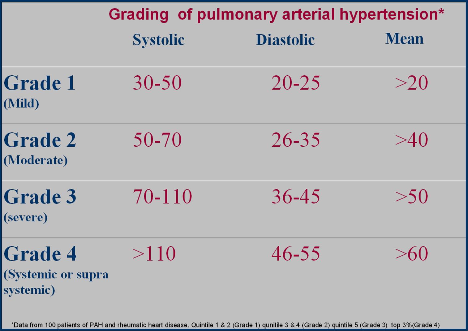
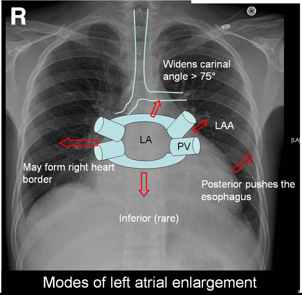
4.Finally, always have a look at the degree of pulmonary hypertension, LA enlargement, and sub-valvular disease before deciding if it is moderate or severe.
5.In pregnant women this one and a half MS is going to be really, really tricky to make a decision to Intervene. A fair rule of thumb is, If the mother crosses 20-24 weeks, whatever be the MVO , it is generally a good hemodynamic sign . (Except for the transient high risk period of the post natal uterine involution push and enhanced preload.) Having said that, we must realize , we are living in a near foolish and unrealistic era of demanding zero maternal complication even in high risk pregnancy. Many of us are compelled to do a seemingly unnecessary & risky PTMC during pregnancy due to the collective anxiety of cardio-obstetrics-patient team or a potential legal threat .
Other options
Dobutamine stress is an option , but many are hesitant to do.Stress testing that can be as simple as leg raising and bending 30 times while doing echo and check the gradient(?>20 mmHg)
What is new in hemodynamics of MS ?
There is something called low gradient severe MS (as in aortic stenosis). One must be aware of this. This often occurs in atrial fibrillation, where LA struggle to generate sufficient reservoir-stretch triggered flow gradient The other reason being presence of sub-clinical LV dysfunction hiking downstream pressure attenuating the gradient.(El Sabbagh A, Low-Gradient Severe Mitral Stenosis J Am Heart Assoc. 2019)
Final message
Though we are used to 1 cm² MVO cutoff , we can’t hang on to it strictly. Mind you, even a small gain in orifice can give a dramatic improvement in functional life. We have come across instances of splitting a mitral valve from a patient .8 to 1.5 cm² (technically they are still in severe MS), walking home briskly with a thankful smile. Same thing may happen for a patient with apparently moderate MS, right !


