Feeds:
Posts
Comments

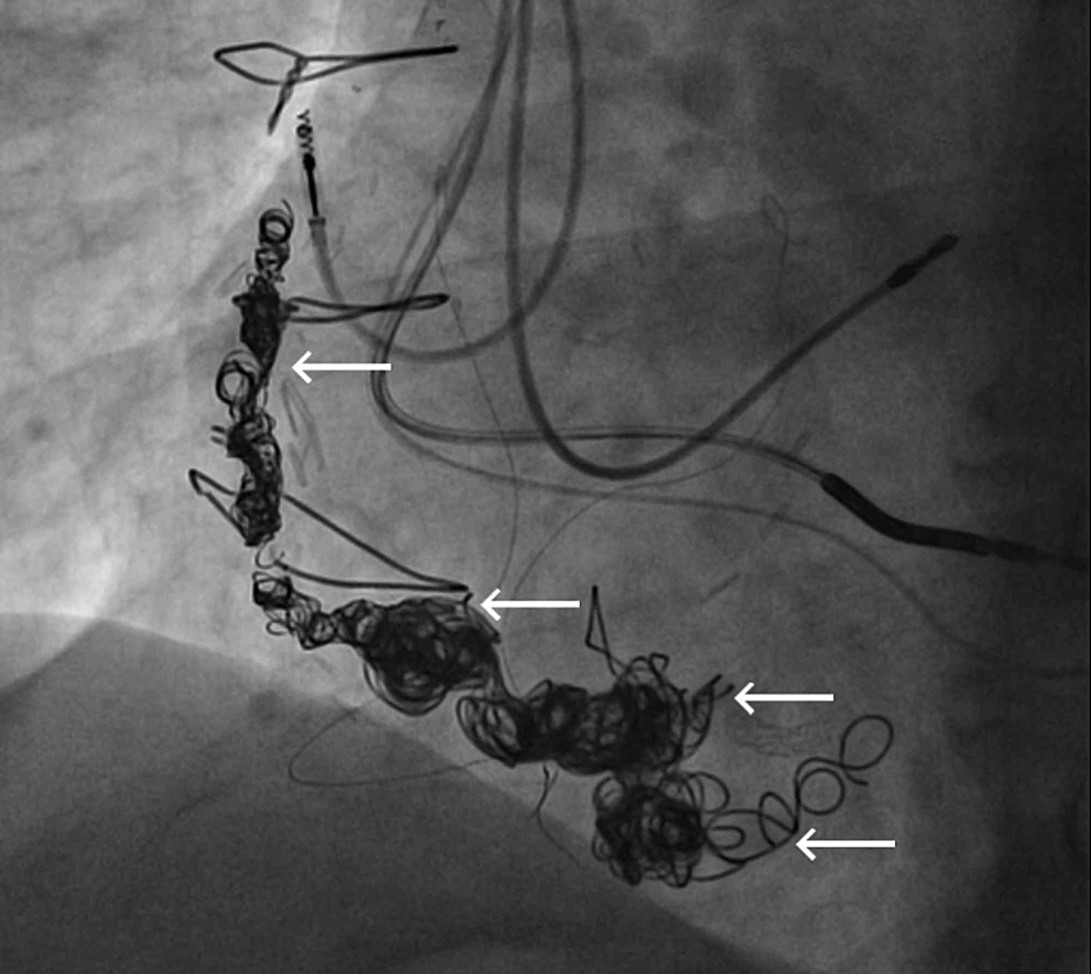
The commonest cause* for repeated entry of right radial catheter to descending aorta is not due to any anomaly. Most times,it is just a skewed angle between right brachio-cephalic artery with Aortic arch, that deflects the catheter to the descending aorta . Just make sure, aortic root is entered with a deeply held inspiration.

*Anomalies of the aortic arch, aberrant right subclavian, Kommerell’s diverticulum, vascular ring must be kept in mind.


Postamble: A true abnormal course

Though, It might appear prudent to avoid the radial route when encountering anomalous subclavian arteries, the reality is different and adventurous. We have acquired great expertise and successful PTCAs have been done through these tortuous vascular highways.

This is a case report from Dr H.S. Isser, Gunjan Garg, from Safdarjung hospital New Delhi. 

A successful PTCA through arteria lusoria : The right subclavian connect to the descending aorta, distal to the left subclavian at the level of ductus arteriosus. and pass retrotracheal and retroesophageal before reaching right arm. Image source and courtesy: H.S. Isser, Gunjan Garg, Arteria lusoria: A challenge for transradial coronary interventionist, IHJ Cardiovascular Case Reports (CVCR), Volume 4, Issue 1, 2020,


One of the topics rarely discussed in heart failure is  CAD as a contributory factor in HFpEF.

This is a copy of the presentation done at the ECHO India Annual scientific meet  2019 at Kolkatta. India.


Will try to find out the recorded version with audio. Here is a GIF run through.

PDF version Download here

Embryology

Sinus venosus ASD (also referred to as SVC ASD)  is a defect in the failure of the sinoatrial orifice to lateralize completely to the right side during atrial septation.Left venous valve, as well as the septum secundum, fails to fuse with the roof of the atria creating interatrial communication. During this process, the developing pulmonary vein overshoot to the right side making PAPVD a mandatory add-on defect. (Harley ,Thorax 1958 ) It can be referred to as embryonal venous migration defect at the level of RA. In the same sense, it is not a true defect in IAS but a defect in septation between SVC/PV. It may also be referred to as unroofing of RUPV. The so-called Inter atrial communication actually is the confluence point of RUPV/SVC/RA.(See TEE images below) 

We know, in SVC ASD-commonest associated anomaly is PAPVC . It is not an ideal term to use though, instead, it is encouraged to use the term PAPVD (drainage) . Technically true PAPVC can not be connected to RA cavity as PV can connect either to cardinal or vitelline vein only. This distinction is helpful when we search for additional PAPVCs cranial to SVC. Sometimes we might recognize this error only after closure of SVC ASD. 

Should we close SVC ASD ? How do we close?

Age, natural history, symptoms, the quantum of shunt will answer an occasional troubling query , “should we close it at all? Surgery is the standard approach till now. What makes device closure popular? Two reasons 1.Patient /or parent’s fear of surgery  2.Cardiologist’s urge for innovative Interventional procedures. (The fact that a simple covered stent will do the job is too tempting to make an attempt) However,please note, the procedure is not at all simple as one would Imagine.

Anatomical prerequisite for device closure 

The defect must fulfill some critical anatomical essentials.  

  • It should be an isolated defect.
  • RA should not be grossly enlarged
  • Re-routing of RUPV to LA should be possible 
  • A significant circumference of RUPV should be committed to LA.
  • There should not be downward extension involving septum secundum making it an SVC + OS ASD 

Technical issues

  • Self-expanding vs balloon expanding stent (Anyone may be chosen)
  • Stent flare-up SVC RA junction crucial
  • Must ensure  RUPV doesn’t get compressed with device.

  • Forces that hold the SVC end of the stent is very important. Sometimes It may require a second proximal stent just to prevent migration of the first stent.
  • Live LA pressure and RUPV monitoring may be critical to recognize PV ostial compromise. For this, a transeptal puncture may be required (Ironically creating another mini ASD !)


Finally, and most importantly follow-up is mandatory with device closure since the stent is on the venous circuit as RA, SVC thrombosis expected. (Anticoagulation protocol not clearly  defined as of now )

Final message 

Device closure for SVC ASD is a good Innovation. A perfectly delivered covered stent at the RA/SVC junction will do the trick. However, In my  opinion, surgeons do a neat(More complete)  job It is time-tested. Single or double patch or warden procedure may be done.(Ref 2)

Reference 

1.Hinnerk Hansen, Phuoc Duong, Salim .M. Jivanji, A. Qureshi, Eric Rosenthal Transcatheter Correction of Superior Sinus Venosus Atrial Septal Defects as an Alternative to Surgical Treatment J Am Coll Cardiol. 2020 Mar, 75 (11) 1266-1278.

2.Warden HE, Gustafson RA, Tarnay TJ, Neal WA. An alternative method for repair of partial anomalous pulmonary venous connection to the superior vena cava. Ann Thorac Surg 1984;38:601-5. 


https://doi.org/10.1016/j.pedneo.2019.06.013


https://doi.org/10.1016/j.athoracsur.2020.03.113

Mitra clip is a small metal device that is delivered percutaneously, to clip the incompletely coapting (closing) mitral valve. It was first introduced to treat degenerative mitral regurgitation. It is an interventional imitation of the famous edge to edge Alfieri stitch repair.This procedure in fact converts the single mitral valve orifice into two. In the process, curtails the regurgitation jet orifice significantly. Though the technique looks nice and simple to hear, lots of per and post-procedure issues need refinement. Conceptually it is ideal in primary disorders of the mitral valve. (Read EVEREST 2 criteria for optimal patient selection)  There have been more than 60000 Mitra clips implanted worldwide wide. Thanks to Abbot.

In secondary MR (due to LV dysfunction) Mitra clip has shown mixed results( MITRA-FR not much benefit, COAPT -Did show benefits)

Now, what about Mitra clip as a remedy for rheumatic mitral regurgitation?

This is the question everyone likes to ask. Now we have some interesting breakthroughs. Dr. Ningyan Wong from the National University of Singapore reports probably the first case (Ref 1) . The videos are reproduced with the creative commons license.


Note the classical thickened AML in rheumatic mitral regurgitation.


TEE showing severe MR


Post Mitra clip : A real surprise to note near-total abolition of regurgitation. (This really is good news for the rheumatic mitral valves )

Technical issues

  • Should be isolated MR
  • P2/A2 scallop clipping is the key to success. 
  • The thickness of the leaflet limits the success (Grasping the leaflet will be difficult)
  • Clip Induced mitral stenosis is a distinct risk.

Potential role and future

RHD forms 90 % of valvular heart disease in a country like India. The incidence of Isolated MR in both acute rheumatic fever and chronic  RHD are substantial. If only we refine the hardware and technique to suit these thickened rheumatic valves, Mitra clip is expected to make an impact in this unique group of patients where surgery can be avoided or at least postponed

Though we would very much like to do such a trial in our place, logistics has effectively precluded it. I wish some large centers like AIIMS New Delhi or PGIMER Chandigarh and others can take this concept to the next level.  


Reference

1.Ningyan Wong, Peilin Cheryl Marise Tan, Zee Pin Ding, Khung Keong Yeo, Successful MitraClip for severe rheumatic mitral regurgitation: a case report, European Heart Journal – Case Reports, Volume 3, Issue 3, September 2019

Two queries that linger in the medical profession for a long. I am afraid they aren’t addressed specifically by the stakeholders.

Question 1 

Foe a moment, let us assume there is no option to answer all three are equally important. Medical colleges are supposedly Godly places where high-quality noble professionals would germinate, let into the community thereupon, to heal the ill and suffering. The teaching faculty has a huge responsibility. They must ensure that students are transformed into responsible caregivers in the first place. They should be made to understand that the knowledge they acquire has a short half-life and medical education is all about continuous learning and unlearning. Unless teaching and research are morally genuine and scientifically perfect, the things we do in the name of patient care is going to be redundant. Hope you got the answer right!


Question 2

Now that, we got the answer to the question 1, here is a more difficult question. 

Out of the four, only one addresses the skill and expertise. The rest of the three generally happen away from the bedside. The answer is strikingly clear I guess.


Postamble

I agree the answers to these queries can be extremely sensitive, and contentious for many of us. Little deeper lies the truth. Hope, the quote from the much-stigmatized father of modern medicine  “Primum non-nocere” will help find the answer. 

Just roll over the virtual marker along the coronary lesion to get the underlying flow ratio. Blue is an absolute normal segment. Green is ok, orange and red slow-moving coronary traffic jam zones. it’s just like drawing a google map showing life traffic. No wire, no adenosine FFR comes inbuilt in every angio shot. Looks great Isn’t it? This is called QFR. Quantitative flow ratio derived from routine coronary angiograms. It can also guide us to find the optimal sites of both proximal and distal stent landing zone in the best physiological manner.

Which company makes this ?

Any studies done with QFR ?

FAVOR 2 study was reported in TCT. This modality is expected to evolve.

Final message

Whenever possible every anatomical lesion in the coronary should be substantiated by physiological parameter and possibly coronary Imaging to know plaque morphology and vulnerability. Though it is wishful thinking, still for all logistic reasons, most of the real world stenting will be based only on the blind anatomical luminogram.

At this point, please let me utter a non-academic hyperbole. Even a casual query to your beloved patients about their true symptoms and exercise capacity shall make these ultra-modern coronary physiology studies redundant in many. A well-performed and well-interpreted stress test is a good, objective, non-invasive indicator of coronary flow across lesions. It is wise to keep this as a basic clinical foundation in the evaluation of CAD, even as we continue to learn and forget half evolved modalities with rapid expiry dates like FFR, IFR, CT-FFR. QFR shows some promise though. Please watch for next in line coronary physiology – OFR, Optical flow ratio from OCT run through.

Reference

Bicaval view is an Important TEE view to visualize, the LA, IAS, and right atrium. I used to have some trouble getting oriented to this view. Hence this post. It is obtained in the 90-120 degree view at the mid esophageal position. Imagine the patient is lying on his left side and the probe comes from above down between the spine and heart to the LA from within the esophagus. This is the best view to see IAS in the profile.(Subcostal TTE can also do it) Note how the LA hugs the right atrium which is actually an ill-defined (In TEE I mean) common meeting point of both IVC and SVC. Also important is the relationship of RUPV with SVC & the horizontally running RPA sitting right over the top of LA.

The relationship between RUPV and SVC is crucial in device closure of large ASD, especially in sinus venous defect.

Clinical Importance of this view

  • Very useful in ASD rim morphology especially in the posterosuperior rim.
  • Delineates clearly the defect boundaries in SVC ASD.

Sinus venosus defect: Image source not known. Thanks to the creator.

  • This view doesn’t miss even the smallest PFO (With Contrast )
  • Can be used to guide IAS puncture in structural heart Interventions.
  • IVC /SVC mass extension into RA well visualized.

RA myxoma attached to septum: Image source -Michael Essandoh from Research gate

Final message

Getting oriented to TEE  planes and images is so useful in structural heart interventions, like TAVRs, mitral clip, LAA occluder, tandem heart, valve in valves, etc. It is indeed a tough exercise and requires re-learning of cardiac anatomy with fluoroscopic overlay*.I wish, I go back and sit with first-year medical school students and start all over again.

*Current hybrid cath labs do provide Echo/Fluro co-registration, still it demands core 3D anatomical Imagination.

Cardiac myxoma is the most common primary tumor of the heart that presents as mitral inflow obstruction/ regurgitation often with a systemic presentation. It can be either familial, syndromic, or sporadic. Excellent imaging is possible and diagnosis has become straightforward. Surgery is the specific treatment,

What information do the Surgeons need? 

Size, attachment to surrounding structures is the key. The myxoma origins most often in IAS and defining its attachment is crucial. Mitral leaflet distortion, Injury ( and even attachment) is possible. It is helpful for the surgeons if we let them know the mechanism of mitral regurgitation prior to surgery. Echocardiogram including  TEE is sufficient in most. MRI may add some more info. The aim of surgery is to remove the tumor mass completely.

Is myxoma a completely benign tumor?

Another issue is our poor understanding of the recurrence of myxoma. Why should a benign tumor be recurrent? If recurrence is a feature to be counted as a sign of malignancy, myxoma can be definitely a suspect. There seems to be a catch. It is invasive, locally recurrent,  still not malignant. (Whether sarcomatoid degeneration happens is not known. Most pathologists deny this) The problem is, still we are not clear about the cell of origin of this tumor. All that we know is its origin mesenchymal stem cell.


Note 50 % tumor mass enters the left ventricle with diastole. No wonder, as the tumor plops with diastolic cardiac cycle a high-pitched sound simulating opening snap followed by an MDM perfectly mimics rheumatic mitral stenosis. An MR murmur is equally common.

Common sites of recurrence

  • Interatrial septum
  • The atrial surface of anterior mitral leaflet 
  • From contra-lateral atrium rarely

The mechanism of recurrence is either due to incomplete resection or due to its multifocal origin.

4 chamber view showing what appears to be a small narrow pedicle attaching to IAS. Please note echo imaging can be deceiving. Surgeons must inspect the mass in toto before taking the decision to excise IAS or not

What the surgeon needs to do?

The aim of surgery is to remove the tumor completely. It’s painful to diagnose recurrence and subject the patient to another surgery. (We encountered a sorry situation recently) So, when we remove the tumor we should ensure sufficient clearance with normal tissue.Biatrial approach is preferred by some surgeons.( Ahmet Yüksel  Braz. J. Cardiovasc. Surg. vol.31 no.4  July/Sept. 2016)

If the tumor is not well delineated, it is better to remove a significant area of IAS along with tumor mass and subsequent patch closure. Recurrences in AML and contralateral atria is unfortunate and can’t be predicted.

Further, the mitral valve is to be inspected and functionally tested. Minimal repair work or even rarely replacement might be necessary.


TEE imaging of LA myxoma. Note how fragile the edges of the tumor looks. It explains the high incidence of tumor embolus in this condition. Also, to be noted is the forceful impact of the tumor mass on AML that predisposes chronic mitral valve damage.

Final message

Referring a patient to a cardiac surgeon is not a bland ritual. (Have seen many single line referrals such as triple vessel disease referred for CABG) A well-informed interaction by the cardiologist with the surgical team and a possible per-operative echo consult, especially in rare surgeries will bring the best for the patient.   

Reference

1.Reber D, Birnbaum DE. Recurrent cardiac myxoma: why it occurs. A case report with literature review. J Cardiovasc Surg (Torino). 2001 Jun;42(3):345-8. PMID: 11398030