Feeds:
Posts
Comments

Posts Tagged ‘nstemi’

T waves attract less  attention in STEMI ,except for the  fact   tall T waves  implies   hyper acute phase of  STEMI.

What is the duration of hyper acute phase ?

  1. Few seconds
  2. Few minutes
  3. An hour
  4. Few hours
  5. Any of the above

Answer

No one exactly knows  .It can  be highly variable .  So , 5  could be  the correct answer .  

 * Most importantly  hyper acute phase  need not occur in all patients with STEMI as suggested in experimental models.

Some  observations in T wave behavior in STEMI

Mechanism of hyper acute  T waves

It is the pottsium channel dynamics.Transient intracellular hyperkalemia  is thought to be responsible.

T wave as marker of  reperfusion

Inverted T wave in precordial leads are a good marker of IRA patency  especially in LAD

Slowly evolving STEMI

This is relatively  new concept . STEMI with a prolonged hyper acute phase  ,  ie ,  T waves ” dilly dallying”  for hours or even few days have been recognised. (This was  refered  to pre-infarction angina in the past )

This sort of T wave behavior makes it difficult to diagnose STEMI.Enzymes will help , still  thrombolytic guidelines  demand us to wait till ST elevation to occur. This is  unfortunate .But as physicians we are  justified to thrombolyse tall T waves with a clinical ACS .The other simple solution is to shift the patient to cath lab to find what exactly is happening in the LAD ! 

Now , what is new about  T waves in STEMI ?

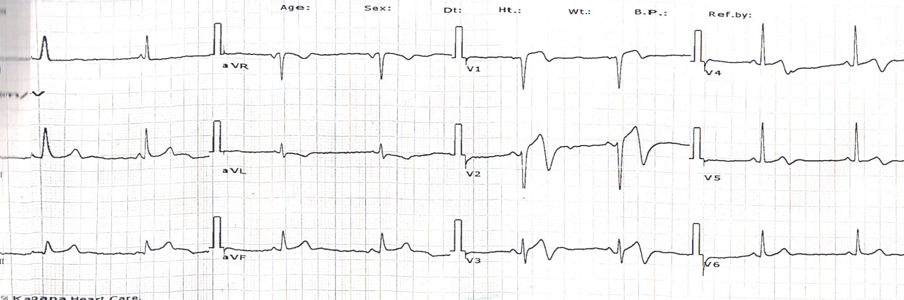
It is  the localizing value  in LAD infarct

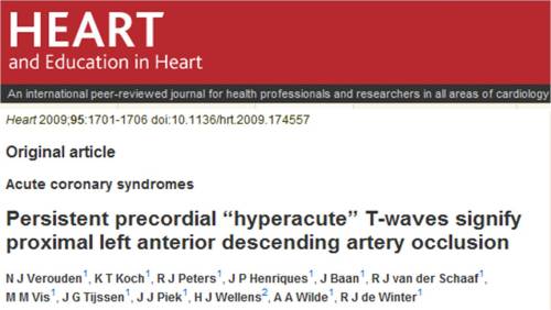
A tall persistent  hyper acute T wave  helps us to localise a LAD lesion .This paper from Netherlands ,  clearly  confirms this observation. The study was done from a primary PCI cohort,   a perfect setting to assess the  T wave behavior  in the early minutes /hours of  STEMI .
Other mysteries about T waves in STEMI

Does hyperacute T waves  occur in infero-posterior STEMI ?

I would believe it is very rare .Our CCU has not seen any tall T waves in inferior lead. Further analysis of the  data from the  above study could answer this question .

How often a  hyperacute T waves transform into NSTEMI ?

This again is not clear.Most of the hyper acute T will evolve as STEMI .But  , nothing prevents it to evolve as NSTEMI a well . After all , a hyper acute T   MI can  spontaneously lyse in a lucky few , ( Who has that critical  mass of natural  circulating TPA )  .If  these natural lytic forces are only partially successful , it may evolve into de nova NSTEMI.

Bi-phasic T waves in ACS.

A benign looking T waves with terminal negativity in precordial leads  can some times be a deadly marker of critical LAD disease.This has been notorious to cause deaths in young men which often correlates with the widow maker lesion in LAD.

What is a slowly evolving STEMI ?

Prolonged tall T wave phase  possibly   indicate , the myocardium is relatively resistant to hypoxic damage .

The most bizarre aspect in our understanding about ACS pathophysiology  is the concept of  time window , based on which , all our  ACS therapeutics revolve !

Does all myocardial   cells  have a same ischemic shelf  life ?  Can some patients  be  blessed with  resistant myocardial cells   when confronted with hypoxia or ischemia ?

                                 It is well-known  , in some hearts ,  the  muscles go for necrosis within  30 minutes of  ischemia,  while some hearts can not be infarcted even after 24 hours of occlusion .So , slowly evolving STEMI is a feature of  myocardial ischemic resistance .This is not  a new phenomenon as we have extensively studied about the concept   ischemic preconditioning .

We wonder there is something more to it . . .  the quantum of preconditioning  can be inherited .Further  , we are grossly ignorant about  the molecular secrets of  non ischemic metabolic  preconditioning  .

Final message

                         T waves attract less  attention in STEMI . Cardiologists are often tuned to look only the ST segment , after all ,  ACS  itself is classified based on  the behavior of this segment.(STEMI/NSTEMI) . We need to recognise ,there is a significant subset of ACS   affecting exclusively T waves.  Shall we call T elevation  MI ? ( TEMI )

Do not ignore T waves in STEMI. It has more hidden electrophysiological  treasures that  is waiting to be explored .

Read Full Post »
Cleveland clinic is a leading centre for cardiac care .Major technological breakthrough occurs from this institute than any other place. Thousands of articles come out every year. Some articles , get global attention and make  a huge impact. These are usually related to a new hi- tech modality like CRT devices or percutaneous aortic valve deployment etc ,etc.

                                                Some articles , which are very important  may not get the due  attention . Journal editorial boards often  have a scorecard called impact factor .That is ,   how  a  journal  is  impacting the practice habits of  medical professionals . Ideally we need to have to grade individual   articles with impact factor .Many articles may not have any significant  impact  however good the impact factor of the journal.

Here is an article,  which excellently depicts the principles of management of ACS.  It was published in 2003 JACC,  by Steven Nissen  from Cleveland,  Ohio .It deserves more attention . Every cardiologist , involved in ACS management should read this, especially the interventionist.

Link to article placed her with courtesey of JACC

Read Full Post »

Acute coronary syndrome  is primarily a disease of blood vessel , which perfuses  the heart.  It can even be a disorder of blood, often called vulnerable blood which predispose  for intra- coronary thrombus .

Mind you  , heart is an innocent bystander ! to the onslaught of  coronary atherosclerosis !

Hence , we  often use two terminologies .

CAD : Pure vascular (Coronary )  disease without  any structural and functional impairment of heart  ( No Angina, No myocardial damage ) Most of the asymptomatic plaques  , non flow limiting  lesions, incidentally detected by the modern coronary imaging gadgets  fall in this category.

When does  CAD becomes CAHD ?

CAHD : Coronary artery heart disease .Here not only the coronary artery is diseased , but it has it’s mission fulfilled   ie target organ either damaged structurally (STEMI, NSTEMI ) or functionally (EST positive , Chronic stable angina CSA )

Does the heart does any wrong to suffer from Acute coronary  syndrome  ?

No, it is simply not .The fault lies in one or more  of the following   .Generally at-least two these factors are enough to impede blood flow )  . They  combine to produce an ACS.

  • Blood defect
  • Vessel wall defect
  • Slowing of flow (Stasis)

This is called as Virchow’s triad   suggested over 100 years ago . Still valid in the era of per cutaneous  aortic valve implantation.

* The concept of de-linking  disorders of  coronary  vascular disease  from myocardial disease  is vital  in understanding the implications of current modalities of treatment. 

Even though we PCIs target the culprit ie blood vessel , it need to  realised , we  always fall short of real target . . .namely the heart . In coronary interventions  the catheters and wires roam around superficially over the heart  and they never even touch the heart .This is the reason PCIs are struggling to prove it’s  worthiness over medical therapy in many CAHD patients , which can reach deep  into the vessel, heart  and even every individual cells of heart.

Many (or . . . is it most ?)  Interventional  cardiologists have a bad  reputation for ” failing to look  look beyond the lesion” .  It is estimated  a vast  number  of cathlabs  and CABG theaters worldwide  are engaged in futile  attempt to restore coronary artery patency after a target organ damage is done .This is akin to building flyovers  to dead and closed highways .

Salvaging a coronary  artery and reliving a coronary obstruction is an entirely unrelated and futile  exercise to  a patient who has a problem  primarily in  musculature .

The much debated concept of  documenting  myocardial viability  , before revascularisation  died a premature death as the concept  by itself , was not viable commercially . (Viability studies   , tend to tie down the hands of device industry further , some  interventional   cardiologists began to see this concept  as an  interference to their freedom to adventure  )

Of-course , now  we have  other parameters  phenomenon  like  FFR estimation by Doppler , epicardial  -myocardial dissociation, slow  flow , no re-flow are  gaining importance.

Final message

ACS is primarily a disease of blood vessel but it’s impact is huge on heart. We need to look beyond the lesion .Restoring  a blood vessel  patency  to an ailing organ (Heart ) is not synonymous with total  cardiac intervention  and protection . There is lot more to cardiac physiology other than it’s blood flow. Heart muscle is a too complex organ to be controlled by few balloons and wires  which beat around the bush.

Read Full Post »

Thrombus formation  and subsequent lysis  either spontaneous or pharmacological is the key events in acute vascular emergencies .We know both STEMI and acute strokes can get aborted  naturally.

The thrombus which initially forms , triggers a natural lytic mechanism and this fights vigorously against the clotting process , and tries to get rid  of the intravascular clot.

The early  minutes are vital. (Like the T 20 cricket )  the win or loss is decided in the first few overs . The mantra is unrelented attack of the ball . . .ie thrombus !  In some patients  the clot  can never grow big to fill the lumen.These are lucky few .The mechanisms are common in both cerebral and coronary circulation. Here is were comes the role of antiplatelet agesnt .An aspirin or clopidogrel administered within minutes can prevent the genesis of  central core of the thrombus .(This is the  secret of aspirin scoring over stretokinase in STEMI in ISIS2 study done three deaceds ago !)

It should be realised,  our understanding about spontaneous lysis is very little considering explosive growth of other aspects of cardiology. It is mediated by circulating  TPA and antithrombin 3  .  Remember  every humans have it in their  blood .But how much ? How to augment it ‘s power at times of thrombotic crises ?

What could be the clinical correlates of spontaneous thrombolysis ?

  1. In brain classically it is TIA .
  2. In heart do we have TIA equivalents ?  .Yes it transient rest angina

Link to video on TIA of the heart

Read this article to get a glimpse of  natural cerebral thrombolysis  and shall we   extrapolate it to coronary spontaneous thrombolysis  .Why not ?

Arch Neurol — Nonocclusion and Spontaneous Recanalization Rates in Acute Ischemic Stroke: A Review of Cerebral Angiography Studies, December 2002, Kassem-Moussa and Graffagnino 59 (12): 1870

Read Full Post »

Chest pain as a symptom in  acute MI is vitally important as it only  brings the patient  to the ER. (Realise ,silent MIs  can never reach the hospital in time ! ). Heart is  located  few  centimeters beneath the chest wall and extend up to  15 cm posteriorly.The location heart within the chest wall  , make it a  three dimensional structure .Theoretically  pain can initiate in one focus and radiate to any direction. Traditionally , when we say  chest   pain , we mean the anterior chest wall on either side .Technically , chest contains a lateral  and a posterior wall .The posterior  surface of the chest is called back of chest , or some times simply the  back .

We know , chest pain can radiate to many sites , of course the  much hyped  (May not be common yet !) being  the radiation to left shoulder , and arm.

The ischemic chest pain , even though described  as classical angina over a century ago . It applies mainly to stable exertional  angina .In    STEMI  or  unstable angina  these rules are   can not be expected to be followed  strictly.

We often think the pain of MI comes only from the myocardium ,  but there are many potential sources

  • The adjacent pericardium
  • coronary artery dissection, plaque fissures
  • Neuralgic pain from the  ischemic  nerve terminals
  • Finally dermatomal  reference pain

What is the quantum of pain signals  arise from each of these  components ?   Obviously ,  myocardial pain should be the dominant one .Here again ,  there is a dichotomy .Whether   the infarct segment elicits more  pain or the surrounding  ischemic   segment is also not clear. The  is an important difference the character of pain infarct pain is a  severe continuous  dull  aching .Some believe in   a fully infarcted segment where the nerve terminals are dead can not carry  pain  signals and pain is absent, while partially dead muscle produce maximum pain.

The somatic nervous system , that mirrors the visceral pain  into the dermatomes that the patient  feels as if the pain is originating from these sites. Heart is a huge middle mediastinal structure , primarily reflects the pain to the anterior chest wall , but  no surprise if it deflects  the pain signals posteriorly  also. Of course , the spine and the thick posterior chest muscle walls tend to  block this transmission.

But , on many occasions  patient who are admitted with ACS in CCU complain pain in the   back of chest

the following things has been observed.

  • Severe back pain in  a patient with large STEMI invariably indicate a myocardial tear .
  • Mesentric and coeliac artery occlusion
  • Aortic dissection

Back pain only STEMI

Every cardiologists would have seen atleast   few cases of STEMI presenting only as back pain.The problem here is they land up in varied departments .We have on instance of  a STEMI landing to a ortho surgeon .He was good enough to suggest an ECG and that showed an extensive infero posterior MI and later shifted to coronary care unit.

Back pain as marker of impending rupture

Severe back pain in an established STEMI is a ominous  sign as it is often a  marker of impending rupture. Here the patient is in extreme distress, and may become violent and restless .(Hypoxia adds to woes!)

Does posterior  MI  more likely to produce back  pain or posterior chest pain ?

Not proven  but distinctly possible.  ( posterior MI -Posterior pericarditis- Back  pain .)We emphasize  posterior  chest leads  in  ECG V7  to V10 in inferoposterior MI .  We  expect  the injury current to  flow to  the back , is it not logical  some of the neural signals would  also  reach the back.

Final message

Never underestimate back pain. We are tuned to think chest  has only one surface that is anterior .This is a gross missense .After all , there is a huge area(> 30X 30 cm )  of chest wall located behind us .

Take an ECG in all patients with  acute  pain  in the back of the chest . Even though this may look a  funny advice   . . . it is  an  important clinical tip   for all those  budding physicians  of this world. If  one life   is  saved per 100 innocent back pain cases ,  this article  acheives it’s purpose !

Read Full Post »

Exercise stress testing(EST)  is one of the common investigation modality in the evaluation of CAD.he indication for EST  generally fall into two broad categories.

  • Diagnostic in patients suspected to have CAD
  • Prognostic evaluation in patients with established CAD .9Many times after a coronary angiogram)

Currently there is a major shift in our thinking,  patients with  classical angina  may undergo coronary angiogram  directly .This is understandable as the stress test  has little to   improve  diagnostic  sensitivity and specificity in patents with clinically obvious CAD.

So , it is now becoming clear , the diagnostic  value  is  increasingly  restricted in the evaluation of  o atypical chest pain .

What is a strongly positive response ?

  • Gross ST segment depression > 2-3mm
  • Occurring in stage one
  • Fall in blood pressure
  • Prolonged angina into recovery

What is the angiographic  correlates of strongly positive EST?

  • Critical left main disease
  • Near total proximal LAD /LCX
  • A severely compromised bifurcation lesion

Morphological correlation

  • These patients  often have eccentric lesions with irregular margins.
  • unstable  lesions
  • Lack collaterals

What is the effect of vigorous  excercise on a critical flow limiting lesion ?

The shear stress over the plaque  increases  with  exercise  and  the  transcoronary gradient can reach a theoretical 60-90mmhg .One can imagine the what this stress can do to the  unstable lipid core .This is the reason unstable angina is an absolute contraindication  to EST.

What does a strongly positive EST imply for the patient ?

  • It indicates he needs urgent CAG and  most likely an immediate revascularisation.
  • Often , these patients have prolonged angina , and mandates admission in a coronary care unit.
  • there has been many incidence of ACS in these  patients  within 24hours of EST.
  • Lives have been lost  on their  way back    ,   as  these patients are sent home , as EST is a  OP procedure .

Final message

  1. It need to be realised a strongly positive response to EST  could  be a  clinical equivalent of  unstable angina .
  2. The common response  from a   physician or cardiologist    after witnessing  a  gross ST depression to EST  would be   “Had  I known this  I would have sent him straight into cathlab instead of EST ”
  3. If only , we give little ear to our patient’s  history we can pick the high risk clue in 9 out of 10 cases !
  4. It can be argued ,  a strongly   positive  EST  by itself  is  “A  clinical diagnostic  failure”  ,   ie  failure  of the physician  to recognise  the likely hood of strongly  positive EST ie a left main disease.
  5. These patients  should never be sent home immediately  after the EST .This is fraught with a risk SCD
  6. Most of them will require observation in step down unit for 24 hours  and if feasible they should be posted for coronary angiogram in the earliest available slot.

Read Full Post »

What is a successful outcome in unstable angina ?

  • Preventing a STEMI
  • Preventing an NSTEMI ?
  • Complete cure of angina and patient becoming pain free
  • A negative stress test at 30 days
  • Converting him/her into  chronic stable angina  subset ?
  • Preventing recurrent ACS (Stable angina allowed >)

Achieving the above goal without a need a for PCI/CABG can be termed the ultimate success

  • UA is the most heterogeneous group of CAD population. The mortality and morbidity widely varies. All of the  above are therapeutic targets.
  • One of them is converting them into a chronic stable angina patient, which imply the plaques are passified, stabilised, and the risk of future  ACS is  minimized.
  • Further CSA patients are more amenable to longterm medical management.
  • It can be argued avoiding a revascularisation  procedure (PCI/CABG) by itself  , could  mean a success in the management  of UA .

This is because any revascularisation (ie meddling with human coronary artery with metals or grafts) confers an added risk of future  ACS* (Than a  naturally stabilised UA) This is because,  every future episode of  angina in a post PCI or post CABG patient  by definition becomes an unstable angina . Further , these patient’s  lifeline is dependent  on disciplined lifelong antiplatelet  protocol.

* Post PCI/CABG patients are  often  under  privileged care ! This may include pseudo emergencies due to non cardiac chest pain . This results in    unnecessary 911 calls , admissions , inappropriate coronary care ,burden of  check angiograms etc .This notonly  increases the cathlab burden but also  the economic burden of the  nation’s  ailing health resources.

Final message

It is suggested ,  the world cardiology community  should consider ” attaining  a medically manageable ” stable angina status is an acceptable therapeutic goal in patients who present with  UA. This is because,  the cost and consequences  of eliminating  angina  in toto , in  these patients  may not be  worthwhile and it is often  futile or some times  even fatal !

Read Full Post »

Persistent ST elevation is the  general technical term for  failed thrombolysis.Regression of 50%  of admission ST elevation is the required criteria for susccesful thrombolysis .

Thrmobolysis fails in about 40-50% .

Main determinant is the timing of thrombolysis – not the thrombolytic agent ! do not get carried away with all those curent hoopla  about Tenecteplase stuff

If we take 100 patients with persistent ST elavation 90-95 will be in anterior LAD territory .

This is a stunning a cardiology secret no book of cardiology address . . . Implication of which could be very significant . Primary PCI  will always struggle to  prove it’s superiority over thrombolysis  in the right coronary artery .(Note LCX STEMI is different , infact it is more tricky than even even LAD .This issue will be addressed seperately in my blog.)

Read the following link  for  answer to the title question .

How common is persistent ST elevation in inferior leads following STEMI ? https://drsvenkatesan.wordpress.com/2008/09/22/why-thrombolysis-rarely-fails-in-right-coronary-artery/

Read Full Post »

Primary ventricular fibrillation is the number one killer in STEMI.It is  believed to occur  ( Rather it occurs really !) in up to 25 % of all patients with STEMI before they reach the hospital and another 4% after reaching the hospital.

What triggers this primary VF  ?

Easily answered : It is the  acute ischemia in majority.

Why it triggers in only in some patients? The  rest reach the ER safely and  some  casually walk in to the  OPD  few days  after a STEMI

This can never be answered with our current knowledge base. Some call this as fate !

Scientists should work hard on this issue, if we know the answer we could  possibly prevent the number one killer of the mankind at bay.

ventricular fibrillation ecg

Many factors are being analysed  to find the reasons for primary VF

  • Extent of infarct
  • Area of infarct
  • Intensity of pain
  • Adrenergic drive
  • Gender
  • Myocardial critical mass
  • Is it the  left main STEMI ?
  • Is it a bifurcation STEMI ?

If nothing  explains the VF it is always safe to blame it on susceptibility and inherited risk for primary VF , which of course is very much likely as the K+ channel  activity and it’s response to ischemia  is largely inherited

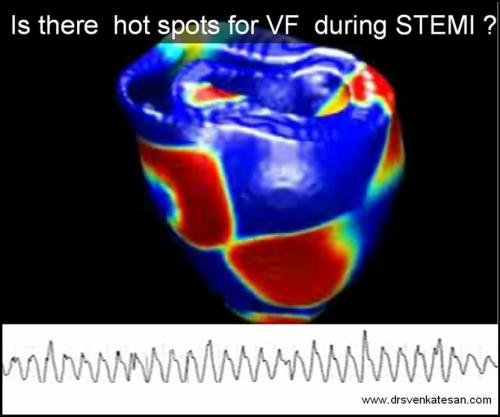
Is there any hot spots in the heart that are hypersensitive to ischemia ?

Some studies have clearly documented increased incidence of primary VF in infero posterior MI , and RV MI

than anterior MI .   J Am Coll Cardiol 2001; 37: 37-43

Why  ischemia of a certain location of heart should be more prone for  primary VF ?

The answer is any body’s guess.

Some intriguing possibilities are ,

  • RV is a anterior chamber , when infero posterior MI occur in association with RV MI  the ischemic zone encircles a almost 50% of heart like a band .This could be one explanation for more incidence of VF in infero postero RVMI.
  • Any MI which involves a  antero -posterior axis  of heart is likely to trigger a VF
  • Some of our patients  who survived a primary VF had a short left main  and early bifurcation with a large diagonal branch.The lesion was noted in the bifurcation.This raises a possibility ,  if a STEMI occur at a bifurcation with two divergent areas of  acute ischemia it has a high chance for precipitating a VF.

Related video by the author

Ignorance based cardiology -You tube

Potential research areas

Genetic susceptibility

Environmental Energy flows and primary VF

Some believe  a role for astrological  forces and  VF

Read Full Post »

  1. Do 64slice MDCT  in all patients who has  a coronary event and follow it up with catheter based CAG.
  2. Use liberally the new biochemical marker ,  serum  B-naturetic peptide (BNP) to diagnose cardiac failure in lieu of basal auscultation.
  3. Advice  cardiac resynchronisation therapy in all patients  who are in class 4 cardiac failure with a wide qrs complex .
  4. As it is may be considered a  crime to administer empirical  heparin, do ventilation perfusion scan in all cases with suspected pulmonary embolism.
  5. Do serial CPK MB and troponin levels in all patients with well  established  STEMI .
  6. Open up all occluded coronary arteries irrespective  of symptoms and muscle viability.
  7. Consider  ablation of pulmonary veins as an  initial strategy in  patients with recurrent idiopathic AF. If it is not feasible  atleast occlude their left atrial appendage with watch man  device.
  8. Never tell  your patients   the  truths  about the  diet , exercise &  lifestyle modification (That can  cure most of the early hypertension) . Instead encourage the  use of  newest ARBs  or even  try direct renin antoagonists   to treat all those patients in  stage 1 hypertension.
  9. Avoid regular heparin in acute coronary syndromes   as  it  is a disgrace to use it  in today’s world. Replace all prescription of heparin with  enoxaparine  or  still better ,  fondaparinux  whenever  possible.
  10. Finally never discharge  a  heftily  insured patient   until  he completes all the  cardiology investigations  that are available in your hospital  .

Coming soon :  10 more ways to  increase cost of cardiology care . . .beyond common man’s reach

Read Full Post »

« Newer Posts - Older Posts »