Ventricular tachycardia is considered as a dangerous electrical rhythm abnormality .It can immediately degenrate into ventricular fibrillation and result in SCD in many.Ironically, it is also a fact , a patient with VT can present silently without any symptom .Some VTs are slow and recurrent without much affecting The hemodyanmics.
In chronic recurrent, beningn VT (Some may consider it , ” height of absurdity ” to call a VT beningn ! but it is a reality , the term beningn denotes – very remote chance of converting into VF) ” Is there any other therapeutic option other than convertng into sinus rhythm. “( Read related topics)
The following paper was presented in the Annual scientific sessions of Cardiological society of India, Kochi , seven years ago in 2002
VENTRICULAR RATE CONTROL IN VENTRICULAR TACHYCARDIA
S.Venkatesan,,. MadrasMedicalCollege. Chennai
Mangement of hemodynamically stable recurrent ventricular tachycardia remains a delicate clinical problem. Reverting to sinus rhythm is considered as the only aim of treating VT.While rate control is accepted as a therapeutic option in atrial fibrillation, it is not so, for ventricular tachycardia.In this context we attempted to analyse the effect of Amiodarone on ventricular rate in stable ventricular tachycardia which fail to convert to sinus rhythm.
The study cohort consisted of 49 patients with stable VT who were admitted in the coronary care unit of Govt. General Hospital between 1998 to 2002.The criteria for inclusion were systolic BP>100mmHg and absence of hypoperfusion of vital organs The mean age was 52 years (range 26-68) with a male female ratio of 4:1. Of the study group 36 patients were either reverted with IV lignocaine , Amiodarone ( 150-300mg bolus ) or DC cardioversion . 13 patients who did not respond to either of these were followed up with Amiodaroneinfusion(1000mg) for 24 hours. The baseline diagnosis were old MI (6)) DCM (3) Arrhythmogenic RV displasia(2). Idiopathic VT was diagnosed in 2 patients.All these patients had VT during most part of the 24 hour follow up.
The pre Amiodarone mean ventricular rate was 152 (124 –196). Post amiadaorne (at 24hrs) mean ventricular rate was 128(88-142). The time taken for 50% heart rate reduction was 6.6h (4-24h). The average systolic blood pressure improved from 100 to 112mmhg . These patients were discharged in stable clinical status with oral Amiodarone and were referred for EP study.
It is concluded that Amiodarone, apart from it’s cardioverting ability , has a distinct ventricular rate controlling effect which can be of therapeutic value in at least certain subset of chronic recurrent VT.
Final message
Some of the patients with VT carry a very low risk of VF and SCD .In these patients , the only other major aim is to prevent tachycardiac cardiomyopathy that can be done with drugs which controls the ventricular rate whenever VT occurs !
Corrrecting the primary cause like cardiac failire , revascularisation ,detailed EP study ,tachycardia mapping , followed by RF ablation and ICD implantation is the state of the art approch in the management of VTs.But this small clinical observation was made to impress rate control could also be an option in patients in whom these procedures are contraindicated or not available .
Anginal pain is a type of visceral pain.It is carried by type C unmylinated nerve fibres.The perception of angina is a complex process.It is a combination of visceral and cutaneous referral pain.
How often is angina silent in diabetes mellitus ?
Presence of diabetes per se does not make an angina silent. In fact, if one takes 100 patients with diabetes , if angina occur in them , it is more often , manifest than silent. So , only few of the diabetic patients who develop diabetic autonomic neuropathy fail to have angina.The exact incidence is not known.It could be around 20%.
If angina can be silent in diabteics , can they have anginal equivalents ?
This again is not answered in literature. Among the anginal equivalents , the most common is dyspnea , which can occur in diabetics.But now , we know dyspnea also needs thoracic nerve signals from the intercostal muscle spindle and colgi organs.This can also be impaired in diabetics.
Can silent and mainfest episodes occur in a same patient ?
Yes.
Once silent does not mean always silent, and similarly once angina is felt it does not mean he is going to feel the next episode as well !
This strongly reminds us medical science is much a complex subject and what we know is very little in pain perception.
How is silent ischmia different from silent angina ?
There is considerable overlap between silent ischemia and silent angina
The questions to be answered are
Which is silent ? Is it the angina or is it the ischemia or both ?
Silent ischemia can occur in any individual , this is also called as silent CAD . When ischemia occurs but fails to generate pain it is silent ischemia .Undiagnosed CAD in asymptomatic individuals is also called silent ischemia or CAD.In this population Exercise stress testing detects CAD which was otherwise silent and masked.These patients may develop angina during EST.
During exercise stress testing many times patient has significant ST depression more than 2mm but still chest pain may not occur.These episodes may either be silent ischemia or ngina. Many times the EST is terminated before angina is manifest .( Chest pain is the last to occur in the chain of events following ischemia- Concept of ischemic cascade )
What are the other situations where angina can be silent ?
Pain perception and threshold level is high , so patient indeed has anginal signals but fails to feel it .
Patients on antianginal medication , fail to feel the angina.
Chronic betablocker therapy can exactly mimic autonomic neuropathy
Is it a blessing for the patient to have painless episodes of angina ?
When their ischemic colleagues , suffer a lot with chest pain it is tempting to think these diabetic patients are blessed!
Scientifically , this could be true in at least in some especially in a patients who’s coronary anatomy is known and devoid of any critical proximal lesions. For example a small PDA lesion can produce severe angina , but may be silent in diabetic and be comfortable .This lesion is insignificant other wise * !
It should also be recalled , pain relief has been an important goal for treatment of CAD .In olden days, thoracic sympathectomy was done for angina . In fact , even in CABG , one of the the mechanisms for angina relief is attributed to cardiac denervation.
Caution: Even a small episode of ischemia can trigger an electrical event .But it is rare.
How common is silent infarct (STEMI) in diabetic patients ?
In a simple questionnaire we asked the diabetic patients in our CCU how they felt their pain during MI.Most felt it normally as do other non diabetic . Diabetes does not make all anginal episodes silent. Severe episodes of ischemia may be painful while less severe episodes may be painless. Diabetic autonomic neuropathy is a least recognized and poorly understood complication of diabetes.Diabetes , involves the vasanervorum of the autonomic nerves.
The other mechanisms postulated in diabetic neuropathy are
Reduction in neurotrophic growth factors.
deficiency of essential fatty acids .
Reduced endoneurial blood flow and
Nerve hypoxia .
Is diabetic autonomic neuropathy treatable ?
Very difficult problem indeed.Controlling diabetes may partially correct the neural dysfunction.Many add on neuro vitamins and aminoacids are having a good market !
If you successfully treat diabetic autonomic neuropathy will my patient start feeling the hitherto silent episodes of angina ?
We don’t know.Logic would answer ” YES”
What is the ultimate effect of cardiac autonomic neuropathy.
The growth of medical science has been phenomenal .It is estimated , the quantum of break throughs and development in the last 50 years is nearly equal to 2000 years of evolution of our knowledge put together. Along with this growth , came the unavoidable misuse , and abuse of medical science. This is mainly due to contamination of medicine with commerce . Federal drug authority (FDA) and it’s variants were formed in all countries to monitor the proper usage of these technologies for the benefit of mankind. It has an authority to ban a drug or device , if it is found to bring more injury or side effects than benefit !
But , unfortunately there is no legal authority to ban an an investigation which is potentially or (really harmful )
or used extensively without any valid purpose .
The list of such investigation is increasing in every speciality
In cardiology
Doing a Troponin assay in patients wuth classical STEMI
MDCT in general population
Pro BNP in all suspected cardiac failure
Routine C reactive protein for CAD
Central venous catheters for all pateints with shock.
Is there a case for banning an investigation (Like banning a drug) for the benefit of our patients ?
Looking superficially , it may seem ironical. But we realise many seemingly innocuous investigations are responsible for uncontrolled misery for many patients.
This especially true in people who throng the wellness clinic (Also called master health check up)
A incidentally high C – reactive protein can lead on to forearm blood flow assessment of endothelial dysfunction and carotid intimal plaque that could lead onto carotid stents ! and life long anticoagulation , and an excess INR and sudden cerebral bleed and death !
This is one sample story in one particular speciality
There is a definite case for banning ( Either total or partial) some of the questionable investigations which are done routinely !
Just because these investigation do not have any physical , visible , adverse reactions like a drug , it should not be allowed to be abused .The consequence of false positive results of these investigations could be terrible and worse than the real disese itself !
Coronary arterial circulation is the life line for the human heart and it’s survival.Typically it is supplied by two coronary arteries, left and right coronary artery.Both, together carry about 250ml of blood every minute.( Approxinately equal to a cup of coke ! ).These coronary arteries generally divide in a predetermined fashion , and have multiple branches . It is a mystery , what decides this branching pattern
Is it like a our palmar crease ? or the cerebral gyri ?
However , it does follow a certain rule, one major coronary artery will follow the four important grooves of heart. In the left side , left main coronary artery (LM) originates in the left coronary sinus (Size varying between 1mm -20mm) and usually bifurcates into LAD and LCX. The left anterior descending artery (LAD) runs in anterior interventricular groove while , the right atrio ventriculo groove carries the right coronary artery(RCA) .Left circumflex artery (LCX) traverses the left atrio ventricular groove.The most inconstant branch is the posterior descending artery (PDA) which runs in the posterior interventricular groove.PDA can arise from either RCA, LCX or both or even from LAD.
The major branches of LAD are called diagonal and septal while the branches of LCX are called obtuse marginal(OM).There can be two to three diagonal and OMs.
What is ramus intermedius coronary artery ? What is the incidence of Ramus ?
The left main coronary artery instead of bifurcating into two , it trifurcates into three vessels.(LAD, LCX, Ramus)
The real incidence could vary betweenn (10% to 30%) depending upon the series.
What course it takes ?
It generally goes in the angle between the LAD and the LCX.It may either behave like a large OM or a diagonal branch.It supplies the lateral free wall of the LV many times.The peculiarity of this vessel is it does not run in a anatomical groove .It simply slides over the free surface of LV.Rarely, a very abnormal course of ramus, criss cross the aorta and pulmonary artery .
How common is atherosclerosis within Ramus ?
We don’t know yet. But it is very likely since it is an early branch from left main, it might have a predilection for atherosclerosis as like LAD or LCX ostium.In fact now we recognise more of trifurcation lesions involving three branches of left main .
What would be the ECG finding if a large ramus is the culpirit vessel during STEMI ?
This scenario could be rare.
ACS in ramus could present as ST elevation in 1/Avl /V5,V6
Lateral MI
Apical MI
High lateral MI
But it is realised , whenever the ECG changes are not fitting with typical ASMI or a lateral MI one should suspect a ramus lesion
What is the significance of ramus for an interventional cardiologist ?
PCI in ramus is a rare opportunity for a cardiologist .The issue here is, if ramus is involved adjacent LAD and LCX is also likely to be involved .So it would logically be a multivessel , complex angioplasty.Isolated ramus lesion could be tackled easily.Another issue here could be ,since this vessel is not within any anatomical groove stent deployment would have a poor support and prone for mobilisation and migration .
Stents are mechanical devices like a spring , used to keep an artery open after a PTCA or PCI.
Bare metal stents(BMS) were found to have restenois rate of about 25%. So it was perceived a stent should have it’s own protective coat , so that it won’t get restenosed.For this the researchers thought anti cancer drugs are ideal as they block cell proliferation and thus neovascualrisation and restenosis.Alas, they were found dismally wrong , after all , neointiaml proliferation is only a part of the problem of restenosis and simple blocking of cell growth is insufficient . The issue doesn’t stop with that, the anti cancer drugs incorporated within the stent simply can not differentiate normal from abnormal cells and
DES effectively blocks the normal endothelisation over the stents and make this highly vulnerable for acute stent thrombosis .
This complication is unique to DES and can result in SCD.Further ,during the last 6 years of DES , we recognised the restenosis rate has increased form the much hyped O % to almost 15% and it’s still growing . These complications has made a huge question mark over the future of drug eluting stents !
The concept of DES may not die , but which drug it should elute should be answered ! This again is going to be a long battle. So it is currently adviced, based on common sense ( With due respects to those RCTs funded by industry )
Whenever you encounter a block within the coronary artery* Ask the following questions in sequence ,
Whether we can leave it alone with medical therapy , if the answer is no , proceed to the next step !
Is there a possibility for plain balloon angioplasty in a given vessel (POBA, Yes ! the concept is not dead yet !)
If you decide a stent is required , Will the bare metal do the job ?
In multivessel CAD , Did the issue of increased metal load on the long term outcome was considered ?
If lesions appear complex, should we notstrongly consider CABG as an option ?
However if we have the habit of ask ing the following question you are likely to deviate from scientific approach
Is it possible to put a stent across the block ?
Yes , will be the answer most of the time ,and the patient will invariably get one or more stents and carry a life long stent related problems.
*The rule does not apply in Acute coronary syndromes
Also read this letter posted by the author published in British medical journal
One of the important principles of post PCI care is, we need to be very careful till the metal struts are fully endothelialised . This is of vital importance as improper endothelialisation is a powerful trigger and nidus for a imminent thrombosis and acute coronary syndrome.
It is a billion dollor irony , the much hyped DES does exactly what we don’t want ! and still it’s usage is increasing world wide . The drugs (Anti cancer agents) which coat the DES are the villains as it prevents the metal struts from being endothelialised and keep the metal surface raw and vulnerable , while the much maligned bare metal stents allow this natural endothelialisation process without any interruption ! So right now it is mandatory to administer dual antiplatelet agents life long( life of the stent !) for the patients with DES.
Just look , at the following image of a stent in vitro at 30 days follow up
Hypertension is the most common clinical cardiovascular entity.Left ventricular hypertrophy (LVH) is an important consequence of HT.In fact, it is considered as a end organ effect or damage. Others being brain, kidney, and peripheral vascular disease.Knowing about LVH is important because it has been linked to increased cardiovascular events.
Though LVH is considered as a close companion of HT it is surprising only a minority (15-30%) show evidence of LVH .Some experienced clinicians (Level C evidence) quote even lower < 10 % .Traditionally LVH was detected by ECG and now it is replaced by echocardiography.
What determines the LVH ?
It will be suprising to note , answer to this question is still not clear .
Is it the duration of elevated blood pressure ?
Is it the absolute level of blood pressure ?
If so , is it the systolic BP , diastolic BP or the mean BP ?
Or is it related to the etiology of HT ?
There has been no significant correlation between the above parameters
When we don’t know the answer to a question in medicine , the answer will generally will be inside the genes !
So in HT also the major determinant of LVH is in the genes that determine the myosin heavy chain response .
and also ACE gene polymorphism.ACE genes are involved in the expression of growth factors within the myocardium.
It implicates , gender, age, race etc in the genesis of LVH
Final message
So , the myocardium does not respond with LVH in all patients with HT.It happens only in a minority* .Duration of HT can be an important determinant , but the major factor is the alteration of genetic switches within the myocytes How this switches are going to behave , is largely inherited .Regression of LVH is also not uniform again implying lesser role for hemodynamics. (Some studies revealed ACEI have maximum regression of LVH , later disputed )
*LVH is more consistently seen in hypertension due to reno vascular or parenchymal disorders .It is also an observed fact , a combination of diabetes and HT is more likely to result in LVH.
The other major issue that needs explanation in HT/LVH is , how much of LVH is due to myocyte hypertrophy perse and how much is contributed by interstitial cell hypertrophy(Non myocytic hypertrophy)
The science of medicine has evolved over 2000 years since the stone age days.It has currently reached a glorious era with cutting edge scientifc technology .Today one can map the entire human genetic blue print and intervene in the disease even before they manifest .One can keep dying people alive for years with multi organ transplantation. Modern medicine has taught us how human sufferings can be prevented and life can be prolonged (with or without purpose !)
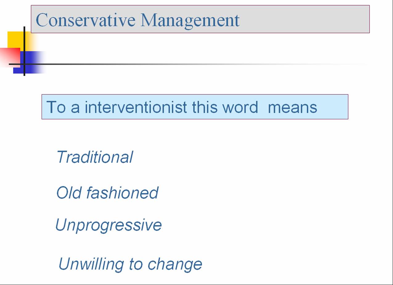
The term conservative management conveys two different
meanings for medical professionals.
For other group of physicians
Ever since the days of application of leech over the head for treating migraine and a crude knife abdominotomy for emergency exit of babies from pregnant mothers in distress , healer’s mind has always perceived “something has to be done urgently when some body suffers” this sort of reaction is probably inherited and is related to the primitive flight or fight response .
This may be true in some of the emergencies but it is untrue in many of the non emergencies.
Unfortunately , our mind finds it difficult to differentiate between these situations . With constant exposure to dramatic medical breakthroughs , modern day physician is made to believe “Some thing is always better than nothing when illness strikes. Human body is a wonderful machine which has it’s own service station ! in the form autoregulation and the meticulous homeostatic mechanisms. Only if the disease process overwhelms, it needs intervention.( Typical example:In the routine viral fever , you don’t adminster Acyclovir or other antiviral for all of them !)
The problem with early aggressive approach is, it fails to give an oppurtunity for the body’s natural defence forces to respond. Further , we will never ever knowhow the administered treatment is going to fare vis a viz the natural response.( With due respects to RCTs). While the field of medicine has so much evolved , our thought process, especially the aspect of clinical reasoning has always been lagging behind .It is now considered as inferior or even unscientific treatment if some one follows a conservative approach to a problem even if it provides same outcome of that of an invasive or aggressive approach ( The classical example is PCI for chronic stable angina The COURAGE study).
The other major issue is the hazards of unwarrnted invesitigations , drugs and procedures
Classical example:No one knows how much morbidity or mortality the routine Swan ganz catheter caused when it was rampantly used for over two decades to monitor central venous pressure .It is estimated that in modern medicine there are at least few drugs or devices in each speciality waiting for the same fate as that of the swan ganz catheter.
No body knows when it will be exposed .Our EBM will take it’s own time . . .Till that time humanity need to suffer.
This thinking is not new The concept “First do no harm is over 2000 years old”
Questions in search of answers
Does law of conservation of energy applicable to human body and medicine ?
Can we defy death with modern medicine ?
Final message
Conservative management is still a great medical concept in many situations and one should not allow it to die by the whims and fancies of the modern scientific forces.
Whatever you do on the patent’s body do it , only if it is going to helpful for him /her. If you are unsure Whether a given treatment is going to help or not ask this question to an expert .
The widely prevailing dogma of aggression is always better than non aggression has absolutely no evidence.
So approach a clinical issue disease by disease , individual by individual.
Now , in this era high tech medicine , It is lot more tougher to choose a conservative path as the pressure to do more and more looms larger ! It is easier to follow the crowd than a path of your own .
Always remember it needs a stronger mind to act according to our conscience !
A normally functioning circulatory system is vital for our survival . We have about 6000 ml of blood, circulating all over the body in an approximate time of 15-20 seconds.The pressure at which this blood moves across the body is called the blood pressure . Hypertension or simply , high blood pressure is an undesirable hemodynamic disturbance in human circulatory system.Systemic hypertension is the most common type of hypertension. The blood pressure is primarily dependent on the status of the blood vessel(vascular resistance) and cardiac contractility. This regulation is under many neural and hormonal factors.Further the blood pressure varies depending upon the blood vessel calibre, and the local milieu.There is a progressive drop in blood pressure from major arteries to the small arteries .The pressure drop is maximum across the arterioles to reach the venules .The venous circulation has the lowest pressure, it ends up at right atrium with a mean pressure of 0- 5mmhg.
Importance of regional variation of blood pressure.
It should be realised , each organ has it’s own regulated blood pressure.The brain perfuses by the intracerebral pressure .The lungs decide how much should be the pulmonary arterial pressure.The kidney not only controls it’s own pressure but also has a major regulatory role in systemic pressure by rennin angiotensin system.The examples are numerous, portal system has it’s unique pressure controlling hepatic hemodynamics. The retinal blood vessels regulate intra ocular pressure. While the human circulatory system has a wide variation of blood pressure across the breadth and length of vascular system, it is ironical a single snap shot BP with a brachial cuff is used to define the normality and if it is normal every thing is thought to be hunky dory !
It is widely acknowledged now , aging of humanity is nothing but aging of our vascular system
So we should have new parameters to assess individual organ’s vascular health as well as the currently popular systemic vascular health.The single important factor that determine coronary endothelial damage is the intra coronary pressure.It is never taken into account in any of the cardivascular mortality studies. This is the prime reason for the widely prevalent conflict in the cardiology literature , namely : Controlling systemic blood pressure has poor correlation with cardiovascular outcome. Many of the so called normotensive individuals have serious hemodynamic injury in their coronary arteries.This was made apparent in the ASCOT LLA study , in which patients with near normal blood pressure also benefited from statin therapy , implying endothelial damage could occur at any level of systemic blood pressure.
What is the normal intracoronary pressure ? When do you diagnose intracoonary hypertension?
The normal intracoronary pressure is around 40mmhg . Intra coronary hypertension as a clinical entity is yet to be recognised . There is no defintion available for intracoronary HT , intracerebral hypertension as well.
It’s still a long way to go , for the cardiology and neurology community to assess non invasively intracoronary pressures and intra cerebral arterial pressure to prevent coronary events ant strokes.
Final message
Simple risk prediction using brachial cuff blood pressure is a grossly unscientific method (Sorry, i really mean it ) to assess one’s vascular health.There has been few attempts like vascular endothelial health assessment by fore arm blood flow , central aortic pressure (Instead of brachial cuff pressure) as an index for risk predictment and assessment for hypertension is suggested.
Angina pectoris , classically occur on exertion and gets relieved on rest .This is called typical chronic stable angina as described by Heberden (CSA ) . Unstable angina(UA), the term originally described by Noble O Fowler in early 1970s. ( Also being referred as intermediate coronary syndrome , preinfarction angina etc).The definition for unstable angina has evolved over the years and currently refers to .
1.All new onset angina of any degree*Some include severe angina only ! New onset angina of very mild degree on exertion could be the onset of the first episode of stable angina.
2.Rest angina of more than >30 mts not relieved by taking sublingual nitroglycerine.
3.All Post MI angina
4.Any angina in patients who have been stented by PCI.
How to recognise a patient who is shifting from stable angina to UA ? UA is to be suspected when a patient develops. 5.More frequent episodes than usual6.Angina occurring at lesser level of exertion than before7.Angina radiating to new site ( Example : Chest pain radiating to jaw rather than to the usual left arm or vice versa)
Why the first episode of angina is given a special status and often considered critica ?
Angina is the clinical expression of myocardial ischemia.The course of the first episode of angina , can not be predicted.It could be a the beginning of a chronic disease process, or it could be a progressive coronary occlusion as in unstable angina /NSTMEI , or the onset of even a STEMI.
In contrast a patient with chronic stable angina has a predictable chest pain , at a particular level of exertion, radiation to same site, same character, and the patient knows for sure the pain would promptly dissappear when he takes rest or nitroglycerine tablets.
What is the underlying pathology in UA ?
Generally it is very rare for a stable plaque to produce a serious episode of unstable angina .It requires an unstable plaque* to precipitate an unstable angina !
Unstable plaque refers to any plaque which is eroded, fissured, ruptured or hanging eccentrically , with
an active thrombus.
What is the significance of post PCI angina?
It is an irony, any angina following PCI is to be considered unstable as sudden occlusion of stent is quiet common.This is a paradox of sorts as one would wonder in a patient with CSA who undergoes PCI with stenting of left anterior descending coronary artery (LAD) all his subsequent episodes of angina will be labelled as UA even if a stable angina occur in his other coronary artery.And these patients would go for early invasive approach and potentially inappropriate interventions even if they are at low risk !
Is all angina at rest can be termed as unstable angina ?
No, but many times , rather most of the times cardiologist believe all rest angina to be unstable.
What are the situations where stable angina can occur at rest?
An episode of angina during mental stress, or post prandial* state are very common in patients with CSA. This gets relieved after the stress. Some times patients with CSA during episodes of fever may get angina at rest .These are considered variants of stable angina.
Post prandial angina , may be considered by some as unstable
How often a diagnostic confusion occur between CSA and UA ?
Generally, this issue is rarely addressed in cardiology literature , for the simple reason it is never considered an issue at all !
According to Canadian cardiovascular society grade 4 stable angina is almost similar to unstable angina , as it denotes angina occurs with minimal effort or even at rest. In fact CCSC grade 4 should be termed as UA.
Can ECG be useful to identify stable angina from unstable angina ?
ECG will some times come to our rescue when one is confused between stable and unstable angina even though resting ST depression can occur in both stable and unstable angina . Statistically , if ST depression is noted during an episode of angina it is more likely to be UA rather than CSA. . Apart from ECG , Troponin T or I levels may be elevated in some of the patients with unstable angina. Rarely stable angina can also show elevated troponin.
In patients with systemic hypertension and LVH or cardiomyopathy resting ST depression may not indicate UA
So differentiation between, stable and unstable angina even though appear simple and straight forward, it requires a diligent appraisal of history , physical examination (Aortic stenosis /HCM may cause stable angina) and ECG, enzyme evaluation.
Final message
In any coronary care unit , admissions with initial diagnosis of ACS/UA/NSTEMI , subsequently turn out to be simple stable coronary artery disese . This error happens because the chest pain or ECG changes are aggravated by non cardiac factors like a mental stress or a post operative stress or fever etc.
There could be another school of thought, that is to err on the side of safety, and manage all rest angina as UA .But the hazards of unwarranted therapy might exceed the risks of leaving these patients alone.
In this context ,there is a need for a new definition for unstable angina .
One ideal version could be . . .
Any angina , of any degree which is caused mainly by the supply side defect (By a acute thrombotic /disruptive plaque occluding the coronary lumen with a imminent danger of myocardial infarction is to termed as real UA.
All post MI and post PCI angina are unstable angina
Rest angina which occurs due to increased demand situations need not be labelled as unstable angina for the simple reason there is neither an active plaque nor a fresh thrombus likely in these patients. They rarely develop recurrent angina or MI . The mechanism of angina at rest here is most often due to a tachycardia and resultant increase in MVO2 .(myocardial oxygen consumption) .Currently they are called as secondary unstable angina.In fact , anti thrombotic drugs are misused in these situations as they satisfy the criteria of UA/NSTEMI.
The contents of the this blog is being published as Kindle E book , as per the request of many of the readers. Every article will continue to be open source in this site. Again I shall reiterate the book format is not aimed at any commercial intent. It is only to facilitate learning in a single book format Here is the link to book https://amzn.in/d/euhL5vu
Click below to see who is watching this website live !
This site will never aim for profit. Still ,this donation link is added at the request of few visitors who wanted to contribute and of-course that will help make it sustainable .
Please Note
The author acknowledges all the queries posted by the readers and wishes to answer them .Due to logistic reasons only few could be responded. Inconvenience caused is regretted.


 A normally functioning circulatory system is vital for our survival . We have about 6000 ml of blood, circulating all over the body in an approximate time of 15-20 seconds.The pressure at which this blood moves across the body is called the blood pressure . Hypertension or simply , high blood pressure is an undesirable hemodynamic disturbance in human circulatory system.Systemic hypertension is the most common type of hypertension. The blood pressure is primarily dependent on the status of the blood vessel(vascular resistance) and cardiac contractility. This regulation is under many neural and hormonal factors.Further the blood pressure varies depending upon the blood vessel calibre, and the local milieu.There is a progressive drop in blood pressure from major arteries to the small arteries .The pressure drop is maximum across the arterioles to reach the venules .The venous circulation has the lowest pressure, it ends up at right atrium with a mean pressure of 0- 5mmhg.


