Feeds:
Posts
Comments

It is tempting to fix the  “Force of cardiac  contractility” ,  to be the  prime determinant  of  systolic  blood pressure* .  Rather ,  it is  influenzed heavily  by   multitude of anatomical  and  physiological factors.

                                        ”  In most  life instances  the primary determinant  blood pressure  is not the state of cardiac contractility  “

                 For many ,  this  would  appear as  shocker of  a statement !

Fellows  should not be  confused with above inference  . What it means is ,  the  heart initiates  the blood pressure by a brief  period of  systole .The pulse wave attains a peak during ejection phase  . This is the peak systolic blood pressure  . There is nothing called  sustained  systolic  blood pressure .  The quantum and  duration of peak systolic pressure  contributed by the LV contraction  is  far less than we imagine .

If blood pressure is to be controlled   primarily  by  cardiac contractility , how is that ,  a  blood  pressure of   about 80mmhg is maintained throughout diastole when the heart is taking rest and the  aortic  valves  are closed  ?
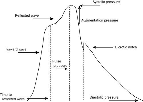
The  major elastic blood vessels  aorta and the major branches use the potential  energy gained during systole  (Like a rubber band )  into   kinetic energy as vessels recoil during diastole . This recoil  imparts an   important component  to the  diastolic blood pressure  augmentation  and maintenance.

It is  prudent to note  since  diastole is  much  longer than systole  , integrity of the vascular tree  become  much more important  to maintain the blood  pressure  till the next systole arrives.

Note

*The cardiac contractility  , might  still be  important  in determining systolic BP  in  patients  with  severely compromised  LV function** For example ,  in  dilated cardiomyopathy  with  LV failure ,  systolic blood pressure will  be directly related to LV  function.  When LV function is critically  low , the elastic  blood vessels  fail  to  amplify the blood pressure  beyond  a limit.

**Still it is not  uncommon to find high systolic blood pressure  recorded in the back ground of with severe LV dysfunction especially hypertensive individuals.

What happens during aging ?

The  aorta and it’s major branches  gets thickened , the  vascular collagen  goes  cracking  with wear and tear of  life.  In effect , these vessels become less compliant . So , when blood is rapidly ejected  from the  left ventricle  into aorta  and their branches  it’s  distensibility   is  reduced  .This  fails to dampen the  pressure  wave  and  hence systolic  pressure spiking occurs. This we refer to systolic hypertension of elderly.

It is  important to  emphasise   major elastic arteries  has a big  say in fixing the systolic pressure. For the same cardiac output systolic pressure can surge in elderly this  is why we have kept the normal  in adults as 16o mmhg.

Another key point to be understood  is  ,  Aortic compliance  has an impact on diastole blood  pressure too ! . The  stiff vessels during diastole bring  less diastolic recoil. Diastolic recoil of large elastic arteries  determines the diastolic pressure . Hence there  could be  a mild fall in diastolic pressure with physiological aging when recoil is attenuated .  Since the  reduced diastolic  recoil ensures diastolic pressure from being elevated  the entity is aptly named as isolated systolic hypertension.(ISH)
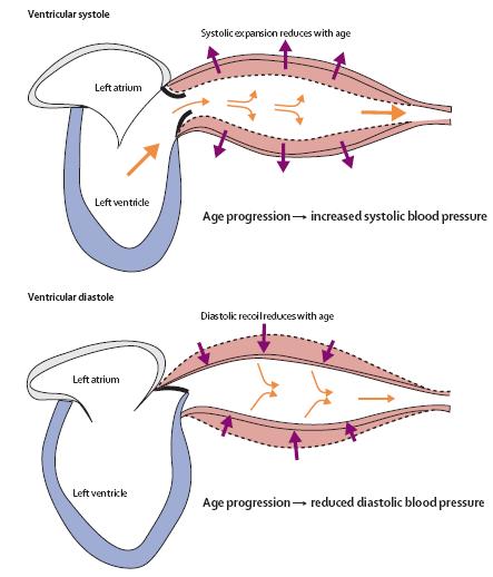
Image courtesey :Norman M Kaplan, Lionel H Opie Lancet 2006; 367: 168–76

Final message

While the traditional  teaching  ramains  as  systolic blood  pressure  would be determined by cardiac contractility  / cardiac out put , while the   diastolic pressure is determined by peripheral  vascular  resistance .This is not an absolute reality ,  rather it is  too simplistic way of teaching circulatory physiology !

The  peak systolic blood  pressure is more often determined by the integrity of  Aortic  and major arteries   rather than cardiac contractility  and stroke volume. Similarly , aortic properties do have a  say in the diastolic pressure as well !

Further reading and debate

 The net effect of aging  on blood pressure :  Is it  physiological or pathological  ?

  Should we  treat  this  raised  pressure due to aging  related systolic hypertension  ?

There is a huge controversy going around ,  regarding the need  of  treating this mild elevation of systolic  blood pressure due to arterial stiffening .This will be addressed separately in this forum .

Reference

http://www.mayoclinicproceedings.com/content/85/5/460.full.pdf+html
Medical science has grown ( and growing )  in an  astonishing pace. Many of the  inventions  which were  considered as  major break throughs   have fallen on the wayside over the years . Of course ,  quite a few  withstood the test of time .

One of  the great inventions  of last century  is per-cutaneous interventions  inside the human coronary artery .

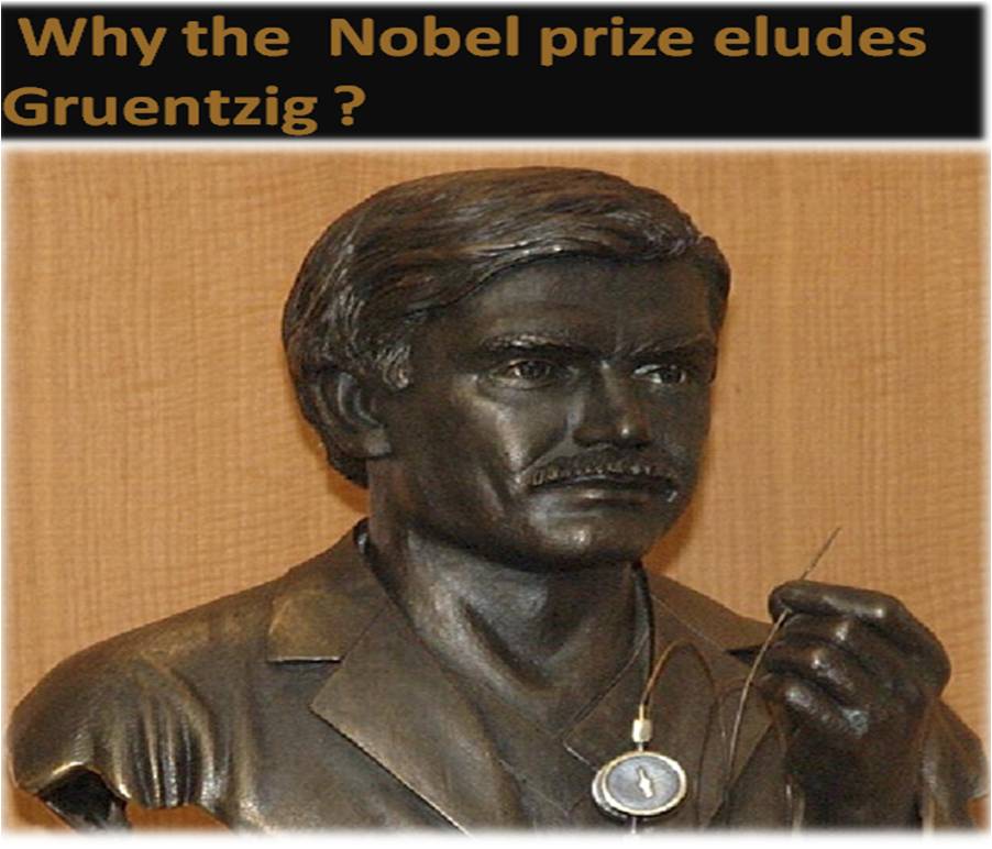
The concept was first conceived and executed by Andreas  Gruentzig  of  Germany in year 1977.  Now , at-least a  million PCIs  are done every year to tackle  CAD  with greatly  improved knowledge base, evidence ,  hardware,  techniques and expertise .

PTCA  is  an  invention worth a Nobel prize . . .well , that’s what we cardiologists feel. The Nobel committee  seems to think otherwise .

What could  be the reasons ?

  1. PTCA is  simply an extension of an old invention. Already the  inventors  of the  cadiac catheterization were  conferred with  Nobel prize (Forssman, Cournand,Richards)  . Hence , it is a sort of duplication of  invention . If Gruentzig is conferred a Nobel prize  the man who discovered the  coronary  stent (A plaque scaffolding device)  will  argue he too deserve  a  Nobel !
  2. What Gruentzig  did  was  in-fact a fundamental  human  response  by Instinct !  .When you encounter  a mechanical obstruction on the road   just try to overcome it . “Here is an obstruction impeding the blood flow , let  me  remove it”  . He did this  with  a wire and balloon . There is not much intellectual  innovation . It was  delivery of a mechanical force through a wire  .   But what the  Nobel  committee should take it to account is , he did this  in live human beating heart  and  cured of his illness most dramatically avoiding a need for surgery.
  3. Finally  comes the vital question. What is the impact of this invention in the health of mankind. ? How  many lives have been  saved when compared to other modalities to treat the   coronary artery disease ? *.This again is not convincingly answered  especially in    stable angina  , for which Geuentzig  originally developed this modality  . One popular argument  is , in terms of life saved and sufferings  relived oral rehydration  fluid  or penicillin  would beat PTCA most convincingly !

* Another possible reason is ,  the  Nobel medical committee is probably well aware of the  perennial  controversy  about  role of  Medicine vs Surgery vs PCI on the outcome   CAD  and  the  superiority of one over the other !

Final message

Whatever  be the reasoning  ,  Nobel committee has to  rethink .  Cardiologists  all over the world   would definitely  agree  if one man who have  made a  huge difference in their patient’s  life ,  it must be  Gruentzig  .

It is well-known  Nobel prize  is given  for path breaking  research that  break  new grounds like  decoding  cosmic mysteries ,  expansion of universe  , cell signalling ,  molecular mimicry  and the  stuff  like that .

Still ,  Gruentzig  definitely  deserves  a Nobel  solely  for  the novelty  in his  procedure  and in the process it helped  avoid  surgery in vast majority of heart patients.

Reference

http://www.nobelprizemedicine.org/

http://circ.ahajournals.org/content/93/9/1621.full

http://www.nobelprize.org/

Video

http://www.angioplasty.org/video/aghero.ram

http://www.angioplasty.org/video/agcomplex.ram

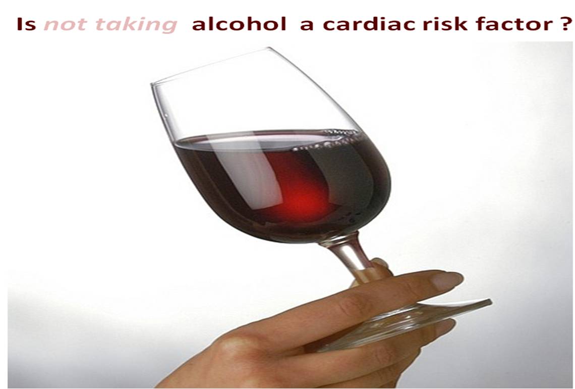
“It seems  certain . . . both  zero alcohol  intake and excessive alcohol  confers cardiac risk   “

I stumbled upon  the above   conclusion    from a respectable source*

*This  is from the  famous  INTERHEART  study published in Lancet.

Can it be true ?   What is the proof ?

Consuming  moderate   quantity of  alcohol  reduces  cardiac risk

  Does it make sense  to  skew   this   statement  like this

   . . . Not taking  alcohol   would   be a cardiac  risk  in other wise healthy individual .

Can we  profess  such a reasoning ?  My colleagues call it stupidity ?

If  it is true ,  are we justified  to use  alcohol as  a primary prevention drug ?

Which type  of alcohol we are  talking about ?

I am struggling to get specific answers .

After reading the INTERHEART study , my conviction is  the  “dangerous suggestion”   may indeed  have a significant  quantum of truth !  Readers may share their thinking .

In a country like India where alcohol is considered as a  killer chemical with a huge social stigma ,  it is  blasphemous to suggest  not taking it can be  cardiac  risk factor !

Reference

http://www.thelancet.com/journals/lancet/article/PIIS0140-6736%2804%2917018-9/abstract#

A. DiCastelnuovo, S. Castanzo, V. Bagnardi, M.B. Donati, L. Iacoviello, G. de Gaetano. Alcohol dosing and total mortality in men and women. Arch Intern Med 166;2006: 2437-2445

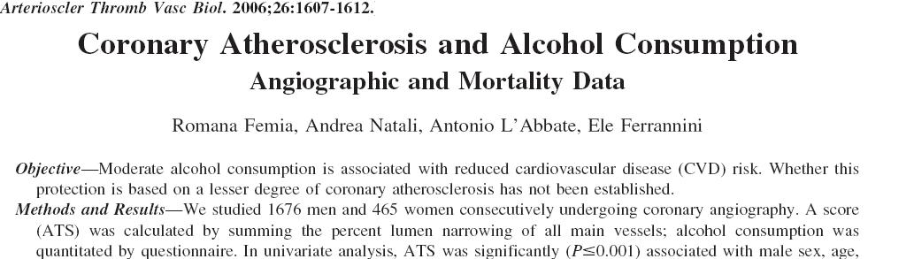
R. Femia, A. Natali, A. L’Abbate, E. Ferrannini. Coronary atherosclerosis and alcohol consumption: angiographic and mortality data. Arterioscler Thromb Vasc Biol 26;2006: 1607-1612

D.L. Lucas, R.A. Brown, M. Wassef, T.D. Giles. Alcohol and the cardiovascular system. J Am Coll Cardiol 45;2005: 1916-1924

How to rapidly  diagnose  significant LV dysfunction  at the bed side ?

Look for

  1. Tachycardia*
  2. Exertional LV  S3
  3. Muffled S1
  4. Weak carotids
  5. Often inconspicuous apical  impulse

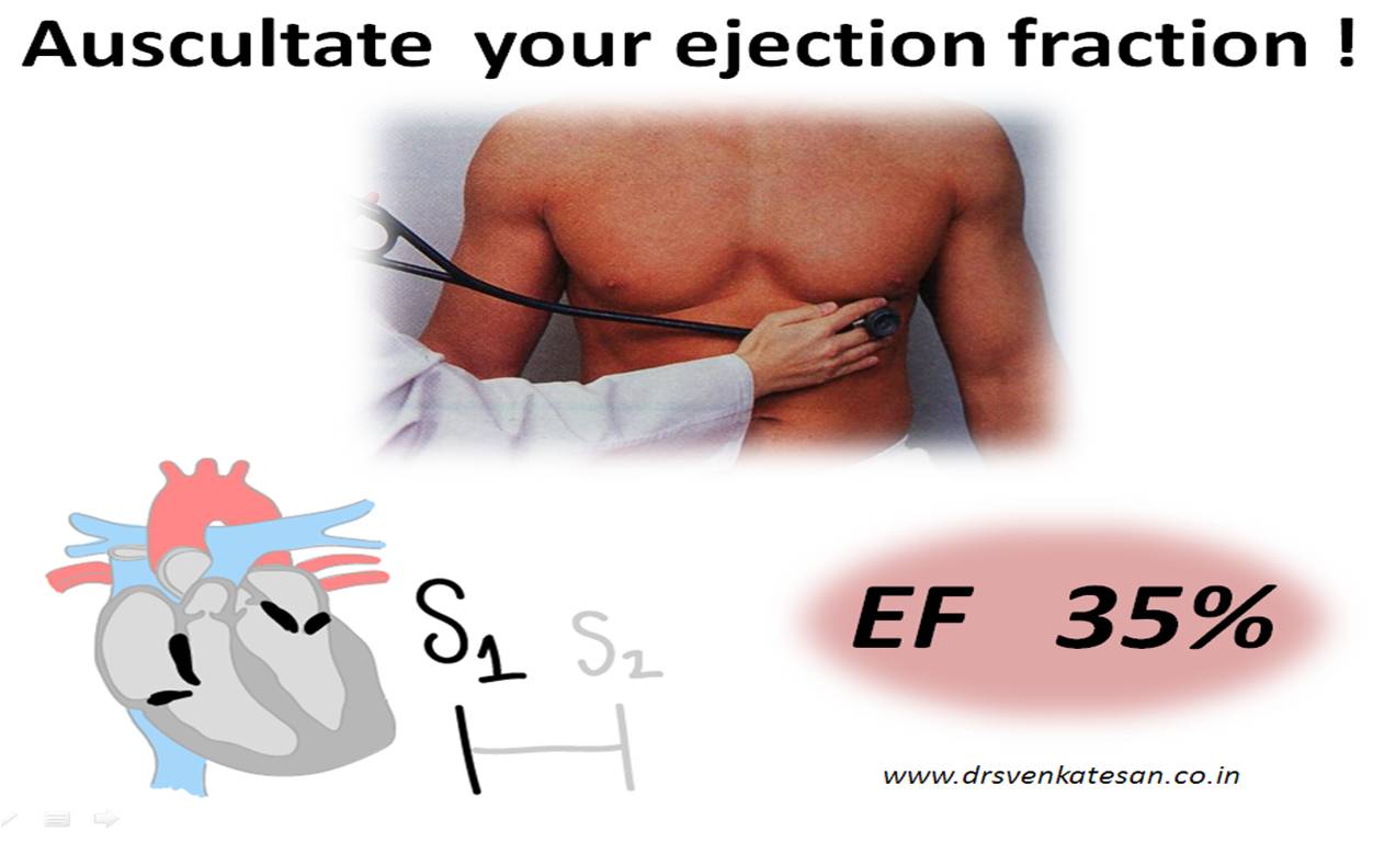
If all these signs are present EF is likely to be less than 35 % with 90 % specificity . If this is accompanied by  true cardio-megaly in X-ray chest,  LV dysfunction can be diagnosed with a  precision  reaching almost  100% .

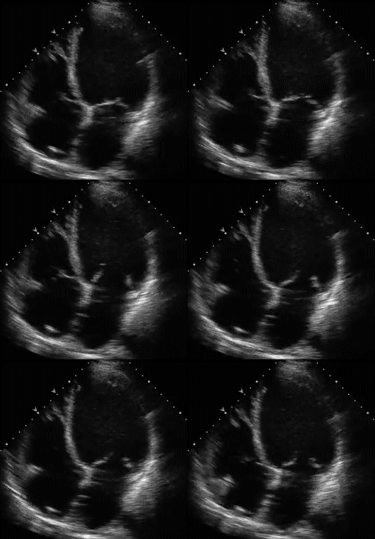
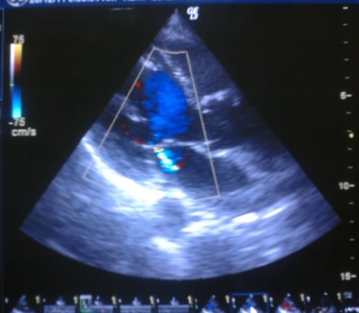
Note the sluggish motion of mitral leaflets and how closely the LV contractility is related to AML movement.This man had a soft S1 and his EF was 30 %

* Tachycardia may be  a non specific finding . Further ,base line tachycardia may  not be present  in all cases of LV dysfunction . When there is a  sudden surge in HR  even with minimal exertion , it  suggests   severe  LV dysfunction.

** The above clues  may not apply  in  valvular heart disease  , and isolated right heart disease  as multiple factors may impact S1 intensity .

*** LV failure must be distinguished from LV dysfunction (Vide infra)

Similarly , a  patient can not have significant  LV  dysfunction if  one  detects any of the following.

  • If the first heart sound is loud
  • If he feels chest thumping as palpitation.(A fluttering and audible   mitral  AML has 100 %  predictive value for normal LV function )
  • If you here an aortic ejection sound (Vascular clicks ) . Ejection clicks need significant force for it’s generation.

Final message

The most mobile structure of the heart is  anterior mitral leaflet . Fortunately it’s closure is  well heard as   S1 . Mind you, the most important determinant of  S1 intensity is  LV  contractility.  If your ear is sharp , and if you are able to  rule out other  reasons for soft S1  (Like obesity, pericardial effusion )  we are fairly  justified in suspecting significant Left ventricular dysfunction.

Further reading :

***What is the difference between LV dysfunction and  LV failure ?

Both these terms are  often  perceived  to convey the same meaning . But it  can  never be used synonymously .Cardiac failure is a clinical entity while LV dysfunction  is  a  derived  technical parameter  by and large an echocardiographic entity. Cardiac failure   is defined classically as a clinical syndrome .(elevated jvp, edema * S 3 rales etc)  Neuro hormonal activation  can occur with both.

A patient with   LV dysfunction    when destabilsed  develops   LV  failure and after stabilisation of   LV failure he is brought  back to  the baseline  LV dysfunction.

A strong willed  person rarely develop syncope.  We know  weak hearted (Or is it weak brained ?)   men and women may  faint  when  the emotions swing unexpectedly  .The  commonest cause of syncope is  neuro-cardiogenic  syncope (NCS) . (Formerly  called as  vaso-vagal syncope  VVS ). Few facts need to be  emphasized  here . There are  many  critical  circuits  and components to  common syncope.

  1. Trigger
  2. Afferent
  3. Center
  4. Efferent
  5. Fall /Near fall
  6. Prompt recovery after the fall.

Trigger can be emotional or mechanical (Prolonged standing ,  dehydration , etc )  . It occurs generally  in an emotionally  charged  environment with a high  basal sympathetic tone .

Afferent for  NCS   is  mostly sympathetic but it can be  para- sympathetic also (Sensitive GI tract ,  Micturition etc )

* Many times a  trigger and afferent pathway can overlap with each other.It is still unclear what exactly constitutes the afferent , since  triggers can be either sympathetic or para- sympathetic .  ( Pain, GI stimuli, vascular puncture etc) .  Further , afferent  can be be same as the trigger and reach the brain  stem directly  or touch  the heart en route .  ( Cardiac axis  in classical NCS)

The  center is  in the medulla  . Both vagal and sympathetic centers  are involved with potential  spill over on either side.

Final efferent  pathway is the strong  vagal surge resulting in bradycardia and peripheral vasodilatation , cerebral hypo-perfusion  and the person usually falls .( Near fall or aborted NCS  is also a common theme )

                                        If stress increases the blood pressure , absence  of stress  will have to  lower the blood pressure . If anxiety cause hypertension  ,  depression is expected to   cause hypo-tension.

These  inferences  may  appear  correct by logic . As  is always in medicine ,  such   logic works only partially ! ( We are told  the  Sadhus of Himalayas rarely record  blood pressure  beyond 100mmhg systolic  !)

There are strong reasons to believe common syncope (NCS)  is primarily related to the state  of mind and  the neural regulation. Dizziness ,  giddiness  near syncope  are closely related  to  psycho-somatic disorders. Strong willed men and women rarely develop   syncope.Their vascular   tone is well in control even in critical times .This fact has been  well observed  in  the setting of   traumatic  and hemorrhagic   shock  in critical care units  ,  where  some  hold their blood pressure well  even in   adverse circumstances  and few sink without any fight .

Is psychogenic , situational , pain syncope  same as NCS ?

Technically it may not be same. But all of  them  share at least 50 % 0f the  circuits of  NCS.  .However  there is no consensus  to call  psychogenic and   pain syncope  as  types of  NCS.

One critical aspect of  the debate is ,  we do not know whether the  cardiac axis is involved in these  syncope or not. It is preferable to call these types of syncope  as neural syncope (NS)

While in the classical NCS  heart has a  central role in generating hyper active sympathetic afferent from  myocardial stretch receptors. In psychogenic and pain syncope cardiac stretch receptors  are not much stimulated instead ,  the  spillover occur  directly from sympathetic to parasympathetic  nucleus in medulla.

In pain induced syncope parasympathetic limb  gets vigorously stimulated in isolation  to cause a severe  vaso-dilatation  . But once the syncope sets in we often observe bradycardia  and cardiac  limb may get activated as well.

* Presence or absence of cardiac limb in NCS and NS is critical with reference to efficacy of  beta blockers in NCS. The current guideline of NCS  management(  ESC 2010) is strongly biased against beta blocker (Class 3 -level A)  which we feel is  incorrect . Bulk of the patients with NCS respond well to long term beta blockers  .

Please realise , beta blocker  is the only drug which  can break the  NCS  circuit at multiple levels .(Sympathetic trigger, sympathetic afferent, cardiac stretch !

So what is the message ?

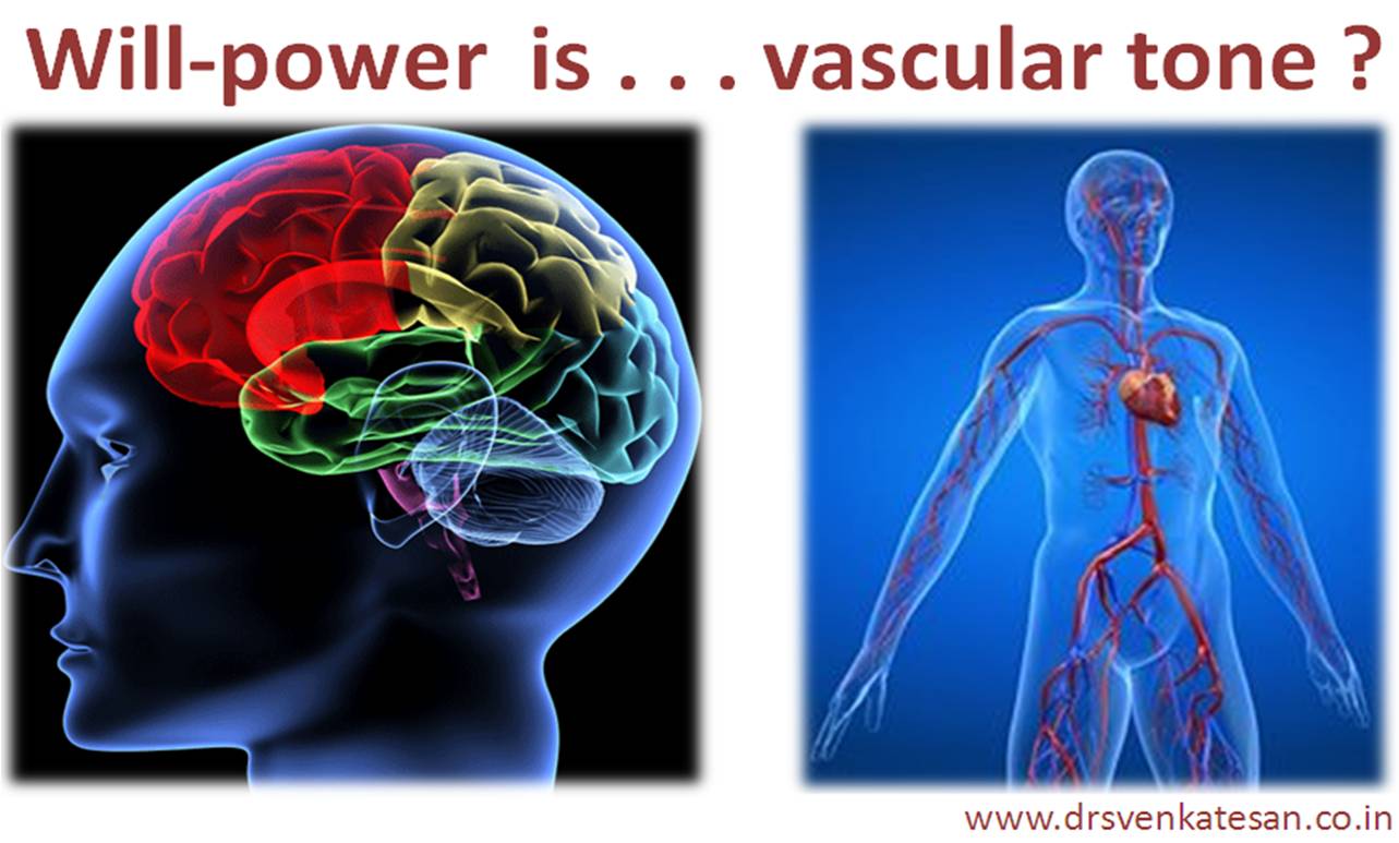
It doesn’t require great brains  to realise  vascular  and neural system are  intimately linked  . We know today,  NCS  is primarily a neural phenomenon  hence the  mental status has a  dominant  control over the vascular system especially at times of stress .

The confusion between classical  NCS and psychogenic  / situational  syncope can be largely avoided  , if  only  we call these entities  as simply neurogenic / neural syncope (NS ) ( Omitting the word cardiac is helpful ,   as cardiac axis is not vital  here  ? Non existent  )

Clarity is still  elusive  in defining the  trigger  and afferent limb for the NCS  , fortunately  the final common  efferent pathway that makes the patient fall is indisputably   vagal  !   .  Medullary  vagal nucleus  though fires independently  , also gets  powerful central  parasympathetic flow  from  cortical areas  . Paradoxically ,   controlling sympathetic outflow (Anxiety ) is often an easier  way to reduce parasympathetic flow. This is referred  to as competitive , accentuated  antagonism.

One can prevent recurrent  syncope  by vigorous  mind  control at times of  extreme stress. This is  confirmed  indirectly , by the fact  reassurance is the key to successful  management  in vast majority of  patients with NCS .We learnt this  simple fact  after trying exotic methods like DDDR pacing  and so on .

Final message

Power of  the mind can never be under estimated even in cardio- vascular hemodynamics .  When  pathologically high,   it can spike the blood pressure and break  few vessels in brain , while  if it  is inappropriately  low ,  may induce a syncope or result in persistent  hypo-tension .

Let us learn to use  our  mind over  body  properly .Yogis do it style  and live for 100 years !

Reference

http://europace.oxfordjournals.org/content/12/4/567.full.pdf+html

http://europace.oxfordjournals.org/content/12/4/466.full.pdf+html


Iam surprised why this post has been looked so negative by the readees. Almost all  rated it  as very poor.

Iam still pondering over it. Realised to lable patients as weak minded could be one of the reasons.

I think what I wanted to convey was there is link between mind and vascular system.

Please let me what is seen as offensive, let me learn and correct in future.


Most  cardiologists  are familiar with  “Circulation” . We know  it  is a top  cardiology  journal with highest impact factor.  Few of us are  aware  of  a journal called  “Circulation  research” ( I wonder  why it is named  like that ,  as if  the regular   circulation journal  does  not carry research stuff  !)

It is one of the  path breaking   journals that regularly  churn out state  of  the art , often  mind  boggling research stuff.  Once in while we should get a feel of  basic science  research  as it  happens.

How else we are going to know an  atrial cell is to be bio engineered  shortly to behave like a  SA node  in patients with sinus node dysfunction. (Biological pacing )

This team from academic  medical  centre Amsterdam   should be credited   for  publishing   this gem of  an  article   from  a  study  involving the  measly mice !

It  deals elaborately  about the embryonic basis of AV nodal  disorders  . Specifically it  explains  the genesis of  WPW syndrome and how AV rings get muscularised  .

(It  is  due to   error in  bio-genetic forces ,which  affect the    incorporation  of AV nodal tissue  in the  fibrous  skeleton .This   results  in ectopic  junctional  tissues appear   any where along  the AV ring . This is the basis of  accessory AV pathway and   clinical  re-excitation.)

Final message

Once in a while  we should develop the habit of reading  tough  journals  like circulation research . After all ,   if a cardiologist  is not reading   these stuff who else  . . . will  ?

Reference

http://circres.ahajournals.org/content/107/6/728.full.pdf+html

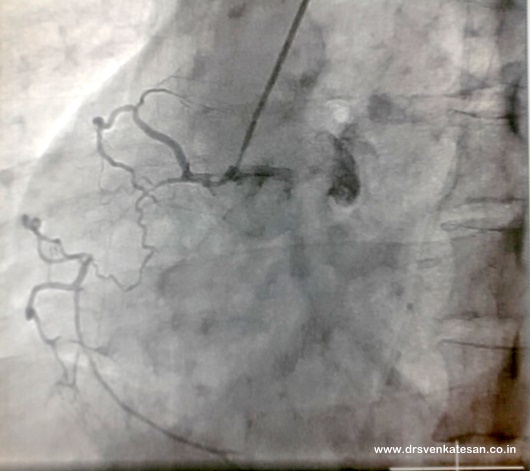
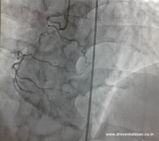
A middle aged man who owns a petty shop in a small  town of south India   came to us for stable angina .His RCA looked like this.

Normally if one coronary artery is obstructed the other comes to the rescue .It seems , this RCA do not trust it’s sibling LAD . See how it  self supports  its own  territory .(The most fascinating and mysterious aspect of coronary circulation is the collateral circulation. LAD  has big brother attitude  . . . it hesitates to help others while   RCA is more philanthropic , we know  it sends prompt  collateral to  LAD  whenever it is  distressed !)

However , there is one advantage of  such   self-sustenance of RCA  (Intra coronary/homo-collaterals ) . If  the  RCA  has to live  at the mercy of LAD  it  runs a risk of   neglect  at times of  distant LAD ischemia as well  !

Management

Single vessel disease , total occlusion , long segment lesion , still  the  PDA  is protected and the vital postero- basal area of heart perfused well ! What to do ?

Scientific  cardiologists  would like to meddle this  RCA with  multi-pronged guide-wires and other weapons  . Non -scientific cardiologists would  send him  home with medicines  . This patient preferred the later ! In the process  he  saved a  lakh ,  which  I  believe was meant for his daughter’s  education . He profusely thanked me for not hijacking his hard earned money for  frivolous  reasons . I said he should thank  his collaterals  and not me , for getting his money back  !

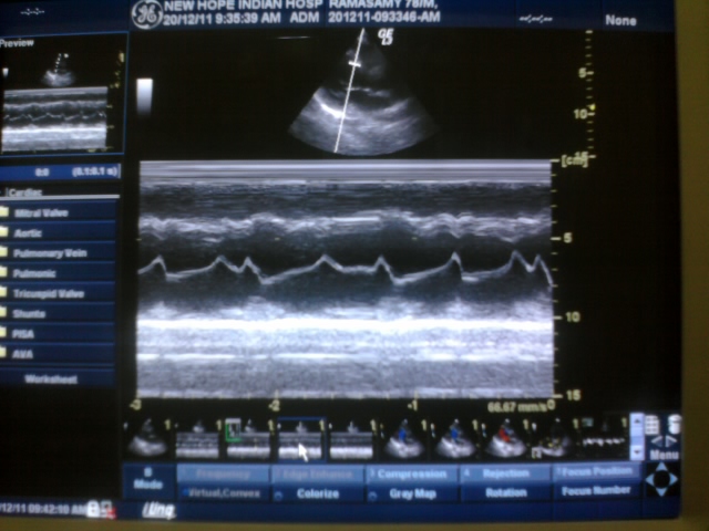
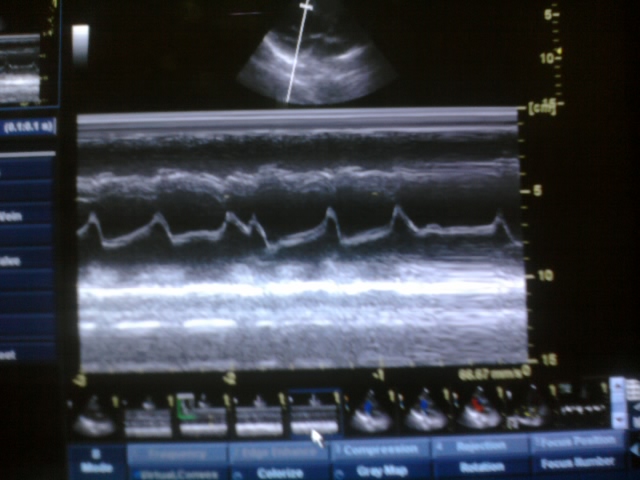
VPDs are the most common arrhythmia  that  confront  us  in  cardiology clinics .While  it can be a totally  benign   manifestation in some  ,  it may signify a sinister condition in others. ECG  is the easiest  and surest way to identify VPD.However  a shrewd echocardiographer can detect the VPDs while imaging the heart.It is often missed if one do not concentrate on the mitral valve motion.

Note :The VPD convert the typical M pattern into a inverted U pattern in mitral valve.

One of the important hemodynamic side-effect of VPD is intermittent mitral regurgitation.

Effect of VPD on mitral valve opening .

By  conventional thinking   VPDs  are expected   to impact  more on the  mitral  valve closure than it’s  opening .In reality it has indirect influence on mitral valve  opening as well. The retrograde  conduction(VA conduction) of the VPD determine the timing of atrial contraction and hence the   mitral valve opening. If the VPD gets blocked retrogradely  within AV node , the normal sinus impulse will activate the atria in an antegrade fashion .Note ,  he atrial activity  occur randomly when multiple VPDs occur.This makes the cardiac cycle too complex to assess especially the diastole. (In fact true  physiological diastole  may  not occur here !)

If  the mitral valve opening  is interfered by a   VPD  (Early diastole is  the  favorite time  for VPDs to  appear  !  )   .When it occurs the AML is    suddenly pushed  upon superiorly  by the premature ventricular activity and hence resets the  mechanical diastole. Please note electrical resetting of atrium is different from mechanical resetting.

It is also possible atria and ventricle contract simultaneously .This is the time , a cannon wave  may occur inside LA .VPDs can result in pulmonary venous canons and may even elevate pulmonary venous pressure   if  this  occurs repetitively .

Another possibility  is ,  VPDs  may not initiate a ventricular  contraction at all .It may be  simply  be an electrical event. That’s why  we changed the name of extra systole  and premature contraction into just   premature depolarisations.

Why is it important to know about M Mode motion of VPDs

Cardiologists  continue to  engage wide qrs  tachycardias   in the  wrong side  of their   brain for many  decades .The ECG debate about wide qrs tachycardia  is expected to  continue  for generations . !  Few smart cardiologists would  rapidly put  the echo probe  over the mitral valve and able to  differentiate  instantly a VT form SVT   with fair  degree of accuracy.

Detection  of regular M shaped mitral AML  will exclude a VT with a high degree of precision .(AV dissociation by echo )*

Even  presence of trivial  MR*  (More often diastolic )   which occur  irregularly  will  definitely indicate it is VT . SVT  hemodynamically   can not result in this  MR is gives us evidence for AV dissociation

* No reference for these observed indices in our lab. (Class 1 Level C expert opinion(  No one calls me as expert though ! )

What is the mechanism  of VPD induced  mitral regurgitation ?

It is well-known VPDs can cause   mitral regurgitation .Not every VPD cause MR.

  • The timing is important .
  • It can be  either systolic or diastolic MR .
  • If VPD occur in early diastole (After the T wave , the MR jet  will collide with  diastolic mitral flow. )
  • Paradoxical septal motion induced by VPDs can alter the pap muscle alignment transiently and result in MR
  • We dot not know how a LV apical VPD  differ from RVOT  VPD in the genesis of MR.
  • Logic would suggest RVOT  VPDs are unlikely to result in MR as there is  a time lag for the impulse to reach the LV base

What is  the effect of  VPD and Aortic valve opening ?

While  every VPD promptly  hits the mitral valve ,  aortic valve may or may not open with VPDs .Again timing and focus of VPD could be  important.This is the reason during  multiple  VPDs  only few open the aortic valve , that  explains  pulse deficit. (The so called missed beat )

Final message

Anterior mitral leaflet (AML) is the most mobile structure  of  the heart . Hence ,  it is not surprising to note  sudden unexpected ventricular contraction will  have maximum impact on this valve .

When VPDs occur in clusters or at random it has a complex effect on the mitral valve motion. This is responsible for  palpitation , minimal mitral regurgitation and rarely trouble some pulmonary venous cannons and raise in pulmonary venous pressure .

Careful analysis of  AML motion can give us useful clues to differentiate VT from SVT during wide  qrs tachycardia

Though heart is  primarily  known  as a  mechanical  organ , in reality   it is a vital  electrical organ as well . The entire mesh of electrical  pathway  from SA node to  Purkinje  fiber  would easily cross  a mile or two .Maintaining and protecting  such a  delicately  woven network  needs lots of  electrical sense  .  It is not surprising to note  , VT or VF  can be induced  virtually  in  every human heart  if stimulated rapidly. Electrocution  induced  by VF is  the typical example.Cardiac surgeons  do it regularly  before  surgery .

So , inducible  VT  in the EP  lab need to be  defined in a strict manner .

  •       VT must be triggered  by a  single stimuli  (or  two )
  •       Multiple sites should not be stimulated(ideally  single site , at most two )
  •       It should be sustained.
  •       Only mono-morphic VT has  significance
  •       Induced  p0lymorphic VT  has no clinical value.
  •       Pharmacological  stimulus  such as isoprenaline   can be used but reduces specificity.

*If a VT  rapidly degenerate  into VF  it  usually  means a polymorphic VT  while   unstable irregular  polymorphic VT   could be  same as   VF )

How do you make sure  what we induce in  EP lab is same as the clinical VT ?

This is the most difficult task for electro -physiologists. In real life setting VT is  often induced by ischemia hypoxia , local  acidosis and electrolytic imbalance. However  rarely mind this issue . In EP lab we induce  it  with  artificial electrodes  . Does it make sense to compare  these two totally different  set of triggers  in real life and a virtual EP life . Ideally  to confirm ischemic  VT  one has to induce ischemia  in EP lab and look for  VT . (Adenosine  stress ? )  Further ,  only re -entrant VTs  can be induced in EP lab by programmed stimulation . Automatic VTs can not be induced by stimulation .

The chances of inducing a VT in EP lab is  directily proportional to the aggression of the electro physiologists and patience  of  the  patient ! One can afford to use  more aggressive  protocols only   if a clinical VT was  recently the   documented .

 Electrical stress testing of heart

It may be tempting  to refer    induction of VT  in EP lab  as  electrical stress testing  for the heart. But fundamentally there is a difference  between this and  the conventional EST . Unlike exercise stress  test the  inducibility of VT highly unpredictable . It has far too many variables . (The surface area of contact , number , Intensity ,  site of stimuli , scar location , irritability of viable myocardium  ,  inertness of scarred myocardium ,  and finally the cellular milieu etc  )

Thoughts to ponder over Is it not  “a fundamentally a wrong concept”  to give importance  to inducible VT  ?

Why should we  treat a clinically non relevant inducible VT ? We do not know yet whether inducible VT in other wise normal LV function  has any long-term significance . Currently it makes   no sense   to intervene in VT  if the LV function is good and the episodes  are not clinical but only inducible.

Note: If there is severe LV dysfunction (EF < 30 % ) one can implant an  ICD without   an  EP study . ( Of course   to state more dramatically   without even single documented VT  !) MADIT 2

Final message .

A VT which is inducible in EP lab has no meaning ,  if the LV function is normal , while  even a  non-existent  (potential  )VT  in the setting of severe LV dysfunction is vitally important !

Though  we  differentiate cardiac function  into mechanical and electrical for academic purposes , it is astonishing to note   how the heart is able to function  as a  single unit  . We know today , the ultimate  outcome of   VT  is  not  dictated  by  electrical status of the heart rather , the mechanical ability  to  with -stand  sudden dis-organized  ventricular  contractions ( A ventricle with good contractile function has inherent  capacity  to extinguish most episodes  of VT .(Myocytes with inbuilt biological ICDs ?)

It is a million dolor question why some VT remain as non- sustained while others rapidly degenerate into  fast VT and VF thereafter

Reference

The two contrasting studies

The MUSTT (1999) trial exposed the limitation of   clinical utility of inducible VT . Multicenter Unsustained Tachycardia Trial (MUSTT) Investigators

While   MADIT 2  (2002)which recommends an ICD in every patients with  severe LV dysfunction following MI without even a EP study .

Murmur of Tetrology of  Fallot is generated at the level of

  1. VSD
  2. RVOT
  3. Aortic root
  4. Any of the above

Answer :   RVOT.

RVOT is the classical site of TOF  murmur , but  there is  a  rider . The murmur of TOF is  inversely proportional to the degree of RVOT obstruction. (Contrary to VPS with intact IVS) .In severe TOF especially during spells the murmur attenuate dramatically and may disappear altogether. Hence a silent and quiet heart do not necessarily  indicate  a mild form of TOF .

Other possibilities also  exist.

  • The VSD in TOF is  large and do not restrict  blood flow on either direction . Rarely  restrictive VSD can generate a murmur across VSD.
  • Aortic flow is increased in all severe cases of TOF ( Highest in pulmonary atresia and VSD)   Hence there is always a possibility of a soft systolic flow murmur across Aortic valve .
  • Other rare  cause for systolic murmur is due to  prolapse of   tricuspid  valve  that occludes the VSD  potentially causing  TR  and in the  process may  convert the  VSD  into restrictive type.
  • One more cause for  systolic murmur is sub Infundibular anomalous  muscle bundles criss crossing the RV body .
  • Peripheral pulmoanry arterial stenosis is recognised cause for distant faint systolic murmur.
  • Diastolic murmurs can also occur in TOF . Absent pulmonary valve and aortic regurgitation can result  in diastolic murmurs .

Question for analysis

What happens to TOF murmur during squatting  ?