Feeds:
Posts
Comments

It is a fashionable topic  in  cath meetings and workshops  to discuss  about thrombus loaded coronary arteries. Still  visualizing a  thrombus  in coronary angiogram is  never a  mean  task !  It needs  lots of  visual  acuity  and  imagination to recognise   intra coronary thrombus.

  • A lesion which looks fresh with multiple layers of  irregularity within the lumen is  often assumed to be thrombus.
  • An   intra luminal filling defect   is the  most often  used  “criteria  to suspect” a  thrombus

A long segment thombus with dye penetrating and coating the thrombus all around

Thrombus  vs plaque ?

Both are  radiolucent .  But a thrombus  or a plaque coated  with  dye  will make it radio opaque. The radio opacity of a thrombus is determined by extent of dye coating ,  the thickness of the dye layer, obliqueness to the x-ray beam . A thrombus plaque  interface can have two different  planes  of densities.

Theoretically dye can not encircle  a plaque  in its entire circumference as it will be attached to vessel wall (Unless  circumferential  dissection is present  )  Hence , dye can not coat a plaque fully ,  at best it can give  an appearance of eccentric filing defect  with over hanging edges .  While a  thrombus  can manifest with a  complete filling defect

Thrombus vs dissection

This is  still more complex   . Both can have a filling defect  .A  flap is a  line like  filling defect To complicate the issues further,  both thrombus and dissection occur together in the same spot .

How confidently  one can  identify a thrombus in coronary angiogram ?

During acute MI there is no difficulty in identifying it ,  as every acute  obstruction  must contain some thrombus* . Some interventionists  have special  ocular   sense   to   detect thrombus. Few others rely  on their intuition  rather than  solid evidence.

Sucking out a thrombus during primary angioplasty  has now become standard concept and is indeed  feasible  in most situations. It is obvious we have a task  on hand to identify thrombus correctly and quickly during primary PCI /UA .

Blind suction,  even though rewarding should be avoided.  Caution is required as blind  suction pulls a plaque with force !

A plaque debri  with a  thrombus, a dissected flap  all can combine together   to produce a complex  “masala”  of  coronary lesion especially after a difficult guidewire cross . This is refereed to as a battered coronary artery .These are the lesions which are prone for recurrent acute or sub acute thrombosis even if the lesion is  stented  properly.

During  primary PCI   thrombus coated dissected plaque  is just tucked  and  opposed behind the  gentle stent struts.The thin layers of thrombus between stent struts and the vessel wall is  missed  , 100 out of 100 times   by coronary angiogram . (IVUS very good in detecting this) .Because of this risk , Intensive anti -coagulation in complex PCI becomes mandatory

* Diagnosing thrombus in a chronic lesion is  much . . .much  difficult !

What are specific  modalities available  to  confirm thrombus

IVUS, Angioscopy, OCT are  hi tech tools to identify intra coronary  thrombus .(Which i feel  have little practical  value in real emergency situations)

Final message

Thrombus  may be a  key  finding  in acute coronary syndrome (Of course  the contribution of fissure, injured ,  plaques to the lesion can never be underestimated . ) Still , we have no simple , accurate method to identify it  ( Forget quantifying it) . Lots of assumption , guess work and gut feeling is at play in the cath lab .

We  expect better online , real time tools to improve out tentativeness inside the coronary artery  .

//
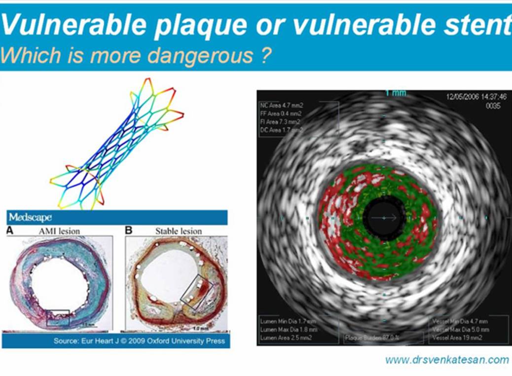
An atherosclerotic  plaque is termed  vulnerable when it’s  future behavior is unpredictable .A vulnerable  plaque has a  tendency to get occluded at any time.

Anatomically  a  vulnerable  is  present  , if the lipid core is more , fibrous cap is  thin  and  a  large lipid  core hanging eccentrically. A plaque with high temperature (Hot plaques ,febrile plaques)detected by OCT/Raman spectroscopy or thermography

Note the T cells and macrophages wage a losing battle against a metal monster !

What is the best method to calm down these vulnerable , hot ,inflamed plaques ?

A stent which scaffolds a plaque is believed to stabilse it  and  make it less vulnerable to rupture. This is the most optimistic view on coronary stenting .

Here comes  a pessimistic view !

A metal inside a coronary artery covering is  additional  threat .A metal  is   perennially  thrombogenic  ,especially the drug eluting stents which suppress the normal endothelial  function .

What  is the realistic view  ?

A stent should be used cautiously and judiciously in coronary plaques  with   high risk features  .Here  a  stent  in all probability  converts a vulnerable plaque  into a  relatively stable plaque

When stenting is done indiscriminately( without application of mind )  in stable non flow limiting lesions  stability is replaced with vulnerability.

Is it not curious to know  any angina  in a patient  who  had   PCI  for chronic  stable angina  is labeled  as unstable angina. 

Vulnerable stents

Following are typical  clinical scenarios   where stents could  carry a vulnerability  tag . 

  1. Poorly deployed  stents
  2. Properly deployed (but unnecessarily deployed especially in chronic stable angina )
  3. All Bifurcation stents
  4. Distal left main stents
  5. Stents with plaque prolapse
  6. Finally and most importantly all  drug eluting stents are considered  vulnerable ! (That’s why  our patients has to  live at the mercy of dual platelet blockers , life long.  Of course , there is no life time warranty   that  drugs do their  job properly)

And now . . .  you answer my  question !

Can  stenting convert a stable plaque  into vulnerable plaque ?

  • If  “yes’ is your answer your patients are in safe hands .
  • If  ” No”   is  your  answer ,  you are  fit to become a leading  interventional cardiologist !

Medical  research can be divided into few  broad  categories

  1. Basic science  research  in animal models
  2. Basic science  research   in Human
  3. Clinical : Bedside-  observational
  4. Clinical:  Epidemiological
  5. Community based long term data analysis
  6. Interventional -Drug /Device/Surgical

*Logically the  top 5  should  constitute  the bulk of research  ,  in reality    last one wins the race with considerable ease . Why ?

The important issues that  confront  today’s medical research  starts  right from the  “Aim” of the research ,  methods , materials statistics,  and  goes on  to   ethical issues , conflicts, futility ,  gimmicks  0f  publication  ,  marketing and ultimately left  for human assimilation .

(Read a related article in this blog   can  Aim of a study be wrong ?)

Data(s)  won’t  lie  . . .humans do  !

Science is nothing but collection of  facts ,  rechecking  the facts , and  finally confirming ,  they are indeed  facts. So medical  data collection becomes vital .  Data,  if  properly collected ,  wont lie.   Bias is always an issue in prospective trials. Further ,  and whenever and wherever  scientifically  motivated  human  beings interact with  data  the later   becomes a vulnerable  target and  get manipulated   for various reasons . (Read the famous article on data torturing  in  NEJM : I will link it soon  ) So blinding  becomes  mandatory   and it should  be total as some studies  tend  to  gain vision half way through !

Image courtesey : Jupeter images

Simplicity of observational studies.

We  give undue importance to RCTs . What we fail to understand is RCTs are required only  in selected situations in medical research (New drugs and interventions ) Meanwhile , we can do wonders with retrospective observational  data. These  data  can not be  manipulated  as the events  have occurred already and those people who collect or record the data  wouldn’t know this data is going to be utilized  for a study (This  , in fact  is  equivalent  to 100 % natural blinding and constitute a  real world study )

Observational  study can involve  patient behavior ,    disease behavior  , community impact, drug action, investigation modality , etc  . . .etc  . Your mind is the limit . Cost of doing a observational study is less but the impact on the society can be great .

Observing skills are the  biggest causality in modern medical times , This was  only scientific weapon of  our ancestors had , which they  used in an exemplary fashion .( Recall how Heberden described angina and Harvey taught us about circulation without even ECG and X RAY chest )

Fraud in medical research

Wherever big money is flowing corruption and fraud is unavoidable . . .at the  least . . .  we  should recognize it

( Many journals  just point out this possibility by simply displaying message of conflicts .They do not bother more than that  . . . just a warning message  )

Now in the modern scientific world  ,   even as the   genuine contributions   from our ancestors  left to  stare  the back of us  , we try to indulge in all sort  of unpleasant things.

In an audit against fraud in medical  research ,  it was found most of the fraudulent research happened with drug and device trials and few in basic science involving genetics and molecular medicine . It  was  rare to identify fraud in research involving purely clinical and  epidemiological  analysis .

Drug trials  need to be prospective . Vested interest can play  havoc in prospective data .There is a  thing called steering committee in all major studies   . . . we do not know what does the  word  steering really   mean .

There has been many  occasions  even well conducted studies turn out be  fraudulent . Now we realise many such studies are struggling to prove its worthiness .

In fact  it is argued every study before getting published   should undergo a  global ,  independent  trial   monitoring  board for genuineness  of the study . (Not the customary  peer review !)

Final message ( Sorry its  a  long one !)

We have a huge problem  here . I am afraid  we  haven’t even  understood ,  what  we  mean by medical  research !

For today’s   youngsters  medical  research means doing sophisticated  tests in nano- labs  , human genome  mapping ,  space age imaging modalities  or  involving a multi- billion dolor drug trials . This is absolute  falsehood.

What we need to do is   “search” , ” search”  ,  search again (That is   why it is called re-search )  for all those elusive  problems  our patients   face .Not only in their body , in their  home , in their community,  etc . Every  patient  teach us  few points,    observing and learning new things  and  publishing is  also an important aspect of  research .One can do  a instant   research in the crowded  OPD of a hospital   , in the wards , (What is the profile  of fever pattern in a winter season in your hospital ? does it reveal a new viral epidemic ?)

An ideal research  should  identify a problem and suggest a practical solution to a given problem .There are millions of such issue waiting for our attention in the bed side.  But what is happening  currently ? Current medical research is largely direction less ,  fueled by vested interest ,  makes  sure it avoids  all genuine problem areas !

Many studies  happen  based on  flimsy scientific   basis  .We are still  wasting our time to increase human HDL levels. ( Not with standing  the famous Torcetrapib fiasco  )   .Hundreds  of thousand of dollars   are pumped into this  research even after realising  only the  endogenous HDLs generated by natural methods like  exercise   are  the really  good HDL !)

While we do million dollar research   with a dubious risk factor called  high sensitive C reactive protein  ,   there is  no takers against number one killer disease of human kind  namely  “The  poverty” (WHO ICD codeZ59.5 )*

Let us prey   God  to instill common sense to all of us  . Patients  suffer with disease and we suffer from irresponsibility  or reduced responsibility ! It  makes us happy at-least few forces  like Lancet  , British medical journal etc are fighting lone war  against this  ailment  medical science is suffering .

*Please note :  http://www.icd10data.com   WHO labeled poverty as disease many years  back without much fanfare ! It is rarely mentioned in  any  graduate student**  medical text  in whom our future lies .  I do not know whether  Wars  and terrorist acts  been included as disease  or not !

**Our students  rattle about  about the  exotic  tick borne  Lyme disease happening once a year in remote hills ,    while  most will stare blank   when asked  how to diagnose and  treat  nutritional  anemia with  which millions suffer  every day !

While anatomical  grading of obstructive  coronary lesions are  quiet easy ,functionalc assessment is always difficult.The famous TIMI grading system had one unique problem .TIMI 1 and 2 grades are relatively easy to grade. TIMI 3 flow  which corresponds to normal penetration  and normal  distal perfusion  . This distal perfusion was entirely optical .

This was an important issue , in assessing post  PCI or thromolysis patients . It was realised much later , TIMI 3 flow is  stunningly  heterogenous group  .It was  ironical  ,  even after a successful PCI ,  restoration of TMI3 flow  could not be relied upon as an index of successful PCI  .

So , the PAMI study group included time as additional factor in grading TIMI 3 flow. PAMI 3 is  essentailly same as  TIMI 3  flow but  with a  condition , complete  distal vessel filling  must  occur within 3 cardiac cycle . PAMI 3 can be termed as a   refined version of TIMI 3 introduced in the evaluation of success of primary PCI . This helps us  define  or  diagnose   slow filling .

What are the other ways  to grade TIMI 3 flow

  • Myocardial blush index
  • TIMI frame count ( < 25 frames )

PAMI : Primary angioplasty in myocardial infarction

TIMI :  thromolysis in acute myocardial infarction

Reference:

http://circ.ahajournals.org/cgi/content/full/circulationaha;104/6/624

When you  encounter  a patient  with shock and  hypotension , the first ( instinct ) response would be to  start  an  IV line and push fluids rapidly . This is more so if  the patient is a child. This is what medicine has taught us for over a century . Now this  NEJM article surprises us with  its conclusion.

The accompanying  editorial in NEJM reiterates  a  fact . . . “In medicine there is nothing called  dictum”   , what you perceive as life saving treatment  will be doing the opposite !

Such is  the fragility of  present day  medical facts.

Please  remember , in medical science  not only  the drugs  have  expiry date even  some of the  break through  concepts suffer from it . 

This study may not have  great implications for cardiologists  but the filed of cardiology is also  infested with  many such false dictum(s ) are waiting to be damned !

In this funny world . when the  scientific methods are  imperfect  ,  we have to realise  two such U turns make  the  original path right .

Similarly ,  some of  those who do  not  make the initial   path correction ultimately  travel  in  the right path !

Message to patients

Many of my patients often wonder how two diagonally opposite  views are expressed  by doctors  for a given medical  condition .  My simple answer to them is do not ever  try to understand your medical condition beyond a point , . .  .  we  our-self  have not yet  mastered it  !

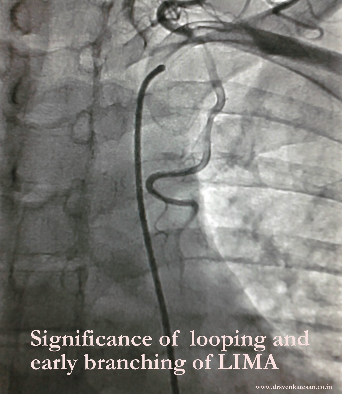
LIMA (Left internal mammary or thoracic )  is an unique  artery ,   incidentally runs close to  heart ,  has  a  privilege  of supporting   of human  heart in its hour of crises ! .  CABG  surgery was started with saphenous grafts in 1967 .  We have  since moved  on ,  from venous grafts to  total arterial grafts .  LIMA as a graft for coronary artery was a  great innovation for cardiac surgery  .Now , it can be stated  ” CABG should not be done without a LIMA graft “

Advantages of LIMA

LIMA   has good anatomical  match for LAD. The 10 year  patency  rate is very favorable (60-80%) .LIMA is also a live graft enriched with nitric oxide , as it has native  communication with subclavian artery  .

Anatomy

The internal mammary artery  originates  from the under surface of the first portion of the subclavian, opposite the thyrocervical trunk. It descends behind  the  upper six ribs at a distance of about 1.25 cm. from the margin of the sternum, and at the level of the sixth intercostal space divides into the musculophrenic and superior epigastric arteries.

The branches of the internal mammary are:
Pericardiacophrenic. Intercostal.
Anterior Mediastinal. Perforating.
Pericardial. Musculophrenic.
Sternal. Superior Epigastric.

There are few Anatomical issues for LIMA

Subclavian -LIMA ostial stenosis : Rare

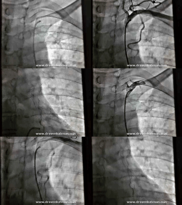
Looping of LIMA is rarely an issue in hemodynamic point of view. But some  believe  a looped up LIMA is slightly prone for graft disease.Complex looping are reported rarely.

A loop and a early branch of LIMA : What is the implication ?

Abnormal  or premature branching pattern  of LIMA  needs clipping as it may divert blood supply to LAD.Terminal branches can be used as a sequential graft to a branch of LAD  usually a diagonal. In spite of all these issues , LIMA is  rarely unsuitable either anatomically or physiologically .It is a safest vessel to graft.

Future of LIMA  graft assessment.

Currently selective LIMA angiogram is the gold standard.

MDCT (64 slice) gives stunning images of LIMA graft , but unfortunately , it has little value for functional assessment .

Functional assessment of LIMA graft By  angiographic frame count  is being attempted in our institute.Will be reported in 2012.

Is hypertension really a major risk factor for CAD ?

    1. Yes it is !
    2. No . . . it is not !
    3. May be !
    4. I don’t think so !

Ans : Any of the above can be a  right response , depending upon our basal and perceived  level of knowledge .

Answer analysis

  1. SHT  is  one of the risk factor for CAD  agreed ,  but definitely not a major one , as SHT per-se rarely precipitate a STEMI
  2. Unless SHT occurs with dyslipidemia, smoking or diabetes it is  rare to cause ACS.
  3. The only  adverse effect of SHT  is  , it has a potential  to aggravate atherosclerosis  by promoting epithelial injury and dysfunction.
  4. Hypertension is a well known  major risk factor for cerebro vascular disease while it is minor risk factor for CAD !
  5. We do not know yet why cerebral vessels are intolerant to high blood pressure while coronaries are pretty happy  with it !

Final comment

SHT is not a major risk factor  for CAD ! At worst , it can propagate chronic CAD. This sort of reasoning  may be considered a huge controversy  . . .but it is really not !

  • One evidence for the above observation is  , we  have  been struggling hard  for over a half a century  to prove a elusive  point that controlling blood pressure  to optimal levels  would  dramatically reduce  cardiac   events !
  • Further,HT’s  relationship with acute coronary syndrome especially STEMI  is vague , it is very rare for patients with accelerated hypertension or malignant hypertension to  present with STEMI *

* Caution :Young doctors should not get confused with this seemingly  controversial observation .This write-up , tries  to convey  a point  , SHT may not be that bad for coronary arteries when compared to cerebral arteries . However BP control remains  vital in  all patients who have  developed a cardiac  event or in patients with multiple risk factors .

Please note ** SHT is still  a powerful risk factor for cardiac failure.(Acute LVF to be precise ) ***SHT can aggravate unstable angina , but very  rare to precipitate unstable angina.**** SHT ./High intra-coronary  pressure can theoretically  dissect or fissure a plaque . (The fact that , HT is so prevalent in a community  but spontaneous  coronary  dissections are not !  should make us think further !)

What  is the   “secret of success”   among current generation  cardiologists ?

A . Strong foundations in cardiology with excellent clinical skills and a rational approach to the given problem.

B.  The  secret lies in the  nimble  fingers  which  acts  almost , like an extension of catheters  in cath lab !

C.  The speed with which he can mobilise a cath lab team in an ” off – office hour”  primary  PCI !

D. It is the the cunning art of  converting coronary  angiograms into angioplasties , by lucid  discussions  with patients and their   relatives  in the the silent cath lab corridors  !

Answer:

When this question was posed to a group of cardiologists ,  D  was considered  most important B,and C came close behind   and   A  was  probably least important  and few thought  “A” character  is rather an  impediment to  become a successful cardiologist !

*Unfortunately a successful cardiologist is defined in India by number of angioplasties he does per month, What  a disgrace to a great medical specialty called cardiology !

What is normal CAG to angioplasty conversion ratio ?

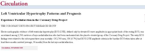
LVH is traditionally believed to be adverse marker for both coronary events and cardiac failure. . (Framingham ) While  this may be  true most of  time  we  also  have evidence to  suggest  patients with LVH  tolerate ischemic injury better.The area of MI is less.  In fact , the coronary drug project was not able to identify LVH as a major risk factor  for MI.  But many other studies continued to portray  LVH  dangerous parameter in ACS.

This paper from Sheba medical center Israel   tries  to answer this  question succinctly !

Summary

LVH  indeed  is an  adverse  predictor  for cardiac outcome  in the  long term.Meanwhile , since the   7 day mortality of STEMI  is well below  3% if  associated with LVH    keeps the controversy alive  ( 5-7% in non LVH group ) .This piece of statistics     gives credibility to the belief   LVH  may  really prevent  early deaths  in STEMI.

This phenomenon about LVH is  consistent with our observation  in  one of the Asia’s oldest coronary  care units (Started in 1972)   .None of the  STEMI patients with LVH  had a cardiogenic shock  in the recent past   !

* It is important to realise not all LVH are same. Inherited LVH, Diabetic LVH and  uremic(  or sub uremic ) LVH  behave differently. Since  the concept  of LVH is  carried  by physicians   in  a single basket ,  we tend to miss the  true benefits of LVH that occur purely due to good exercise training or a mild HT !

In other words , LVH that do not involve interstitial proliferation  is  probably  good for the heart !

Final message

With due regards to Framingam study  , presence of LVH in ECG in any form of acute coronary  syndrome  should  bring a sense of comfort  in the coronary care units .I agree  , it may increase risk of sudden death in some of the population but still it has some unique and definite  advantages at times of  ACS.

Reference

1 Levy D, Garrison RJ, Savage DD, Kannel WB, Castelli WP. Prognostic
implications of echocardiographically determined left ventricular mass in the
Framingham Heart Study. N Engl J Med 1990;322:1561–1566

2.The Coronary Drug Project Research Group. Left ventricular hypertrophy patterns and prognosis. Experienced post infarction in the Coronary Drug Project.Circulation 1974;49:862–869.

3.Behar S, Reicher-Reiss H, Abinadar E, Agmon J, Barzilai J, Friedman Y, Kaplinsky E, Kauli N, Kishon Y, Palant A. Long-term prognosis after acute myocardial infarction in patients with left ventricular hypertrophy on the
electrocardiogrm. Am J Cardiol 1992;69:985–990.

*Coronary drug project (A old study done in early 1970s has more credibility when LVH was not considered as pharma target !)

We presume  ECG  fails miserably against echocardiography for assessing hemodynamics , while  echocardiogram  has  little value  when it comes to studying   electrophysiology .  Ironically ,  we often  ignore  the fact  ,   ECG can  provide  important long-term   hemodynamic  data . The pattern of  chamber enlargement  give us  vital clues to the prevailing hemodynamic  stress and loading conditions. While echo  can be termed as an  anatomical and  physiologic   modality  , ECG  apart from  its unique capacity to record cardiac  electrical finger prints ,  it  provides  useful ,  anatomical ,  hemodynamic information too !

While Doppler is a  fascinating modality to measure hemodynamic data in a moment to moment fashion it can never ever tell us  , what has been going around in the preceding months or years. This  is were chamber size helps which  give us chronic physiological information (Chronic  Doppler ?)

A simple E:A reversal  in  mitral inflow doppler can be a  innocuous  finding in isolation  . If it is associated with even   minimal grades of  LAE  it gains huge importance. That is why left atrial size is  funnily referred to as HB A1C of diastolic dysfunction ( A marker of chronicity  of  diastolic dysfunction)

If LAE is so important to diagnose diastolic dysfunction , why  we are so  obsessed  with doppler filling profiles  of mitral valve ,pulmonary veins, mitral annular tissue Doppler and what not ! .Many of these sophisticated doppler methods are extremely operator dependent  and are  subjected  to technical and mathematical errors. Especially , with  tissue doppler where we  magnify the errors as we  filter  extremely  slow tissue motion .

For  many  decades  we  have failed  to impress ourselves  , about the importance of subtle P wave abnormalities in the  ECGs   of  hypertensive patients.

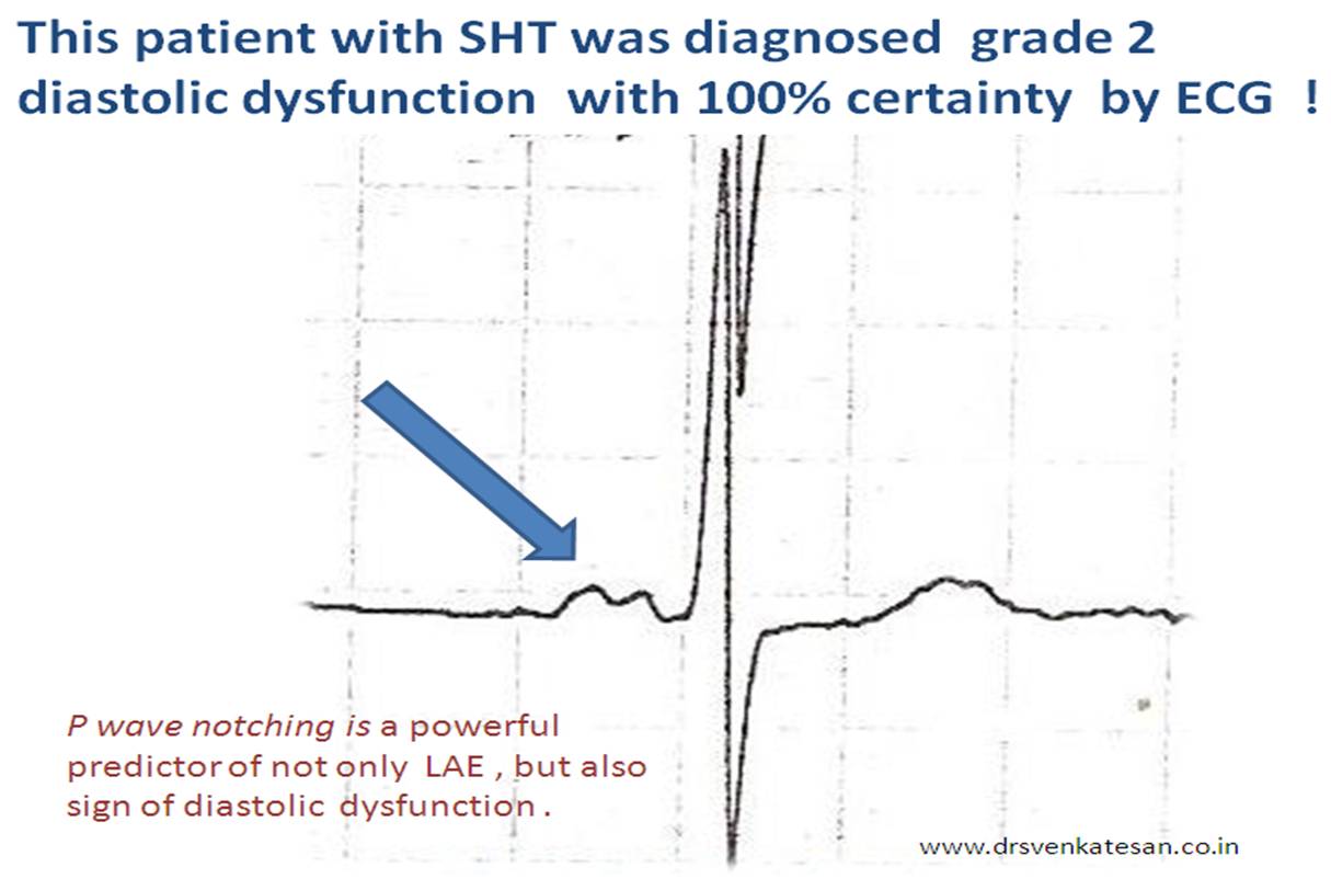
In fact those  innocuous looking  slurs and notches   in P waves ,  suggest the left atrial  stress and a definite marker of underlying LV diastolic dysfunction .

P wave is the only electrical wave that occur in diastole .Hence there is no surprise  ,i  gives us enormous information about this phase of cardiac cycle .

If only we look  at them carefully, zoom it (Now it is made easy with so many softwares)  analyse critically we can find a wealth of information about the atrial behavior in hypertension.

Experience from our hypertension clinic  with periodic echocardiograms suggest ,  the following  ECG  findings   can be   good markers  of significant  diastolic dysfunction .

  1. Notched P wave
  2. Wide  P waves
  3. Slurred  P wave
  4. Bi-phasic P waves

* Surprisingly  , these abnormalities correlated with at least grade 1 diastolic dysfunction even in the absence of  for LAE or LVH by echocardiogram.

** In an  occasional patient  P waves  can widen due to inter atrial block or conduction delay. This a rare exception for wide P waves without LAE.

Final message

A well recorded and   analysed   ECG can  predict diastolic dysfunction  with fair  degree of accuracy .This fact need to be emphasized  by every one  .  Next to ECG ,  LA size and volume  by 2d echo are excellent parameters  to assess diastolic function in a long term fashion. Sophisticated  but  error prone ,  momentary doppler parameters are getting too much attention  at the cost of simple ,  shrewd ECG and 2D echo  !