Feeds:
Posts
Comments

A well researched article on a difficult topic. By Kim et all from Cornell university , New york.

A must read by all cardiologists . The link is placed with the courtesy of Jacconline

http://content.onlinejacc.org/cgi/reprint/49/20/2035.pdf

After reading this article   one  should be able to answer the following questions.

  1. What is Gallavardin VT ?
  2. Classification of RVOT VT
  3. How a non sustained VT becomes a sustained one ?
  4. Why some VTs cause syncope ?
  5. What is the association  between idiopathic VT and Idiopathic VF ?
  6. How does exercise  trigger a  VT ?
  7. What do we mean by structurally normal heart ?

Readers are encouraged to post link to good articles on this topic.

How to name  a ventricular tachycardia ?

This  continues to be a  favorite past & present  time of  modern-day cardiologists. Especially ,  VTs associated with structurally normal heart  suffers with this  protracted problem .Widespread use of  EP study has not solved the issue as yet.

The   VTs that arise from the left ventricle in an apparently normal heart   has been referred  by  various terms.

  • VT with structurally  normal heart
  • Idiopathic left ventricular tachycardia
  • Verapamil sensitive  VT
  • Septal VT (It can be either myocardial /non myocardial origin)
  • Narrow qrs VT
  • Fasicular VT
  • LVOT tachycardia
  • Hemodynamically stable VT
  • Belhassen VT

Now we have still more exotic VTs like Cuspal , mitral annular , etc . All of the above can mean anything , or same thing   in different centers  ,  different cardiologists in different times .

* RVOT tachycardias also have  many synonyms.( Adenosine sensitive, Adrenergic,  Gallavardin, Parkinson Pop, etc )

VTs associated with CAD , valvular , myocardial diseases generally devoid of  nomenclature problems. Ischemic VT  is yet to be classified in a proper fashion.

The confusion in classifying VT is  not due to the complexity of heart disease. It is due to  the general  comprehension failure as  every VT can be described with reference to clinical , ECG morphology, hemodynamics and presence or absence of underlying heart disease. A simplified and clinically useful VT classification is being prepared in this forum .Will be published shortly .

Coronary artery lesions can be classified by many types . The popular ones are by Ambrose and Ellis  .They are adopted by ACC and SCAI  .While various  terms  are  used to   describe a lesion. (diffuse, discreet , eccentric , long , tubular  etc) A tandem lesion is the one which has special significance , but is not well discussed in the literature .

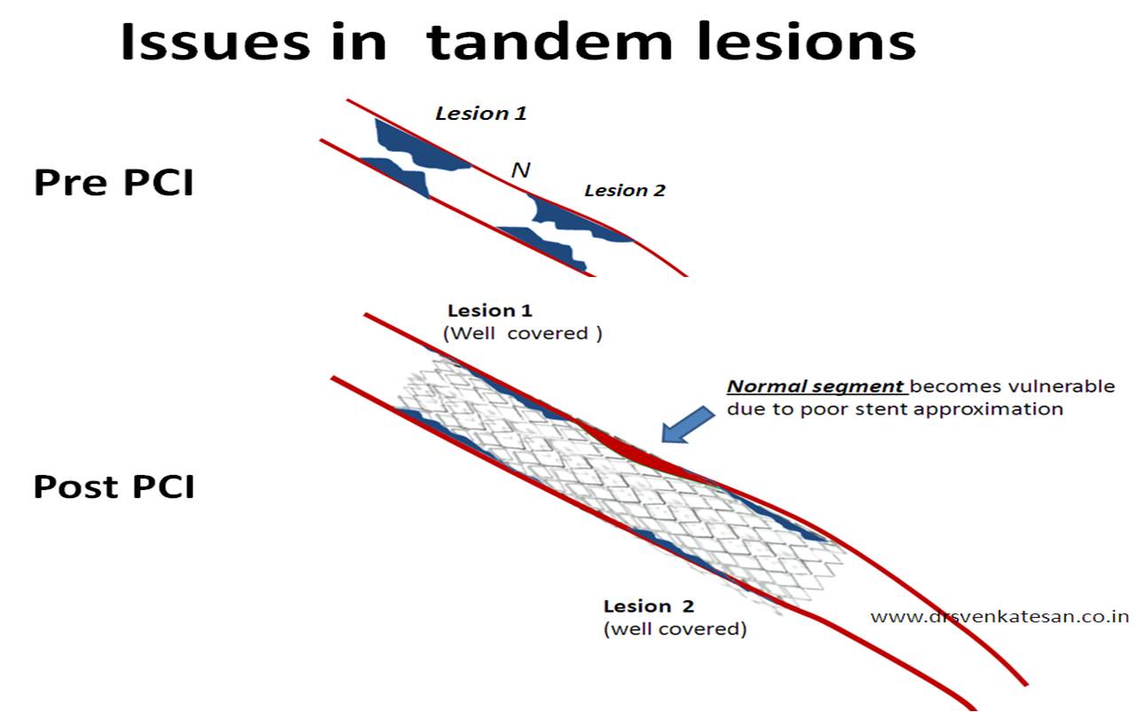
A tandem lesion is  diagnosed when  two lesions  closely abut  each other one behind the other  with an  intervening normal segment. (Like the bullets loaded in a  tandem fashion  in a  gun )

Generally there will be at least  few millimeters of normal intervening  coronary segment.This is  referred to  as  connecting segment.

Clinical importance of tandem lesion

Tandem lesions  carry  the  same significance  like  any other lesion. But ,the primary aim is to tackle the two lesions with a single stent. We know stent edges are rheological culprits.  Two stents  have   4 edges. It is better to cover  the tandem lesions with one long stent* even if we have a sufficient  connecting segment.Geographical miss is less likely with a long stent.  In the strict sense one wold require an IVUS (Intra vascular  ultra sound ) to confirm the normality of the connecting segment. Tandem lesion is  a  marker  of diffuse atherosclerosis  and  the connecting segments often   show ectatic changes.

* This is a ironical as  the conventional wisdom would  tell us , lesser the  metal load it is better for our coronary arteries.But once we embark on a complex  intervention we just can’t restrict the use of stents. The more you put the more it will demand.There are some interventional cardiologists who convert the entire coronary artery in to a metal tube (With or without realising the consequences !)

Illusions of  tandem lesion.

Many  times ,  spiral folds  from a single  atherosclerotic   lesion mimics  a double lesion .This need to be differentiated from true tandem lesion.

What is the hemodynamic significance of  tandem lesions ?

Rules of hemodynamics  would  dictate ,  in a linear and laminar flow  model across a tube ,   immediately after an obstruction there will be a significant  drop in resistance.

This  forms the fundamental   phenomenon  within the coronary artery  . This explains the biggest mystery in cardiology . . .  How  the  TIMI flow is  maintained till 90 % of the  lumen is narrowed. This  also  explains the concept of flow limiting lesion .(Why  a coronary lesion do not obstruct the flow  till late stages  ?)

Does this rule on  hemodynamics  apply in tandem lesions ?

When a lesion is followed  by a lesion with little normal segment in between what happens ?

The blood gets a double jolt every time it traverses a tandem lesion. There  may not be sufficient time and anatomy for the mandatory pressure drop to occur. So for a  given degree of obstruction ,  tandem lesions  is likely to be   more thermodynamically significant than a single lesion.

Pressure recovery after  an obstruction is also incomplete , as the forward head of blood column encounters another hurdle even before it recovers from the initial turbulence.

Which lesion is more important   in tandem proximal  or  distal  ?

The distal lesion determines the thermodynamics of proximal lesion while the distal lesion as  such is  less influenced by proximal.

Long lesion vs tandem lesions


Some times it may appear ,  it is better to have a long lesion than  a two lesion  in tandem. This is because the stent will approximate more evenly .Further there is less likely hood of in -stent restenosis in long lesions as the   edge effect can occur  right in the middle of  the stent in tandem lesions .

Now it is increasingly realised, many of the sub acute thrombosis  are due to po0r stent approximation in tandem lesions or long lesion.


http://www.springerlink.com/content/g063752436617n51/

http://www.ncbi.nlm.nih.gov/pubmed/8789675

Inter-ventricular septum is generally in parallel alignment with Anterior aspect of root of aorta.

Have a look at this echo form a 65year old man

Note the classical bend of basal IVS encroaching the LVOT. Aortic valve opens normally.


The basal septum Projecting into LVOT

Pseudo LVOT gradient. It was about 25mmhg which is not significant.


  • Sigmoid shaped IVS can  cause a unique  LVOT  profile .
  • Present in elderly . The exact mechanism and  mechanical and hemodynamic   implications are not known .
  • It can rarely cause  dynamic LVOT obstruction.
  • When these patients develop Infero posterior MI there could be further collapse of IVS into LVOT .

Relationship between Aortic angle , Hypertension, HCM

Many of the HCM patients may show similar features .When ASH is confined only to basal IVS. HCM should not be diagnosed instead it is  often a defect of aorta /IVS alignment .

Abnormal  aortic angle  with that of IVS  may  make the IVS appear sigmoid.

If  patients  with abnormally angled aorta develop hypertension ASH and sigmoid septum is  more pronounced .

IVS dragging by mass effect  on aortic root is possible and Aortic regurgitation may ensue

Reference

Time is muscle .This  may sound as  an old fashioned statement now ,  for many of us. But the fact remains. Every minute following  STEMI ,  myocytes  keep  losing it’s life one by one unless , the  intervened.

The prevention of myocyte death can be accomplished by three ways

  1. By early thrombolysis
  2. By Primary angioplasty
  3. The  one  that happens naturally by a process called spontaneous thrombolysis *

* Most have a  strong belief  that the  natural forces are incompetent to lyse a  small thrombus within our coronary  arteries  ( While  , we  fully  realise   natural  forces  like  the Tsunami can wash out  the entire ocean floors  ) . Never under- estimate the force of  nature !

Balloons are not privileged !


It is widely accepted , a time window of up to 12 hours is optimal for reperfusion. Beyond that time , there is no point in reperfusing  the muscle  as   it  might have died. While ,  the majority of cardiologists agree  to this and they  promptly  refuse  to thrombolyse ,   if the patient comes  12 hours after an onset of STEMI  .They are labeled  ” late on  arrival”  and  coded  as ineligible for thrombolysis.

The moment they are labeled as ineligible for lysis , a dangerous thought process runs across  the minds of  many cardiologists. It is  possibly  the most important paradox (Shall  we call it as sense failure ? )

Such lysis ineligible  patients    become  automatically eligible for primary PCI . . . It is curious  to note , the  time window for primary PCI is also less than 12 hours is strangely forgotten.

It has become a prevalent  practice  by all unscientific means  , most  cardiologists extend  the time window for primary PCI well beyond 12 hours  , some even up to 36-48 hours.  No wonder . . . then why open artery trial (OAT) miserably failed . Even a  novice  can predict the out come when  one tries  to resuscitate the  dead muscle .

Final message

Myocardium  does not behave in a privileged  manner  during a STEMI.  It  simply does  not bother  about the way  by which  it is going to be rescued and reperfused  .All it needs   is a timely help. It can not extend its   life just because it is being rescued by a  sophisticated modalities like pPCI.

If the patient is late for thrombolysis ,  he is late for  primary PCI as well .

Please do not change the time window in STEMI  according to  our  whims and fancies . It is  an  unscientific and unprofessional  way to practice cardiology .

  • It is a complex PCI procedure meant for  high risk  bifurcation /Trifurcation lesions
  • Two stents are simultaneously  deployed.
  • It aims to prevent sudden acute occlusion of one of the major  branches .
  • It is not an easy procedure , and be used only in rare circumstances .
  • Distal left main and ostio proximal LAD/LCX  is a  classical  example.
  • Navigation can be difficult , only well experienced operators should attempt it.

*Is there a ready made two lumen stent available ?

The image is meant for concept purpose only !


It is one of the techniques available to stent unprotected left main

An excellent review  in  ACC intervention journal for unprotected left main .

Click on the Image to reach the article


Medical imaging technology is the fastest growing  sub specialty in medicine .Now,  we can image almost every organ in detail in our biological system.With capsule  endoscopy one can visualise deep interiors of intestines,  we can get into the coronary artery  by angioscopy.We can even image the molecular metabolism within the cells.

While  , we   feel elated about all these technologies , every emergency  physician  will awkwardly recall how difficult  it is  to  visualise a structure  , located  just few centi- meters  beneath our throat called vocal cords  .

I am sure more  lives are lost in this world by delayed and difficult  intubation than any thing else  in medical emergencies. Cardiac arrest can be tackled lot easier as the defibrillator is largely independent of  rescuer expertise .Intubation of airway is vitally  dependent on expertise.

Click on the Image to reach Airtraq web site


When we are able to visualise the  deep segments of bronchus  with  fibro-optic scopy ,  why there has not been a  simple , practical ,cheap optical solution to the   superficial structure namely the larynx ? Human mind is too  funny ,  if  only larynx was deeper  our scientists  would have discovered such a  device very early .  Since it is just below the oral  cavity  no body  thought it is  worth to  discover a  scopy for that !

Now  the long wait is  over ,  we have a tool called Airtraq .There needs to be further refining of this device .

Is it possible for the tips of endotracheal tubes to  have  cameras  that  transmit  live images wireless ?  , which  can guide the tube   straight to the trachea .

We have twin cameras for our fancy cell phones while the life saving tracheal tubes are as blind as ever !

It is argued more such devices should flood the market . Every doctor and paramedical  worker should acquire one  ( Preferably  integrated to their phones ) Imagine if they  can beam the live pictures of the vocal card into their trendy  3 inch  screens of  i Phones . This could be the greatest revolution to occur in cardiopulmonary resuscitation .

(Airtraq please note this . . .I do not need a  royalty for  the  Idea !)

Final message

It  is vital for the   emerging technologies  , to be intelligently used  for the   betterment of our patients. When video  phones  calls transmit  live action  to  our mobiles  across the continents  ,   Is it not  funny we struggling    hard to get a good image of human vocal cords   that are  sitting  just few cm below the throat !

Let us think simple for complex problems .  Many breakthroughs  will automatically  happen.

Three cheers to the developer of this Airtraq device !

The review published in the prestigious NEJM seems to suggest

PCI  , the most  commonly  performed  therapeutic cardiac intervention  may  result in  more  myocardial  infarction in the community  than  the deadly atherosclerosis itself.


Can it be true in any  stretch of imagination ?

Yes , it seems so . But the only issue  is the  criteria   used to define MI  .

Comments are welcome on this article .

You won’t get the full text article free  .Try to get it from your library .It is worth the time spent  !

http://www.nejm.org/doi/full/10.1056/NEJMra0912134

This is one of the wonderful corporate initiatives to assess the coronary angiogram and reporting . This calculator and teaching material was created by Boston scientific and Syntax study team . This  was used primarily during the  SYNTAX study.  This scoring system ,  though  appear  elaborate,  is a very  useful ,  objective way to assess coronary angiogram.

http://www.syntaxscore.com/calc/start.htm

Final message

It is encouraged to use this scoring system liberally . This will help us  to take more scientific decisions .


  • Coronary collateral circulation continues to be a poorly understood phenomenon.
  • It reduces the impact  of ischemia , salvage myocardium, keep it viable, and  can  even  be  life saving during a STEMI
  • It can support either the same coronary artery  or the contra lateral coronary artery (Like the above patient )
  • The usefulness of  collaterals  at times of exertion is controversial .Most interventionists do not believe in  it . (Facts are opposite of course !)
  • Bridging coronary  arterial collateral often indicate hardened total occlusion and success of  PCI is reduced

Here is  the  angiogram  which shows classical intra coronary bridging  collaterals.