Feeds:
Posts
Comments

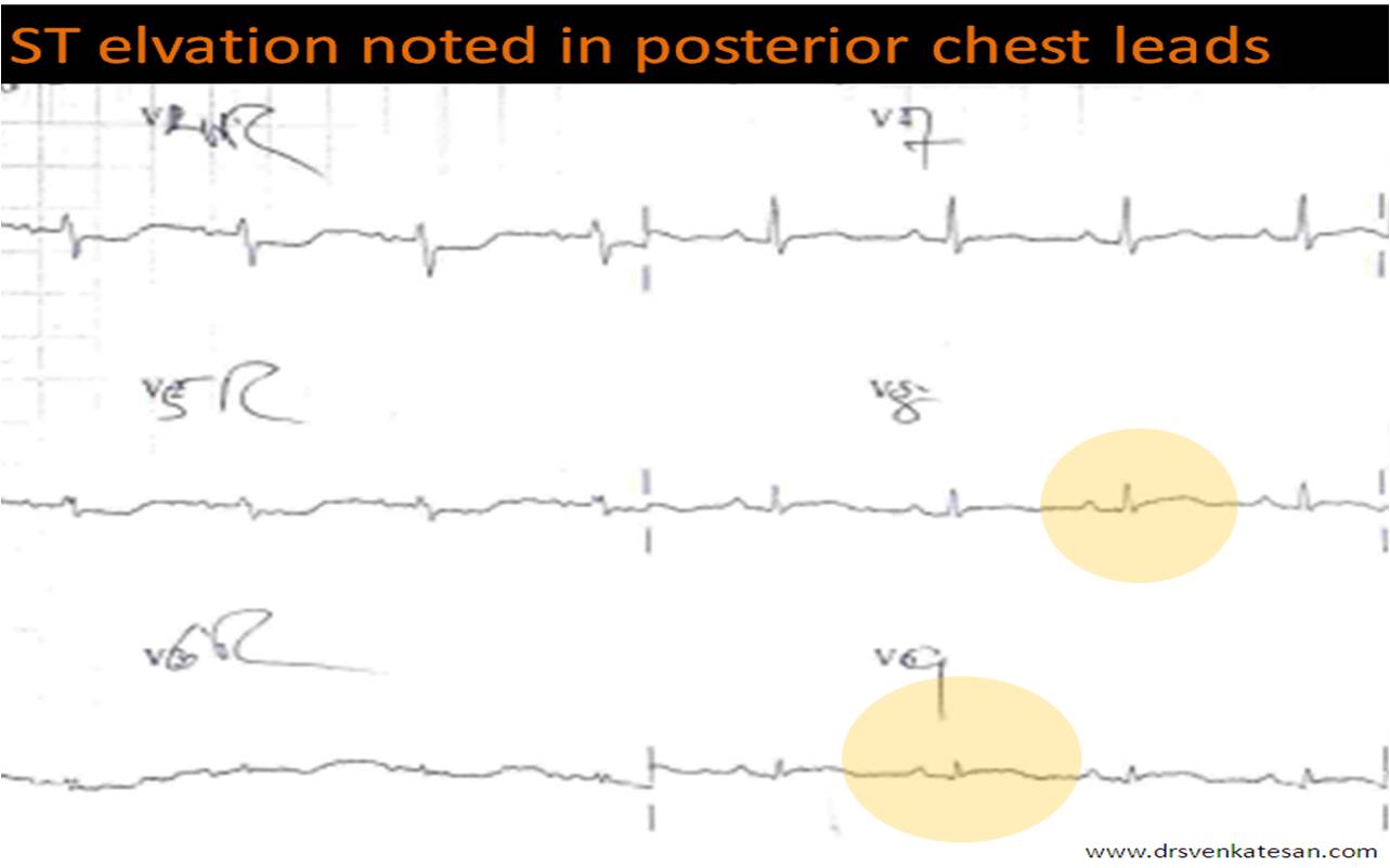
Thrombolysis is specifically indicated when there is  ST elevation in ECG. ST elevation is a surrogate marker for  total coronary occlusion. It occurs due to current of injury flowing   towards* the lead  facing the  infarct territory . There is only one situation where you can safely and effectively administer thrombolysis in the presence of ST depression ie

Note : There is no accepted terminology  to label a MI as  ST depression MI . Here it is used to emphasise ST elevation is not the only indication for thrombolysis .In posterior MI there is infact ST elevation but it is failed to pick up by standard 12 lead ECG.

NSTEMI is a different entity altogether and  thromolysis is never indicated.

Isolated ST depression in V1 V2 V3 .It almost always indicate isolated posterior STEMI. This can be confirmed by posterior chest ECG leads V7-V10 .

*One will be surprised, to know  the mechanism of ST elevation in STEMI is still not fully elucidated .Technically speaking the net movement of current is away from electrode as  there is only a baseline  diastolic shift  which  gets neutralised in systole  mimicking an ST elevation .(Electro-optical illusion !)

How sensitive is these leads to detect isolated posterior STEMI  ?

Fairly sensitive. Both scapula and  para spinal muscles can be a  significant electrical  barrier that can prevent ST elevation from inscribed .In case of doubtful ST elevation in posterior leads , mit is always better to rely on the clinical presentation.Acute chest pain , consistent with ACS and a new onset ST depression >2mm V1 to v3 is a definite indication for thrombolysis .

Link between posterior MI and RV MI ?

They are closely linked entities .In fact posterior surface of heart is contributed significantly by RV.

What is the angiographic correlation of  isolated ST depression in V1 to V3 ?

It almost always localise the lesion to left circumflex artery . If it is dominant , it can involve lateral and RV territories.

Is isolated posterior MI  less dangerous ?

May be yes , but only after the patient reaches the hospital as electrical risk is same in every STEMI .

The area of infarct  is less , LV failure is less common. While conduction disorders and ischemic mitral regurgitation   can occur  significantly.

Also read ,  Why thrombolysis is contraindicated in UA/NSTEMI ? in this blog

Mitral valve can be termed as  the most important valve of heart . The reason  for this  : It is the only valve that is dependent on the  Left ventricular function (The  parameter  which   determines the  ultimate outcome in any form CAD ! )

So , indirectly mitral valve function will invariably be affected by some degree  at least in  most  patients with LV dysfunction. (After all LV free wall , is a component of mitral valve apparatus.)

While , we have numerous modalities to assess mitral valve function  ,  the one that has fascinated the surgeons during  mitral  valve surgery is the intra operative TEE.

Many believe TEE provides live  images  of mitral valve   which are not possible  even under  direct vision ! The eye of the  TEE sees the mitral valve  from a  posterior location , (of -course It can see at any angle !)   while surgeon can see in one angle . The  types of repair , the adequacy  of repair, the annulus status,  even a trial mitral run ,  can be done with the help of TEE.

The TEE probe silently does  this job sitting inside the esophagus   , without  obstructing the surgeon’s operative field .

The success of TEE as an investigative tool did not come easy.Decades of  observation , innovation and learning( Especially from  department of cardiac science  Mayo clinic USA , where they standardized the views. )  are involved .

Now we have omni plane, real time 3D TEE probes .

The books  which are  considered the best for  TEE aspects of mitral valve  and it’s  repair are

Cardiac myxomas are rare tumors. But they present in a dramatic way. It can have  severe  systemic symptoms and present even as  fever of unknown origin ! While , physicians  of  previous era were struggling to make a ante-mortem diagnosis we are blessed  to make a instant  diagnosis with echocardiography !

Want answers  for all these  from the original researchers ?

  • What is the pathology  myxoma ?
  • What  are the  classical Locations ?
  • Difference between Sessile Vs pedunculated
  • Soft vs Hard
  • Benign vs malignant / Locally invasive
  • Recurrent myxomas
  • Vascularity  of myxoma
  • Calcification (RA myxoma> La Myxoma)
  • Non atrial  myxomas(Valvular  papillary myxoma)
  • The  Cell of origin (Stellate , polyhydral )

You  will not get a better reference than the following article , including extensive illustrations . An 1980 article from  Mayo clinic published in American journal of pathology

http://www.ncbi.nlm.nih.gov/pmc/articles/PMC1903582/pdf/amjpathol00223-0227.pdf

LA myxoma  : A Video

A case reported from my hospital Hosted as Video presentation  Follow the link

http://www.youtube.com/watch?v=SD2LrK1mdic&feature=related

Belgium SOAP  wants a  knock out punch to  Dopamine in shock !

Vaso constrictors are  the mainstay  drugs  in the management of shock  syndrome. While ,the ultimate outcome depends on the primary  cause for shock, these vaso- constrictors  have  a critical role in sustaining life , till the organ function is recovered.

The physicians world  over,  differ  in their choice of  vasoconstrictor support .They  are almost divided   equally in their usage between  dopamine and norepinephrine  .

Surprisingly,  there  has been no one to one comparison trial till  this study   in 2010 .This trial  is called SOAP 2 published from  Brussels , BELGIUM .It  compared the usage of these two drugs in variety of shock  syndromes. It favors norepinephrine use ,  that includes  cardiogenic shock as well.

The disadvantages of dopamine noted in this trial was

  • Increased  risk of arrhythmias
  • Increased rate of death  in cardiogenic shock

The implication of this trial may force the ACC/AHA guidelines , which  advices  dopamine as the first choice in shock syndromes especially in cardiogenic cause.

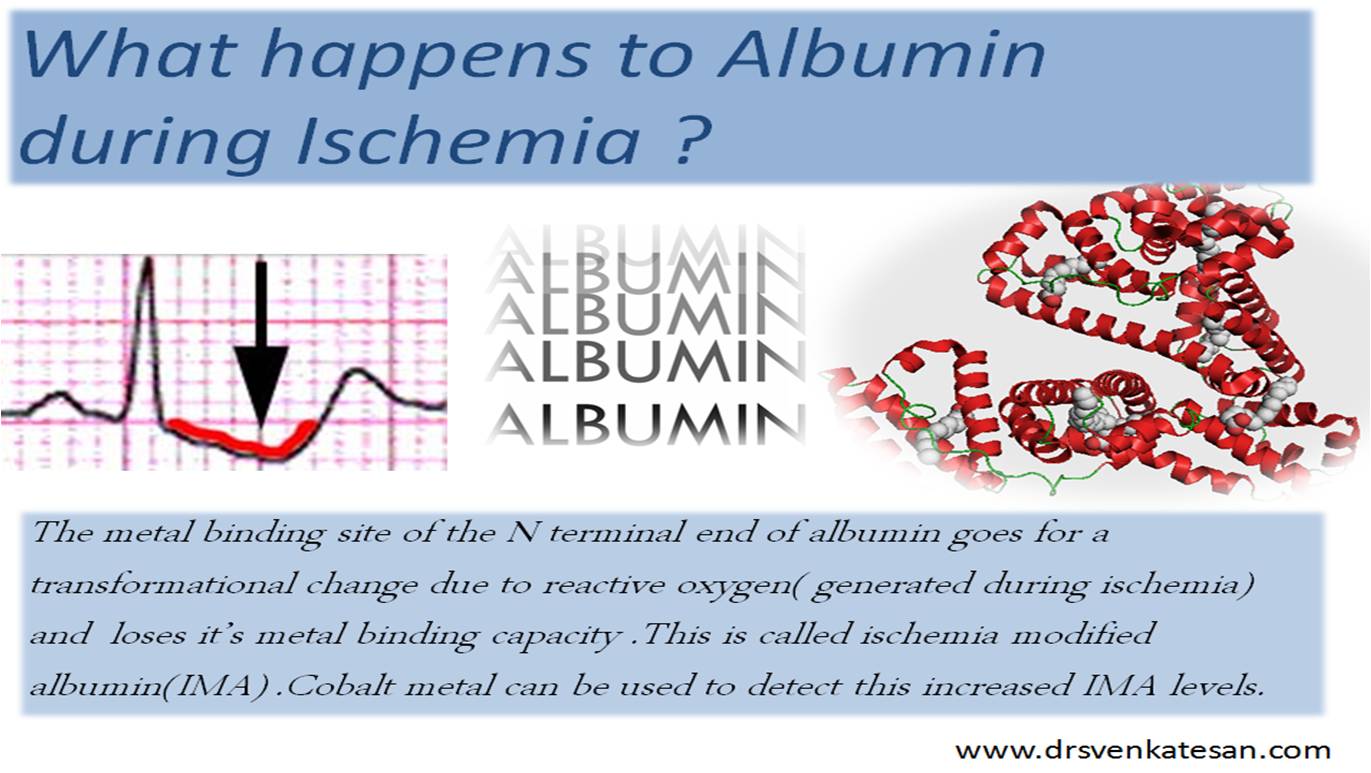
In the diagnosis of ACS, we have definite bio  markers for acute MI . Further, ECG  has a good  spcecificity  for STEMI . While ,clinical and ECG features of ischemia are not perfect. A bio marker for ischemia is the ultimate dream of cardiologists  and  emergency room physicians. . In this context , the IMA -ischemia modifed albumin  has come  with   great expectation .

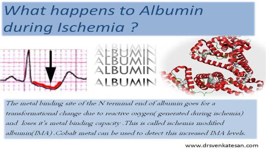
Ischemia modifies what ?

Normal albumin moleule has  a metal binding site (Copper ) .There are few free binding sites available .During ischemia this metal binding capacity  reduces .  A cobalt containing  reagent when added to ischemic blood  finds binding sites scarce , and hence  excess free  cobalt  will color the sample  and a  posiitve test for  ischemia is diagnosed.

*Normal human albumin  may contain  2%  of ischmia modifed albumin which is expected to increase up to 6% during ischemia

IMA raises not only during ischemia it can also  raise during oxidative stress

  1. Stroke, 
  2. Chronic kidney disease 
  3. liver disease,
  4. Maligancy

It can also  be elevated following

  • Routine coronary angiogram
  • PCI
  • DC shock

Increased lactic acid for example in sepsis may reduce the IMA level and can miss an episode of true ischemia

Final message

IMA can be a useful tool to identify  ischemia early .But lacks senstiivty . New improved immunoassays may be more sensitive and specific

Reference

1 .Medscape review  

2. Circulation article

3. PRIMA study which  was done in ER in risk stratifying ACS proved IMA is not vey useful  http://emj.bmj.com/content/23/10/764.abstract

A drug with a peculiar mode of action is recently making waves across the globe (At least some parts of it !) Basically , when the world was looking for an  anti anginal drug without much hemodynamic effects Trimetazidine fitted this slot .While it’s popularity soared  in Europe and Japan , the Americans thought there was a  need for a similar drug but supposedly  different mechanism of action . Thus  Ranalozine was born.It is a piperazine derivative.

How does Ranalozine act ?

There are three important actions for Ranlozine  (  4th one a real surprise !)

  • Antianginal
  • Anti arrhythmic
  • Possible lusiotropic effect
  • Anti diabetic*

*It is  surprising to note Ranalozine was  reducing Hb A1 c . This is incidental or a simple statistical fairy tale we do not know !

Anti anginal

  • It acts on the late Na ion channel  in phase  2 of action potenial .
  • This facilitates  excess Ca  to be removed from the cells by the NA/Ca exchanger
  • Calcium exit improves  ischemic  myocyte’s   metabolic performance as  cell injury is prevented .

This makes this drug an effective anti anginal.

The MERLIN TIMI 36 in NSTEMI  population became a  big dampener against the enthusiasm for this drug.But it did not affect much the sale of the drug !

Antiarrhythmic

A drug , which acts on phase  2  Na channel , it is not at all a  surprise to have anti  arrhythmic properties .

It can reduce phase 2 reentry like EADS

pro-arrhythmic action

Paradoxically  by blocking Na channels it stretches  the phase 2 and hence  QT interval is prolonged

Lusiotropic action

By preventing  the calcium from accumulating  from the cytosol it has a  theoretical ! lusiotropic action.

It is funny , even  theoretical action  is  enough ,  for  many drugs  to enter  the standard cardiology literature !

What is major advantage of  Ranalozine  over other drugs ?

It virtually has no hemodyanmic effects .

Unlike other anti-arrhythmic drugs it acts , only  if the late Na channels are abnormally active .

(Which is a case during episodes of ischemia .)  This prevents  QT interval to prolong in normal persons

In spite of all these meaningful actions and less side effects , FDA approval why cardiologists in the back of the  mind think this drug is not  based on strong scientific principles , and it could  simply  represent  an industry sponsored  placebo ?

I do not know the  answer  to this question ,  but I do share the same feeling.

Will it succeed the test of time ?

In this context , I recall a famous statement   by my professor “A drug , which has least side effects , is very unlikely to have any desired effect also”

But  with  world  ,  taking a commercial avatar  a success of a  drug  lies ,  not in  having a desired action but in  how many million packs are ultimately   sold  in the market. It is akin to a bad movie succeeding  phenomenally  in box office while  a good one lying silently   unnoticed

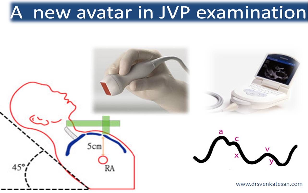
Looking at the neck veins for hours  together  has been a favorite pastime of our cardiology ancestors.Thanks to those sharp intellect , that has led us to this height of cardiovascular revolution. Measuring JV pressure by itself was considered a big science. Putting a patient in 45 degrees , marking the sternal angle, identify the  oscillating venous column,  measuring   the vertical distance  etc . . .

Now in 2010 , with bedside hand-held echo one can rapidly  rule out an elevated  central venous pressure by imaging the jugular vein directly . Here is an article from American heart  journal

http://www.ncbi.nlm.nih.gov/pubmed/20211304

Simon MA, Kliner DE, Girod JP, et al. Detection of elevated right atrial pressure
using a simple bedside ultrasound measure. Am Heart J 2010; 159:421–427.

Soon , your mobile will double up as ultra-portable  echocardiogram

Read this related article in this blog .

Coronary care in your pocket

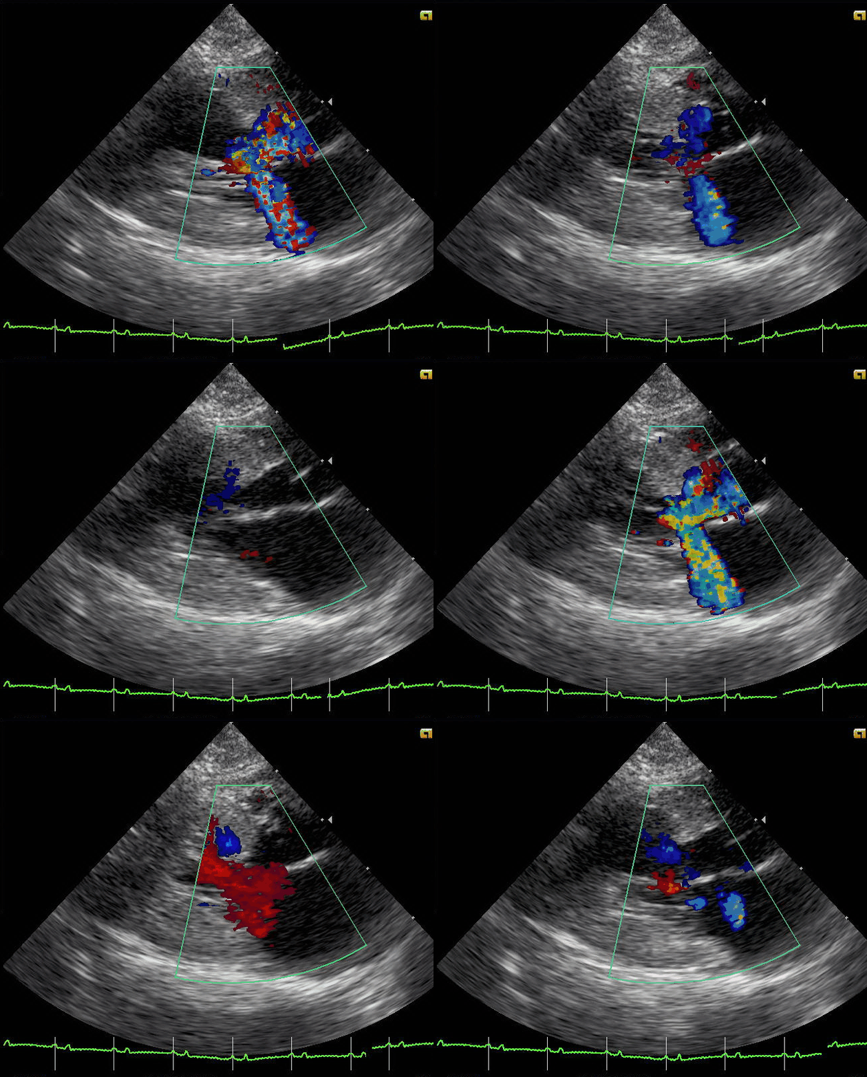
Hypertrophic cardiomyopathy (HCM) manifests   with or without obstruction. Obstructive HCM ,  (ie HOCM)  is more often symptomatic .However , the risk of arrhythmias, sudden death, and some degree of diastolic dysfunction are common in both.

ECG, clinical examination are generally  not sensitive to identify obstruction in HCM  .Echocardiogram is the easiest  way to identify  the obstruction (gradients> 3o mmhg across LVOT are considered significant ).LV angiogram ,MRI, CT scans are rarely necessary today.

However , the following clinical clues will help us  to suspect significant obstruction in HCM

History

  • Class  2 or  3 dyspnea.
  • Exertional syncope
  • Exertional angina

Pulse

  • Pulsus bisferiens (Two peaks in systole )

LV apex

  • Sustained , double apical impulse  often indicate obstruction.
  • Presence of Mitral regurgitation ( 20% can have  MR without obstruction due to intrinsic abnormalities of  mitral valve )

* It should  be realised , valsalva induced MR /LVOTO  may occur in many of the non obstructive HCM.

What happens to  clinical signs of obstruction with medical therapy(Beta blockers etc)

One would expect these signs to regress or disappear, but it rarely happens. The pulse , the  murmur show  little change .  This implies , the main mechanism of beneficial effect could be in  heart rate  reduction , and  improvement in the   diastolic properties of left ventricle.

It is traditionally believed  , renal blood flow is critically determined by the  luminal diameter  of  renal artery.But in reality  there are more important factors  other than renal  arterial diameter  that determine the glomerular  blood flow.  As in any vascular bed, it is the arterioles that determine the resistance and hence the flow . These arterioles  form the  critical  resistance  points and acts as   check  valves in this  “vascular  highway”  flowing across the renal terrain !

Unlike other micro-circulations ,  the kidney has a  special job to do ,  ie  filtering  the toxic  molecules.  Hence,   for the blood entering the kidneys  , even  nourishing the kidney seems ,  a less important  function !  The nephrons  of the kidneys are probably the most  “high – tech” cells in human body (Of course ,next only to brain cells ) .The vascular  tuft of glomerulus located within the bowman’s capsule  is perfused  by afferent arteriole and drained by efferent arteriole .

The entry of blood into glomerulus is regulated both by afferent and  efferent arteriolar  tone .These  two micro-circulaoty units  are under the  sensitive control of both neural  and humoral  signals. Glomerular circulation is meticulously  regulated by renal juxta glomerular apparatus.It modulates the glomerular  blood flow by secreting renin which  acts through Anigiotensin 2  on the   efferent arteriole .

The tone of the  efferent  arteriole  is thought to be the single important factor in this servo control mechanism.

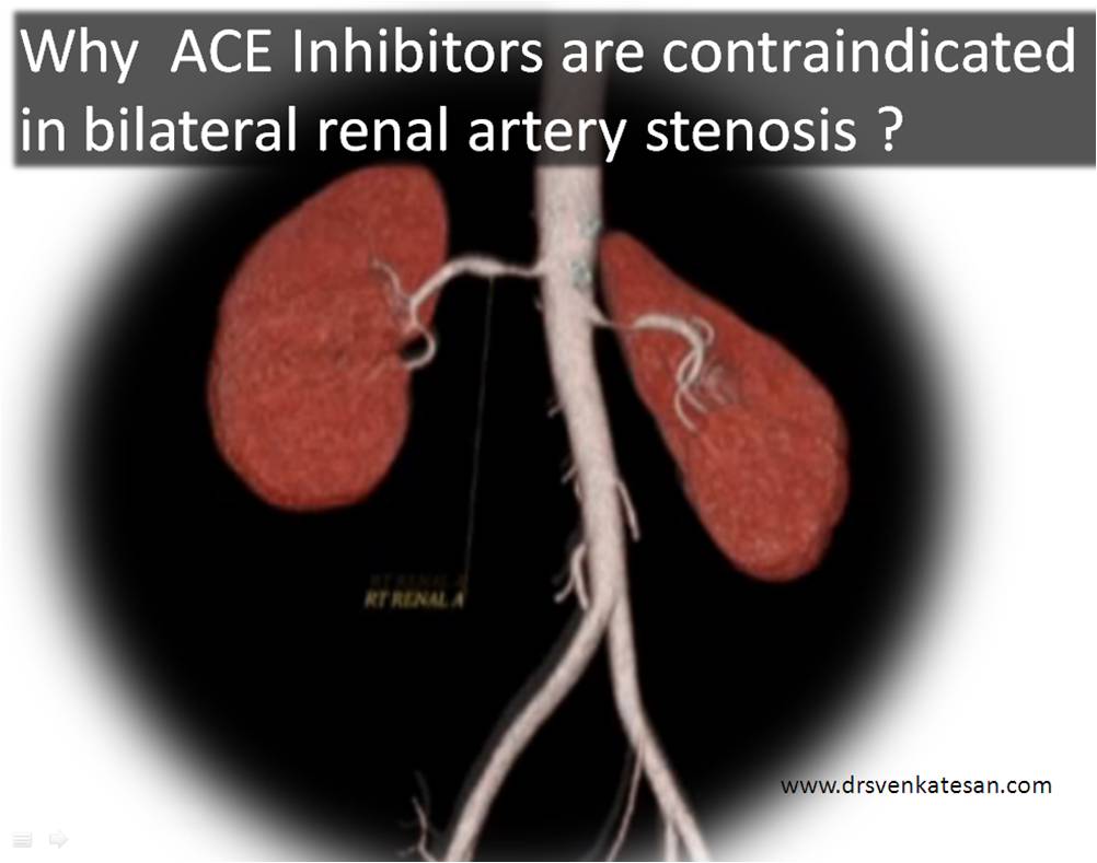
What happens in bilateral renal arterial stenosis ?

When there is bilateral renal arterial stenosis the effective renal blood flow is not  significantly reduced , but maintained at  the cost of increasing the efferent arteriolar constrictor tone. It  is  like a  check valve at  the  exit point of a dam , which is partially closed to maintain the adequate pressure head (Here , intra-glomerular  pressure head )

What happens when ACEI are introduced ?

Once ACE inhibitor  is administered , the efferent arteriolar   tone is removed , this triggers  the intra glomerular pressure to drop  suddenly and filtration pressure reduces .

Note: ACEI does  not reduce the renal  blood flow  directly  but  the glomerular  perfusion pressure drops hence precipitating acute renal function deterioration.

What is your comment about the reno-protective effects of ACEI ?

The medical science’s  most  crucial  moments  occur  , when we confront  two diagonally opposite views  are  debated  and both  suggest , there is definite benefit for the patient ! Cardiologists and nephrologists were always  made to believe  ,  ACEI are  unfriendly to kidneys . But ,we now have  evidence , ACEI is not an untouchable molecule in renal  dysfunction.

This is based on  the observations made , over the years that  excess Angiotensin 2  is  ultimately a liability for the kidneys !

Looking at a  long  term perspective  , AT 2  increases the intra -glomerular hypertension and ACEI inhibitors reduce it.This  pr0tects the  nephrons from  hyper-filtration  mode ,  that accelerates the  glomerular  injury . So , the  current thinking  is  ACEI has a definite role in arresting the progress of  renal cell injury .

This is akin to beta blocker story in CHF which was initially contraindicated in CHF later became a definite indication

The only issue for ACEI is , it should not be continued if an ARF like picture sets in .(Acute deterioration ). Otherwise ,  in CRF at any  basal level of serum creatinine  , ACEI can be continued . Some think even an  increase by few mg of creatinine  can be allowed .

So the following can be a working guideline *

  • ACEI can be started  or continued at any level of creatinine in stable CKD with or without dialysis

But ,ACEIs need to be stopped in all of the following 

  • Acute renal failures
  • Acute on chronic renal  failure
  • Accelerated elevation of  creatinine  (As in bilateral renal artery stenosis)

How much elevation of creatinine is allowed in CKD  with ACEI  ?

This is   not answered yet .

*Caution : The above conclusions on ACEI and creatinine was  derived  by me , based on  with  personal discussions with my  Nephrology colleagues. It may  be subjected to correction.

//

The much fancied criteria   “suspect secondary  HT” if the  onset of  hypertension  is   before 30 years   later than  55  years ,may be useful  .But a caution about this criteria  : It does not mean you should not hesitate to  diagnose renal HT  between 30 -55 years.  The  real onset may be   < 30years , but  the patient may report to the physician  late  in his /her  40 or 50s !

  1. Diastolic blood pressure > 120
  2. Sudden acceleration blood pressure
  3. Blood pressure which is  resistant to control with three or 4 drugs ,that shall typically include a  diuretic.
  4. An episode of left ventricular failure (Often referred to as  called flash pulmonary edema)
  5. Presence of  Hypertensive retinopathy
  6. Para umbilical bruit
  7. HT associated with significant CAD
  8. Marked LVH in echocardiography
  9. Finally , most importantly , worsening of renal function with ACE inhibitor is a  strong clue the kidney is under perfused  and  the   renal circulation  is dependent on  elevated angiotenisn 2 (Which ,if blocked worsens the GFR ).This implies every physician should take a baseline serum creatinine  and urea before starting them on ACEI.(Which is rarely followed , as far as my country is concerned !)

Is there any simple way to  differentiate  reno vascular from renal parenchymal HT ?

It is very difficult to differentiate between these two clinically. It makes things more difficult , as  combination of both occurs. Prolonged renal ischemia can result in parenchymal damage as well.

The simplest way is to do a rapid ultrasound imaging to assess kidney size and texture (Loss of cortical-medullary differentiation indicating early renal contraction phase ).Of course , our nephrology colleagues are always there to help you out .

* It need to be remembered the functional renal HT -Renal tubular acidosis,  Adrenal HT (Conn’s /chromo-pheocytomas  has to be ruled out , as these entities also occur in the same age group ).The combination of hypokalemia and mild alkalosis is a  good clue to rule out many of these  defects.

* The CT scan image used in the above illustration  courtesy

http://www.ajronline.org/cgi/content-nw/full/189/3/528/FIG21