Feeds:
Posts
Comments

Posts Tagged ‘drsvenkatesan’

Recurrent myocardial infarction following an ACS is a fairly common clinical problem. Many times this is not recognised because it is difficult to establish the diagnosis.

The issues relevant here is

When does the first infarct (Index infact) process end ? and when the second infarct process start ?

Can the first infarct be a STEMI and the reinfarct be NSTEMI ? ( Dual acute coronary syndrome )

The only way to confirm a diagnosis of reinfarction is to document raising titres of cardiac enzymes and second peaking of CPK MB . New fresh ST elevation after a succesful thrombolysis is also a useful sign. But ST elevation in a q lead simply reflects a wall motion defect . So it requires enzymes to confirm it.

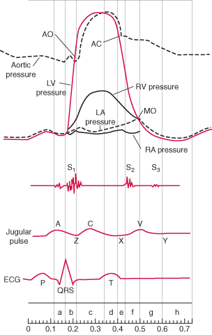
When there is tachycardia the ST segments tend to elevate following MI.

Other confounders are Infarct expansion and infarct extension .

These are macropathological entities almost impossible to dignose with surface ECG. What we diagnose as re-infarction could be an infact a infarct expansion.The modern terminology for infarct expansion is ventricle remodeling .The extreme remodeling results in ventricular aneurysm .Adverse acute ventricular remodeling can closely mimic a reinfarction .

What is clinical relevance of diagnosing reinfarction ?

Nothing great !

In modern day cardiology it is not a bother whether the infarct is expanding, extending or reinfarcting !All one has to do in a patient with chest pain ,showing a fresh ST elevation following STEMI is to take him/her to cath lab .

The only issue here one has to remember there are mechanical cause also for ST elevation following STEMI .

Dr.S.Venkatesan,Madras medical college, Chennai.

Read Full Post »

Pulse deficit is a clinical sign wherein , one is able to find a difference in count between heart beat (Apical beat or Heart sounds ) and  peripheral pulse .This occurs even as the heart is  contracting , the pulse is not reaching the periphery.This can occur in few clinical situations .

1 . Atrial fibrillation.

2. Very early diastolic  ventricular  ectopic beats

3. Some patients with Pacemaker.

The mechanism is  , the ventricular contractions are  too weak and unable to open the aortic valve  (Or opens feebly*)  , but at the same time they are good enough to close the mitral valve. To open the aortic valve it has to generate atleast 60-80 mmhg pressure , while mitral valve closes even  as LV generates  8-14mmhg  .(LV/LA pressure cross over). So intermitently the  second heart sound  is missed while S1 is retained,  producing more heart sounds and less pulse count in the periphery. The S1 is either felt or heard at the apex but the corresponding pulse is missing . Further , this intermittent absence of  S2  results in totally irregular S1 /S 2 relation.


Why some of the contractions of LV is too weak to open the aortic valve ?

Because the RR interval varies , the ventricular filling also varies , diastole duration is constantly changing some of the diastole are too short  and LV hardly gets filled , as the LV force of contraction is directly decided by the LVEDV and LV  fibre length these contractions are too weak.

Other published studies

There has been some doppler observations ,where there is a midventricular LV blood flow reversal in atrial fibrillation that could  explain the pulse deficit . Mechanism of production of pulse deficit in atrial fibrillation: assessment by blood flow dynamics

Second Department of Internal Medicine, Kagoshima University School of Medicine.

 http://www.ncbi.nlm.nih.gov/pubmed/3221309

What is the clinical significance of pulse deficit ?

Currently there is no great clinical significance of this phenomenon. But an astute clinician will pick up this sign and it may indicate underlying LV dysfunction. In patients with PPM,  pulse deficit  suggests  pacemaker malfunction .Some patients with cardiac tamponade &  pulsus paradoxus  systolic  blood pressure falls too low to make the pulse feeble or not palpable in the periphery .This situation may mimic a pulse deficit if not recognised.

Dr.S.Venkatesan ,Madras Medical College , Chennai, India

* What is the evidence for intermittent absence or feeble Aortic valve opening in Atrial fibrillation ? I could find this from the book written by Harvey Feigenbaum. whom we consider Father of Echocardiography


Read Full Post »

To download complete presentation click on the slide

“ WAVE RED FLAG” FOR ANTICOAGULATION, WHEN YOU ENCOUNTER MOBILE LV CLOT !

Venkatesan Sangareddi , G. Gnanavelu ,M.A Rajasekar, V.Jaganathan

Department of cadiology , Madras medical college , Chennai.

Formation of LV mural thrombus is one of the important sequel of STEMI. The natural history of LV clot is variable. Spontaneous dissolution often occur . Stroke and peripheral embolism, are other natural events by which left ventricle get rid of the clot. The morphology and the behavior of LV clot is determined by endogenous procoagulant and fibrinolytic mechanisms. Drugs administered in the peri infarct phase also play an important role. In current thrombolytic era ,the incidence of LV clot has come down. Once the clot begins to form over the raw area adjoining a dyskinetic segment, it follows the local hemodynamic factors , that determine the shape , size of the clot which varies from linear , layered , projectile or pedunculated.

Administration of oral anticoagulants remain the standard practice in patients with LV clot. It is prescribed , in the hope that it will prevent the progression of clot and prevent thrombo embolism . Whether, long term warfarin dissolve , regress or dislodge the thrombus is not known. We have observed the incidence of CVA is high in the first few weeks following introduction of oral anticoagulants . We report our experience in 8 patients, with LV clot in Acute MI . All patients were male . Age range 22-58 .All had anterior MI. The mean EF was 38%(28-43%) the mean size of LV clot was 1.4cm (7mm -24mm) mobility was graded with reference to independent movement parallel or perpendicular to the LV. 3 had highly mobile clot. 5 had relatively fixed clot. All were put on titrated warfarin. Two patients who had large LV clot with a stalk got dislodged after starting anticoagulation. The CVA occurred on 12 th and 14 th day after starting warfarin .The pedicle is probably the vulnerable point and is exposed to greatest risk for dissolution . On the other hand the 5 patients who showed relatively stable clots are attending to our cardiology OPD without any events . One patient who had a mobile clot , which got organized at 4 weeks , incidentally this patient had discontinued anticoagulants.

We conclude, oral anticoagulation has a potential to destabilise and dislodge a mobile LV clot in the early days following STEMI .Existing anticoagulation protocol recommends, oral anticoagulation for all patients who have LV clot. This need to be redefined. If surgery is not an option , temporary withdrawal of anticoagulation may be indicated in selected patients with LV clot, to facilitate organization of clot.


!

Read Full Post »

Transient ischemic attacks are not exclusive to cerebral circulation.

Many such episodes can occur in coronary circulation also .

TIA of heart PPT presentation

Click here to download  tia-of-heart

Read Full Post »

LV clot formation is one of the important complications of acute myocardial infarction. Preventing this is difficult and managing this problem is still more difficult.Some of these clots are linear and laminar along the shape of LV apex and carry less risk of dislodging.

 While mobile LV clots , even if it is small can cause a embolic episode. Most of these patients have a significant LV dysfunction and they are candidates for early CAG and revascularisation. Even If the coronary anatomy is very ideal for a PCI these patients are often sent for CABG and physical removal of LV clot . If  only ,we have an option to remove these LV clots by a catheter based modality, we can offer them a totally non surgical cure.

This is not impossible,  considering  we are in the era of percutaneous implantation of prosthetic valve in Aorta ! The only issue is potential embolism into carotids and periphery .A temporary distal protection at the level of aortic root will prevent that .

Device companies shall produce one such exclusive catheter system to remove LV clot.

Dr .S.Venkatesan, Madras medical college, Chennai,India

Read Full Post »

Peer review of an article even in major journals never scrutinise the “Aim of  a study ” . However big is the journal,  they seem to bother only about the authors, materials, methods, and statistical analysis.  If only they peer review an article , right from the “Aim of the study” like ,

  • Who asks the research questions?
  • Who  defines the aim of the study ?
  • Who decides which drug to be compared with which drug ?
  • Who steers the steering commitee of a trial ?

If only , we could answer these questions without bias , pharma industry and their  regulators  would have ,  far more better image than what they have now !

A typical example for , the aim of the study  to be  wrong  , is  the “ONTARGET’ study on telmisartan.

Here they ( Who ? ) raised an inappropriate  question of     “Non inferiority” of one drug with other  without any  valid reason to compare these two drugs that will benefit the man kind !

Read Full Post »

Atherosclerosis is  the number one killer of mankind  .It involves all medium and large sized blood vessels.Any intima and media can be invaded by the disese process.Most common to involve are  cerebral, carotid, coronary, aortic  and it’s branches, renal, and peripheral arteries. But how pulmonary artery is missing in this list ? Is it really true (or) are we missing it ? One logical explanation is pulmonary circulation is a low pressure circulation and the maximum presssure is less than  30mmhg . This pressure may be insufficient to induce endothelial injury that predispose lipid mediated injury.

Other explanation could be a structural difference in the media and intima compared to aorta .But in patients  with primary or secondary pulmonary hypertension where,   inspite of PA pressure being high ,  still atherosclerotic changes is very uncommon . or Is it the Heath Edwards pulmonary vascular sclerosis  grading  reflects nothing but pulmonary atherosclero-thrombosis !

If this is true there could be a major role for HMG Coa reductase inhibitors in altering the natural course of pulmonary obstructive vascular  disese . Statins might be tried in PPH  a disese with no specific  treatment !

Read Full Post »

CCU’S can also save  patients with cardiogenic shock

Many of us would say ” never” or some may say “rarely” but in reality the answer is “yes it can ” slightly lower than  Primary PCI . One could save atleast  few  lives every month by  intensive medical  management alone (Inotrope, vasodilator,pacing if needed ) in any coronary care unit.

So the message here is, not offering or doing  a primary PCI in a patient with cardiogenic shock is not  synonymous with  inferior treatment or death.  After all, in the much hyped SHOCK  trial a significant no of patients survived in medical limb .

Read Full Post »

Thousands of dissections happen in cath labs  all over the world every day  very rarely it is painful . The answer is not clear. Both have rich vasa nervorum. Aortic dissection  involves media and smooth muscle . Coronary dissection may also be a  equally painful  , probably we are not recognising it ! or we attribute   all  chest pain in ACS  to ischemia .

Deep dissections into the smooth muscle should be painful.  Type c nerve fibers carry pain signals from heart

Answers welcome.

Read Full Post »

Plaque fissure ,rupture and subsequent thrombois is the hallmark of acute coronary syndrome . Are these events painful ? We always attribute any chest pain in an ACS patient to ischemia of myocardium.Is that always true? Coronary artery also has a rich vasa nervorum that could be activated by plaque disruption.

Why  we need an answer to this question ?

We are triaging patients for early invasive apporach based on chestpain .

Many patients may be subjected to revascularisation process for an non ischemic coronary pain !

Read Full Post »

« Newer Posts - Older Posts »