Archive for the ‘Uncategorized’ Category
What is the greatest medical breakthrough since 1840?
Posted in cardiology-ethics, Uncategorized, tagged Add new tag, bmj, COMMON SENSE, ethics, INTERHEART on October 14, 2008| Leave a Comment »

Is atrial fibrillation a benign arrhythmia ?
Posted in Cardiology - Electrophysiology -Pacemaker, Infrequently asked questions in cardiology (iFAQs), tagged atrial fibrillation, cardiac arrhythmia, cardiology, drsvenkatesan, ECG, electro physiology, heart rhythm, nejm, pace, ventricular fibrillation on October 7, 2008| 1 Comment »
Ventricular fibrillation is invariably fatal if not treated . When can atrial fibrillation be fatal ?

Atrial fibrillation is relatively a benign arrhythmia especially when it occurs in isolation with structurally normal heart.This is sometimes referred to lone atrial fibrillation . Even otherwise, atrial fibrillation is rarely fatal except in few situations.But AF commonly destabilises the patient who have baseline valvular or myocardial disease.(Post MI, dilated cardiomyopathy etc)
There are few situations where AF can be life threatening
- In patients with WPW syndrome*where , AF enters into a electrical short circuit , downhill to enter the ventricle and make it fire at the same rate as that of atria . ( ie 400-600) and result in ventricular fibrillation.Note , even here it is the VF that kills not , AF per se.
- AF in acute MI often precipitates LVF , but rarely fatal.
- In patients with critical aortic stenosis, or hypertrophic cardiomyopathy, sudden onset of AF can result in acute cardiac failure.
- AF is often a terminal event in primary pulmonary hypertension
While atrial fibrillation is less likely to cause death , it is a highly morbid arrhythmia .It is one of important cause of stroke in elderly as well as young !
What determines hemodynamic stability in ventricular tachycardia ?
Posted in Cardiology - Electrophysiology -Pacemaker, cardiology- coronary care, Infrequently asked questions in cardiology (iFAQs), tagged bmj, fasicular tachycardia, heart rhythm, Hemodynamics, ischemic vt, jama, lancet, lvot vt, myocardial VT, nrjm, pace, ventricular tachycardia, verapamil sensitive vt on October 3, 2008| Leave a Comment »
Ventricular tachycardia is considered as one of the most dangerous cardiac arrhythmia .Rather , it is the label VT that spreads more fear than the arrhythmia itself. It is a fact many patients with VT walk into hospital , still VT will always be a sinister arrhythmia as long as it carries a risk of degenerating into ventricular fibrillation.
What determines hemodynamic stability in VT ?
- Origin and location of VT
- The ventricular rate
- Presence or absence of AV dissociation
- Impact on mitral inflow pattern
- Associated left ventricular dysfunction or valvular heart disease.
- VT in the setting of acute coronary syndrome.(Ischemic VT)
- Inappropriate drug selection
Origin and location
VTs originating high up in the ventricle( High septal VT,Proximal VTs) have more organised ventricular contraction and they are more stable.Distal VT originating in the myocardium away from the conducting system has chaotic myocyte to myocyte conduction.These are very unstable.
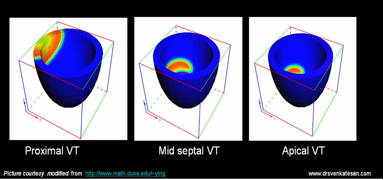
The term fascicular VT is nothing but VTs originating in the His bundle and it’s branches( Can also be termed Septal VT ).These VTs are also stable and some of them respond well to calcium blockers indicating that they are very close to the AV junction and carry the properties of junctional tachycardia. QRS width gives a rough estimate about the location of VT. Narrower the VT higher it’s origin.( But remember even in VT , qrs can further widen on it’s way downhill !)
LV dysfunction.
This is probably the most important determinant of the outcome in VT. Patients with severe LV dysfunction (EF <30%) fare badly .Hence the land mark concepts from MADIT 1& 2 demanded ICDs in these patients.The most common clinical setting is dilated cardiomyopathy.SomE of them have bundle branch re entry(BBR).This particular VT can be stable for many hours.
Ventricular rate.
Usually VT has a rate between 120-200.Higher the rate of VT more the chances of instability .This rule is also not always true as fascicular VT can be well tolerated at high rates.So location of VT focus and LV dysfunction usually over rides the impact of ventricular rate.
Mitral inflow pattern
Proper left ventricular filling is the key to hemodynamic stability in VT. In proximal, septal,fascicular, LVOT VTs doppler studies suggest (ACC /AHA Type C evidence : Personal observations in CCU during VT) near normal preservation of bi modal filling of mitral valve inflow.In ischemic myocardial VT the mitral inflow profile is critically affected . There is no distinctive forward filling was observed .In fact at rapid rates a short pulsatile MR jets are noted instead.
Associated valvular diseases
It is obvious, aortic and mitral valve disorders can aggravate the hemodyanmic instability.
Final message
The clinical behavior of ventricular tachycardia is widely variable and dependent on multiple factors.
Associated LV dysfunction and structural heart disease ultimately determine the outcome.
Why syncope is rarely fatal ?
Posted in Cardiology - Clinical, Infrequently asked questions in cardiology (iFAQs), Uncategorized, tagged bmj, cardiac arrest, cardiology, cardiology lancet, drsvenkatesan, epilepsy, head up tilt test, holter, hut, hypoglycemia, jama, loop recorder, loss of consiouness, nejm, neuro cardiogenic syncope, stroke, sudden cardiac death, syncope, vaso vagal syncope, ventricular tachycardia on September 30, 2008| 1 Comment »


Syncope by definition is a transient loss of consciousness due to cerebral hypo perfusion and loss of muscular tone, and the patient falls but recovers fully and gets up either assisted or spontaneous.The cardiac and vascular counter response to syncope is most often intact .This makes syncope characteristically transient . If a patient does not recover from syncope it could either be a prolonged loss of consciousness( Stroke etc) or if he never gets up he will be called a victim of cardiac arrest or a SCD ! (Sudden cardiac death ) . So technically by defintion , all patients will have to survive the syncopal episode.
But the following questions need to be answered
- How prolonged a syncope can be ?
- Can syncope lead onto sudden cardiac death ?(SCD)
- What are life threatening syncope and non life threatening syncope ?
What is the link between, syncope and SCD in patients with ventricular arrhythmia’s ?
Some case of long QT syndromes could be life threatening especially in children as they inherit sudden death. A patient with a non sustained VT may develop syncope if the VT becomes sustained especially if there is underlying heart disease and LV dysfunction . Among this few , may degenerate into ventricular fibrillation and patient may die.
How common is syncope in acute myocardial infarction ?
Syncope is a very rare presentation of acute myocardial infarction.
Can syncope precipitate or precede a cerebro vascular accident ?
Prolonged syncope , TIA, stroke in evolution and completed stroke can be a continuous spectrum in patients with carotid and cerebrovascular disese . But when a syncope evolves in to a stroke the patient is not considered to be a victim of syncope but they enter the stroke protocol.
There is a big list for the causes of syncope
But to put it simply
A.Cardiac
- Purely electrical ( Arrhythmic- Brady, Tachycardia)
- Mechanical( Valvular obstruction, and other structural heart disease etc)
B. Non cardiac
- Vasovagal (Commonest 90% of all syncope)
C. Metabolic*
- Anemia
- Hypoglycemia
- Hypoxia
How to work up a patient with syncope ?
First , one need to confirm it is indeed a syncope . If the initial examination is not clearcut one need to go back to the history and ask for circumstances under which the syncope occured and details of prodromal symptoms if any . Patient’s family members who witnessed the event can give useful information . It is the most cost effective ( Comes free of cost infact !) investigative tool available .Cardiac syncopes are usually sudden, vasovagal often have environmental or emotional factor. Apart from routine investigations , ECG, Echocardiography, holter are done generally, head up tilt test, Loop, event recorders may be reuired in few.
Final message
Syncope is one of the common symptoms in cardiology and general medical practice. Many times the diagnosis is easy . Common syncope is never fatal but , ruling out dangerous tachy and bradyarrhythmias is a key aim. In a significant number (20-30%) identifying the cause could be really difficult and may never be made in spite of the modern diagnostic tools. These syncope of unknown origin is grouped along with the neurocardiogenic category.
The one, positive thing about syncope is (unlike chest pain) , it is rarely fatal in it’s first episode , gives the physicians to investigate and correct the underlying problem.
In a patient with renal failure and critical coronary artery disese , who is being planned for CABG and renal transplantation which should precede what ?
Posted in Cardiology - Clinical, cardiology -Therapeutics, cardiology- coronary care, Uncategorized, tagged cabg, cardiology, cardiomyopathy, combined cabg and renal transplant, dialysis, drsvenkatesan, end stage renal failure, kidney international., nejm, pci, renal transplant on September 7, 2008| Leave a Comment »
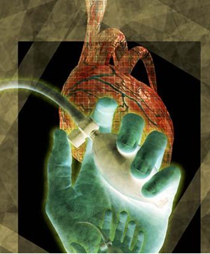
Chronic renal failure and CAD are common companions.Severe CAD in patients with renal failure pose an imposing task on the treating physicians.CABG and kidney transplantation both are major interventions.When a patient requires both the decision making becomes much more difficult.
The possible choices are
A. Do CABG first follow it with renal transplant .
B. Do renal transplant first follow it with CABG.
C. Do CABG first and defer transplant & advice life long dilaysis
D.Do renal transplant and offer medical management / PCI for CAD if feasible.
E.Simultaneous CABG & renal transplant is a remote possibility .
F.In terminally ill , combined cardiac and renal transplantation is the ultimate option. (Possible in very few centres in the world)
G.In severe co-morbid condtions avoid both and support life. Success is not in completing the procdeures but in providing useful life !
Among the options the most prefered worldwide is option no 1. This has a caveat. If angina is dominant CABG should precede transplant. If cardiac failure is dominant the issue need further scrutiny.
Given a situation ( DCM & End stage renal disese) , your patient could undergo only one procedure, which will you prefer ?
This again is highly emprical but logic could still be applied. Never do CABG with a sole aim of improving severe LV dysfunction in ischemic DCM .It happens only in journal articles & major clincal trials!.Of course mitral valve correction and LV reduction surgery might help.But in a patient with renal failure prolonging the CABG on table time , with add on surgery is highly risky. So it would be logical to think intensively for postponement of the CABG in a patient with class 4 cardiac failure and renal failure. Do only the transplant .
What is the impact of end stage renal failure on LV dysfunction ?
End stage renal failure has a great adverse impact on LV function. Many times it is reversible.We will never ever know, if you do a CABG first on them. So always think twice or even thrice before voting on this vital issue . Correction of renal impairment can improve the cardiac status dramatically in some.
Ironies in cardiology – Sinus rhythm loses the battle with atrial fibrillation in cardiac failure !
Posted in Uncategorized, tagged AFFIRM, amiodarone, atrial fibrillation, cardiac failure, drsvenkatesan, heart rhythm, lancet, nejm, pubmed, RACE, rate control, RF ablation, rhythm control on August 26, 2008| 1 Comment »
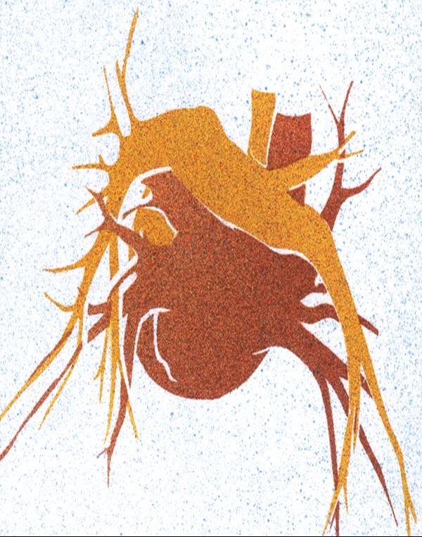
The debate of rate control verses rhythm control in atrial fibrillation goes on and on. The AFFIRM, RACE,PIAF, STAF the HOT CAFE all went against sinus rhythm in the last 10 years . This was one of the settled contoversies in cardiology . The conclusion was ventricular rate control was no way inferior to rhythm control in patients with SHT, CAD population .This made the electrophyiologists wonder how can a natural rhythm fare bad ! . But the findings were consistent .Rhythm control neither improved the quality of life nor it reduced the incidence of stroke. The later finding was very surprising but the explanation was convincing as stroke in elderly was more related to SHT, CAD, DM etc than AF itself. The source of emboli in ischemic stroke could come any where distal to LA .The big assumtion that all strokes in elderly should come from LA appendage or the body of LA was premature and wrong. What prevented stroke in AF was not restoration of SR but administration of oral anticoagulants with adequate INR.(2-3)
Having failed to document superiority in elderly population , the logic machine strongly suggested restoring SR in patients with CHF, will atleast provide hemodynamic and also survival benefit .
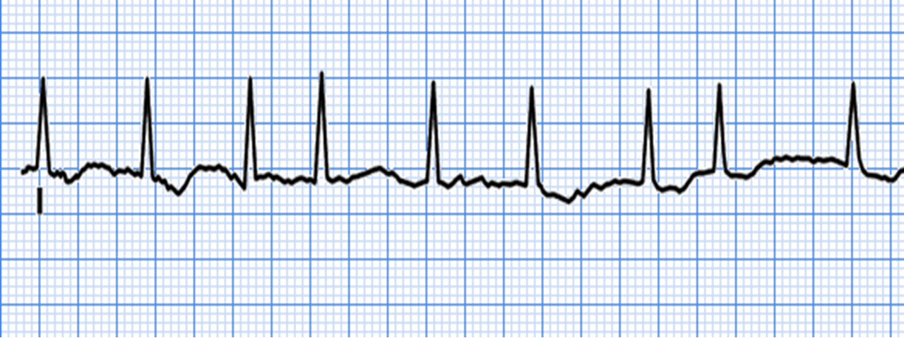
And thus came the AF-CHF trial published in NEJM 2008
Alas ! AF-CHF also found there is no useful purpose of restoring sinus rhythm in patients with atrial fibrillation and cardiac failure. In fact patients in SR fared little worse !
Why . . . why . . . why ?
Should we ask the seemingly absurd question !
Is sinus rhythm poorly tolerated by cardiac failure patients ?
It is some times possible atrial fibrillation by itself could be a mechanism to amplify the cardiac reserve by which it provides a relatively high ventricular rate to improve the cardiac index . Even though the optimal ventricular rate in AF is around 80-90 at times of need it has to increase to 120-130. Patients in class 3 CHF and AF often achieve this in times of demand .This is not possible in patients who are getting rhythm control drugs and further patients in SR can not increase the HR suddenly from 80 -130 .
So is this a wild imagination ! AF could be a safety valve mechanism in CHF to increase the HR . Where the atria come to the rescue of ventricle like a rate adaptive pacemaker .
The other logical* ! argument is that there is nothing wrong with restoring SR , but the methods to achieve and maintain SR is too cumbersome and results in adverse outcome .The currently available drugs are too toxic for the purpose .
If we have a simple and safe way to restore SR in these patients it should always be superior to AF .
But it is a well known fact that , whatever be the rhythm or rate the ultimate outcome will be dictated by the LV function, mitral valve function etc.
Read abstract of AF-CHF
Rhythm Control versus Rate Control for Atrial Fibrillation and Heart Failure
Denis Roy, M.D., Mario Talajic, M.D., Stanley Nattel, M.D., ., for the Atrial Fibrillation and Congestive Heart Failure Investigators
<!–
Rhythm Control versus Rate Control for Atrial Fibrillation and Heart Failure
— Denis Roy, M.D., Mario Talajic, M.D., Stanley Nattel, M.D., the Atrial Fibrillation and Congestive Heart Failure Investigators* –>ABSTRACT
Background It is common practice to restore and maintain sinus rhythm in patients with atrial fibrillation and heart failure. This approach is based in part on data indicating that atrial fibrillation is a predictor of death in patients with heart failure and suggesting that the suppression of atrial fibrillation may favorably affect the outcome. However, the benefits and risks of this approach have not been adequately studied. Methods We conducted a multicenter, randomized trial comparing the maintenance of sinus rhythm (rhythm control) with control of the ventricular rate (rate control) in patients with a left ventricular ejection fraction of 35% or less, symptoms of congestive heart failure, and a history of atrial fibrillation. The primary outcome was the time to death from cardiovascular causes.
Results A total of 1376 patients were enrolled (682 in the rhythm-control group and 694 in the rate-control group) and were followed for a mean of 37 months. Of these patients, 182 (27%) in the rhythm-control group died from cardiovascular causes, as compared with 175 (25%) in the rate-control group (hazard ratio in the rhythm-control group, 1.06; 95% confidence interval, 0.86 to 1.30; P=0.59 by the log-rank test). Secondary outcomes were similar in the two groups, including death from any cause (32% in the rhythm-control group and 33% in the rate-control group), stroke (3% and 4%, respectively), worsening heart failure (28% and 31%), and the composite of death from cardiovascular causes, stroke, or worsening heart failure (43% and 46%). There were also no significant differences favoring either strategy in any predefined subgroup.
Conclusions In patients with atrial fibrillation and congestive heart failure, a routine strategy of rhythm control does not reduce the rate of death from cardiovascular causes, as compared with a rate-control strategy.
Don’t panic when you diagnose a left main disease in routine coronary angiogram : It may cost you a life !
Posted in Cardiology -Interventional -PCI, cardiology- coronary care, Uncategorized, tagged acs, atherosclerosis, cardiology, cath lab, chronic stable angina, coronary artery disese, drsvenkatesan, europcr, intervenional cardiologists, left main, left main disese, pci, ptca, tct md on August 25, 2008| Leave a Comment »
Left main coronary lesions are fairly common during routine coronary angiogram.These may be a critical or a innocuous lesion.The word “left main” triggers a sort of alarm reaction to many cath lab staff as well as the cardiologists and surgeon.Many times, these left main lesions are detected in patients with chronic stable angina who have stable symptoms. Left main disese has not been graded clearly in literature . Often it is perceived , any lesion in LM is serious.
There is an unwritten rule, rather a medical compulsion to take a patient with left main disease for emergency CABG ( Now some centres ,emergency PCI) .Some institutions make it a rule these patients are posted in the next available slot in the theatre.
The basic question we raise here is “Should we consider all left main disease as an emergency”?
Not really , especially when it occurs in a stable angina .One can wait , buy some time to fully evaluate and prepare the patient and may be the patient can be posted as an elective case. It is a well recognised fact that, CABG carries adverse outcome when done as an emergency procedure. This is primarily due to inadequate pre op work up and resultant complications. It is also well known , surgical back up team may not be available in full strength in odd hours .
This post is to convey the message , that left main is a serious disease but that doesn’t mean it should elicit a panic reaction and be taken as an ultra emergency . There has been many morbid and fatal outcomes in many hospitals due to this apparent pseudo emergency !
Note* 1.Left main disease during acute coronary syndrome is to be seen in different perspective.2.Some of the proximal LAD lesions are so tight and could be more significant than left main lesions.
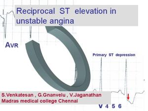
Reciprocal ST elevation in unstable Angina
Posted in Cardiology - Electrophysiology -Pacemaker, cardiology -ECG, Uncategorized, tagged ECG, electro physiology, myocardial infarction, nstemi, reciprocal changes, reciprocal st depression, stemi, unstable angina on August 10, 2008| Leave a Comment »
Is reciprocal ST segment changes occur only in STEMI ? Can it occur in UA/NSTEMI ?
Even after 100 years of electro cardiology the electrophysiological mechanism of ST elevation in STEMI and ST depression in Unstable angina is still in the hypothetical stages. One popular theory says that the current of injury as we see as ST segment elevation in surface ECG is actually an illusion. It’s apparently due to constant negative current pushing down the rest of ECG segments. Ironically the concept of reciprocal ST depression in patients who have ST elevation is well debated for over 3 decades and is considered a settled issue. It probably represents , a purely electrical phenomenon where the tail end of the lead picks up the opposite vector. Even as conflicts continue to confront the basic electro physiological concepts management strategies of acute coronary syndromes is witnessing great strides.
Aim
We hypothesized if ST depression occurs as response to ST elevation it’s logic to expect strong ST depressive forces should possibly elevate The ST segments in the reciprocal leads .
In fact we have seen this phenomenon in three distinct clinical situations.
1) ST elevation in posterior leads: Patients who present with isolated ST depression in V1, V2 , V3 and ST elevation in posterior chest leads V7, V8 .These patients were initially thought to have isolated posterior MI. But later the cardiac enzymes were found to be normal indicating no myocardial necrosis echo evaluation revealed wall motion defects in anterior segments rather than in posterior segments. CAG revealed critical LAD disease . This we believe a pure reciprocal ST elevation in the posterior leads to a ST depressive forces in anterior leads.
2) Inferior ST elevation with ST depression in V4- V6 : Few patients who present with infero lateral STEMI later do not evolve into Q MI but as a NSTEMI .The initial ST elevation was found be transient and disappeared much earlier, while the ST depression lateral leads persisted.
3) ST elevation in AVR in high risk unstable angina :As already reported in the literature, we have seen ST elevation in AVR in patients with high risk unstable angina. This was more often observed when there is > 3mm ST depression in V4-V6. The AVR ST elevation possibly represents the reciprocal vector.
Conclusion
ST elevation in certain specific leads in some of the patients with ACS, could be a pure reciprocal electrical phenomenon to dominant ST depressive forces in Opposite leads . And hence ST elevation in the surface ECG during early hours of ACS should be interpreted more cautiously. The sanctity assocociated with ST segment elevation could be opened for debate.
To down load full PPT click on the slide
//
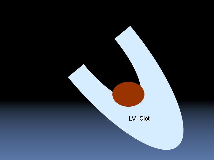
Can we suck out left ventricular apical clots with a suction catheter device in post MI patients?
Posted in Cardiology -Interventional -PCI, Infrequently asked questions in cardiology (iFAQs), Uncategorized, tagged acs, anti coagulation, cabg, cardiology, drsvenkatesan, LV clot, madras medical college, myocardial infarction, primary pci, stemi, thromboembolism, warfarin on August 4, 2008| Leave a Comment »
LV clot formation is one of the important complications of acute myocardial infarction. Preventing this is difficult and managing this problem is still more difficult.Some of these clots are linear and laminar along the shape of LV apex and carry less risk of dislodging.
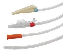
While mobile LV clots , even if it is small can cause a embolic episode. Most of these patients have a significant LV dysfunction and they are candidates for early CAG and revascularisation. Even If the coronary anatomy is very ideal for a PCI these patients are often sent for CABG and physical removal of LV clot . If only ,we have an option to remove these LV clots by a catheter based modality, we can offer them a totally non surgical cure.
This is not impossible, considering we are in the era of percutaneous implantation of prosthetic valve in Aorta ! The only issue is potential embolism into carotids and periphery .A temporary distal protection at the level of aortic root will prevent that .
Device companies shall produce one such exclusive catheter system to remove LV clot.
Dr .S.Venkatesan, Madras medical college, Chennai,India
Is pulmonary artery atherosclerosis possible ? Are we under diagnosing it ?
Posted in Infrequently asked questions in cardiology (iFAQs), Uncategorized, tagged atherosclerosis, cardiology, drsvenkatesan, pulmonary atherosclerosis, pulmonary hypertension, statins on July 26, 2008| 1 Comment »
Atherosclerosis is the number one killer of mankind .It involves all medium and large sized blood vessels.Any intima and media can be invaded by the disese process.Most common to involve are cerebral, carotid, coronary, aortic and it’s branches, renal, and peripheral arteries. But how pulmonary artery is missing in this list ? Is it really true (or) are we missing it ? One logical explanation is pulmonary circulation is a low pressure circulation and the maximum presssure is less than 30mmhg . This pressure may be insufficient to induce endothelial injury that predispose lipid mediated injury.
Other explanation could be a structural difference in the media and intima compared to aorta .But in patients with primary or secondary pulmonary hypertension where, inspite of PA pressure being high , still atherosclerotic changes is very uncommon . or Is it the Heath Edwards pulmonary vascular sclerosis grading reflects nothing but pulmonary atherosclero-thrombosis !
If this is true there could be a major role for HMG Coa reductase inhibitors in altering the natural course of pulmonary obstructive vascular disese . Statins might be tried in PPH a disese with no specific treatment !
Categories
-

-
The contents of the this blog is being published as Kindle E book , as per the request of many of the readers. Every article will continue to be open source in this site. Again I shall reiterate the book format is not aimed at any commercial intent. It is only to facilitate learning in a single book format Here is the link to book
https://amzn.in/d/euhL5vu Archives
- September 2026 (6)
- August 2026 (8)
- July 2026 (6)
- June 2026 (9)
- May 2026 (6)
- April 2026 (11)
- March 2026 (10)
- February 2026 (8)
- January 2026 (8)
- December 2025 (11)
- November 2025 (7)
- October 2025 (8)
- September 2025 (7)
- August 2025 (9)
- July 2025 (10)
- June 2025 (8)
- May 2025 (9)
- April 2025 (7)
- March 2025 (10)
- February 2025 (4)
- January 2025 (9)
- December 2024 (11)
- November 2024 (8)
- October 2024 (10)
- September 2024 (5)
- August 2024 (5)
- July 2024 (6)
- June 2024 (5)
- May 2024 (4)
- April 2024 (7)
- March 2024 (4)
- February 2024 (8)
- January 2024 (6)
- December 2023 (8)
- November 2023 (13)
- October 2023 (14)
- September 2023 (5)
- August 2023 (6)
- July 2023 (10)
- June 2023 (5)
- May 2023 (5)
- April 2023 (4)
- March 2023 (5)
- February 2023 (2)
- January 2023 (7)
- December 2022 (3)
- November 2022 (5)
- October 2022 (5)
- September 2022 (4)
- August 2022 (3)
- July 2022 (9)
- June 2022 (2)
- May 2022 (1)
- April 2022 (2)
- March 2022 (1)
- February 2022 (3)
- January 2022 (7)
- December 2021 (3)
- November 2021 (5)
- October 2021 (8)
- September 2021 (4)
- August 2021 (6)
- July 2021 (6)
- June 2021 (7)
- May 2021 (5)
- April 2021 (4)
- March 2021 (3)
- February 2021 (6)
- January 2021 (8)
- December 2020 (4)
- November 2020 (5)
- October 2020 (7)
- September 2020 (7)
- August 2020 (10)
- July 2020 (6)
- June 2020 (9)
- May 2020 (9)
- April 2020 (5)
- March 2020 (7)
- February 2020 (3)
- January 2020 (4)
- December 2019 (4)
- November 2019 (6)
- October 2019 (3)
- September 2019 (6)
- August 2019 (3)
- July 2019 (1)
- June 2019 (3)
- May 2019 (2)
- April 2019 (2)
- March 2019 (2)
- February 2019 (4)
- January 2019 (2)
- December 2018 (2)
- November 2018 (2)
- October 2018 (2)
- September 2018 (1)
- August 2018 (2)
- July 2018 (3)
- June 2018 (1)
- May 2018 (3)
- April 2018 (1)
- March 2018 (3)
- February 2018 (3)
- January 2018 (1)
- December 2017 (3)
- November 2017 (3)
- October 2017 (3)
- September 2017 (2)
- August 2017 (2)
- July 2017 (2)
- June 2017 (2)
- May 2017 (4)
- April 2017 (3)
- March 2017 (3)
- February 2017 (5)
- January 2017 (3)
- December 2016 (2)
- November 2016 (5)
- October 2016 (4)
- September 2016 (3)
- August 2016 (5)
- July 2016 (3)
- June 2016 (4)
- May 2016 (3)
- April 2016 (6)
- March 2016 (4)
- February 2016 (3)
- January 2016 (5)
- December 2015 (6)
- November 2015 (5)
- October 2015 (8)
- September 2015 (2)
- August 2015 (5)
- July 2015 (7)
- June 2015 (4)
- May 2015 (6)
- April 2015 (5)
- March 2015 (7)
- February 2015 (15)
- January 2015 (8)
- December 2014 (5)
- November 2014 (9)
- October 2014 (7)
- September 2014 (9)
- August 2014 (5)
- July 2014 (11)
- June 2014 (5)
- May 2014 (4)
- April 2014 (5)
- March 2014 (8)
- February 2014 (8)
- January 2014 (5)
- December 2013 (7)
- November 2013 (7)
- October 2013 (14)
- September 2013 (12)
- August 2013 (15)
- July 2013 (15)
- June 2013 (15)
- May 2013 (15)
- April 2013 (15)
- March 2013 (15)
- February 2013 (15)
- January 2013 (15)
- December 2012 (15)
- November 2012 (15)
- October 2012 (15)
- September 2012 (15)
- August 2012 (15)
- July 2012 (15)
- June 2012 (15)
- May 2012 (15)
- April 2012 (15)
- March 2012 (15)
- February 2012 (15)
- January 2012 (15)
- December 2011 (15)
- November 2011 (17)
- October 2011 (17)
- September 2011 (17)
- August 2011 (21)
- July 2011 (20)
- June 2011 (17)
- May 2011 (15)
- April 2011 (17)
- March 2011 (25)
- February 2011 (20)
- January 2011 (20)
- December 2010 (18)
- November 2010 (21)
- October 2010 (21)
- September 2010 (25)
- August 2010 (20)
- July 2010 (10)
- June 2010 (11)
- May 2010 (19)
- April 2010 (16)
- March 2010 (14)
- February 2010 (22)
- January 2010 (18)
- December 2009 (20)
- November 2009 (20)
- October 2009 (3)
- September 2009 (21)
- August 2009 (19)
- July 2009 (12)
- June 2009 (12)
- May 2009 (11)
- April 2009 (15)
- March 2009 (21)
- February 2009 (4)
- January 2009 (12)
- December 2008 (13)
- November 2008 (9)
- October 2008 (22)
- September 2008 (20)
- August 2008 (16)
- July 2008 (14)
- June 2008 (7)
Blog Stats
- 6,742,126 hits
Please give your feed back .
Click below to see who is watching this website live !

- This site will never aim for profit. Still ,this donation link is added at the request of few visitors who wanted to contribute and of-course that will help make it sustainable .

Please Note


