Feeds:
Posts
Comments

                                                     Every day thousands of  hearts  end their life   due to terminal heart failure . Much more  lives are  confined  to their bed rooms.In refractory cardiac failure and severe LV dysfunction the only  long-term option is cardiac transplantation.

Medical therapy has reached its saturation point.  Neuro- humoral modulation shows some promise. The other modalities like cardiac resynchronisation ,LV assist devices ,  ventricular  reduction surgeries ,  restriction devices , mitral valve splinting  are  still experimental .
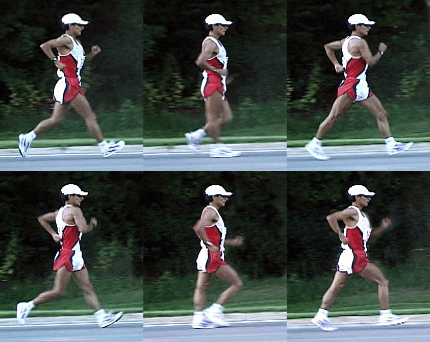
Simply watch this Image : Your heart will get Energy

Modern day  cardiology is trying to add life to these dying  hearts  .

There are two aims

  • To prolong survival
  • Improve functional capacity (Make them at least take care of daily activities and live a fairly independent live)

This is the purpose of the  mushrooming heart failure clinics all over the  world . These clinics , though started with  good intention , ultimately   become  feeding  centres for so many experimental  bridge modalities  , sometimes  with an  infinite wait for  a potential donor  or at the mercy of their insurance companies  . (Many time it turns out to be a  bridge to heaven as the patient fails to cross it !) .

Even though there is strict criteria for terminal  heart failure ,  in practical terms it has many issues .Temporary functional deterioration is misinterpreted   often .

Premature  dependence on LV assist devices and  indulgence  in inappropriate  mitral valve reconstructive  procedures are the currently most important pseudo cardiac interventions .( Myosplint/AV groove tying etc)   Some where along   the  academic  corridors ,   we failed to realise many patients can bridge themselves  to a  transplant (or even   self de-list  from transplant programme  )  provided we are willing to wait and take few   risks  .

It is observed exercise training  programme is awfully inadequate in most centres  who deal with late stages of cardiac failure.

The hidden link  between skeletal muscle and  cardiac muscle

Skeletal muscle  function is impaired in cardiac failure . This impairment is attributable  to both  dis-use and low cardiac output.  Proper training of these muscles can not only improve the functional capacity  but also  sets in  a positive hemodynamic cycle  that   ultimately improves cardiac function as well.
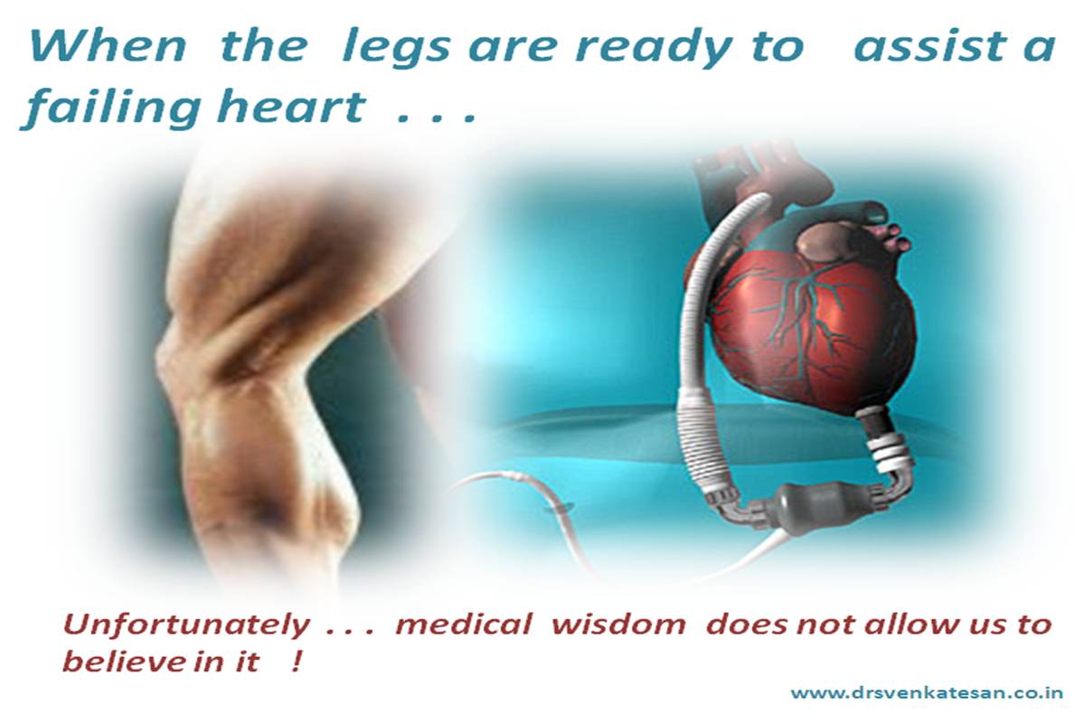
In  our  country we have data  of  thousands of patients  with severe LV dysfunction living with the much ridiculed  digoxin   ,   diuretics ,  ACEI  and minimal exercise living a comfortable life for over 10 years .  It is often said in  cardiology class rooms ,  do not whip a tired horse  as the   failed heart needs rest  .This statement  has  truth  in it even in  this  space age cardiology !

Whipping  a failing heart with electrodes in the name of CRT   could be as  bad as  whipping with inotropic agents . This is not a  personal joke ! This fact has been repeatedly  proved by various inotropic  studies in terminal heart failure(Dobutamine to be specific ) Even CRT  is a suspect .These patients walk for 30 meters  further  with  no convincing survival  benefits .(Of course it requires a ICD -Combo to prevent sudden deaths ) Zero impact in non sudden deaths ?

Can  we propose a  new therapeutic  concept to our  patients   ?

Do you  want to   live with a  low functional capacity (Restricted  life   still  happy  )    for 5 years   or live  apparently unrestricted  life   and die prematurely ?

                         In simple terms,  for all those patients with severe  grades of  heart failure   the  best advice could be . . .to  avoid the levels  of exertion that cause dyspnea / Modern gadgets  may help relieve  exertion for a short  while  , but it  can cut short your longevity * (* This is not a threatening message. This applies to near terminal stages of cardiac failure .All other minor grades of CHF are encouraged to exert up to 70 % of their limits.)

Peripheral mechanism in cardiac failure.

We know cardiac  failure is not a simple  mechanical failure of heart , it activates a complex neuro endocrine system which makes it a systemic disorder .Many of the current research is aimed at favorably modify this. It is now certain Skeletal muscle function is a  major determinant of  cardiac failure outcome and hence a therapeutic target .

If you have good muscle mass ,  good diaphragm and intercostal muscles one can  compensate the compromise inflicted by the heart to a large extent.  We know,   the entire vascular tree has a mechanical function  to do . The stiffness and compliance of aorta , other  major vessels, the muscles  through which these vessels  traverse determine the  ultimate  efficiency  of  circulation.We know  the pulse wave , as it  travels to the periphery , gets amplified. This amplification is not without any significance. It aids in muscle  blood flow . This agumnetation is missing in poorly trained cardiac failure patients. Further muscle respiration is synonymous with  functional capacity . Numerous defects (Both structural and functional )  in skeletal muscle mitochondria are reported.

This is why meticulous  exercise training  becomes an important   intervention in  cardiac failure . There are very good studies that document   muscle respiration defects  getting reverted  with  proper exercise training and  muscle  care  .  Among all muscles the   calf  and thigh muscles show great promise.   We have observed  cardiac failure patients  with good calf muscles ,  outperform others with identical ejection fraction.(Will be published shortly )

Strangely there is no comparative  studies between calf muscle  efficiency   and other available modalities  in cardiac failure .

The concept of  Venous pump vs  Arterial pump

Skeletal muscle mass acts not only as venous pump  it also has a modulating  effect on the arterial pulse transmission .A good venous  pump will activate  vascular  tone . In congestive heart failure  a the RV filling pressure is raised,  blood tends  to  move sluggishly  in right heart chambers .  A proper venous tone  can alleviate this . Well trained  calf muscle  can exactly do this  by a controlled elevation  of  IVC pressure at times of exertion . 

 Dyspnea  of muscular  origin (Peripheral dyspnea)

The symptomatology of cardiac failure has an intimate  realtionship with skeletal muscle integrity  !

Lactate in blood and  hypoxia  in   exercising muscles  can trigger   non hemodynamic dyspnea . Further , there is strong reason to believe  the sensation of dyspnea   is perceived at the chest muscle level  (By muscle spindle length/tension   mismatch ) .It is not known whether lower limb  muscles can generate a feeling of dyspnea  !

But , one thing is certain   by altering the tone of the muscle  spindle and the  optimising the  stretch signals the peripheral component of cardiac dyspnea can be significantly neutralised . This  is what  happens in well-trained   cardiac  failure patients .

How to train the skeletal muscles ? ( In to heart friendly  muscle )

  • Passive stretch
  • Simple 6 minute walking three times a day will help .
  • Muscle massage and toning
  • Drugs like Trimetazidine may improve muscle metabolism by better ATP utilisation
  • Diligent use of diuretics (Excess diuretic can make your muscle exhausted )
  • Chest exercise for improving intercostal muscle function


Final message

Skeletal muscle training  in cardiac  failure  could be as important as  the  digoxins  , diurteics   and ACEI .When a 300 grams of heart muscle is struggling  , God  is willing to  help  it with huge muscle mass that lies elsewhere , we should read the silent  signals of nature . Many cardiac failure patients  realise this and live  a happy live without artificial assistance .This applies  in all grades of cardiac failure .

For  all those physicians  out there in modern hospitals who treat cardiac  failure , spend at least  few minutes  for prescribing a good exercise  program with a specific  mention about calf muscle function  . After all , it  may turn out be the most efficient  RV/LV assist device !

References
                                                                     http://content.onlinejacc.org/cgi/content/abstract/30/7/1758
 http://www.uptodate.com/contents/skeletal-muscle-dysfunction-and-exercise-intolerance-in-heart-failure

In this world of  evidence based medicine  the  funny bedside vocabulary of  medical statistics   has withstood the test of  time. The following words are liberally used by physicians of all walks of life.

We never bother to find what these words mean to our patients  !

Here is a crude and  wild   numerical attempt to  decode  these words.

  • Always                                    99 %
  • It s a rule                                  95-99%
  • Almost always                       90-95%
  • Very common                       > 90 %
  • Common                                  > 75%
  • Uncommon                            < 30 %
  • Rare                                          < 10 %
  • Very rare                                < 5 %
  • It is an exception                2 -5 %
  • Remote                                   < 2 %
  • Never                                     < 1%

Apart from the above   there two  hugely popular  medical words used over  million times every day in all walks of medical practice.

They are  ” May” and “May not”

The greatness of these words lies  in the fact   it can convey any of the above  10 meanings in a single phrase without any fuss !

Further ,  the words may and may not are numberless un-quantified statistical  jargons that   can convey a deep meaning or  . . . no meaning  depending upon the circumstances !

Doctor ,  is there a possibility of  my stent getting occluded    as i have skipped  the  clopidgrel ,and aspirin for the past two weeks

You may  be at risk  . . . but you may not develop  an heart attack immediately . I would advice you start the drug immediately .

Heart is mainly perceived  as  a pumping organ but it need to be realised it  also has a   reservoir function  (Temporarily though , for  about .5 seconds every beat ) . Contrary  to the popular belief heart is not  continuously and tirelessly working  .For every contractile  beat it takes  a brief period of rest .This is called diastole. But , even here it is not a complete rest , as  it has to receive the blood from the atria and get filled and be ready for the next beat.

Many think diastole is an active energy-consuming process . . . but it can be debated still ,  as passive elastic properties may contribute substantially to cardiac relaxation blunting the energy requirement

God is so scientific (Greatest scientist !)  he  made it sure   the resting phase(Diastole)   is slightly more  than the contractile phase (Systole ).

This makes the organ relax a bit more than it stresses  in its entire life time . At  any  given heart rate diastole will be slightly  more than systole  , peculiarly  for the same reason  during tachycardia  diastole suffers more than systole.

What happens in diastolic dysfunction ?

Pathologically the ventricles become stiff  and rigid and the filling of the  ventricle is impaired . The commonest cause for diastolic dysfunction are  hypertension, diabetes, and  ischemic  CAD some forms of myopathies  .In systole ,the calcium  is pumped into actin myosin complex  while in diastole the  same calcium molecules  (Or different !)   are ejected back into the cytosol and sarcoplasmic reticulum. The later process is impaired in many situations of diastolic dysfunction.

It should  also be realised not every one with diastolic dysfunction  has a  calcium release /unloading defect .Many  have structural diastolic dysfunction  like interstitial fibrosis  .Here the mechanism goes beyond  calcium kinetics.These are the patients who get maximum  benefit out of heart rate reduction.

It is all Time  . . . Time as a  lusiotropic  drug !

If the ventricle finds difficult to relax  (or slow /sluggish to relax )  we have  two  options to tackle this .

  • To make relaxation  faster( ie positive lusiotropism )*
  • To  prolong the diastole  itself  .

Prolonging diastole makes it certain , the LV relaxation process is completed   as the excess time compensates for  the slowness of calcium reuptake into the sarcoplasmic reticulum . In fact , we have observed at slow heart rates (<60)  it is very difficult to document diastolic dysfunction  by doppler .

In many of  dilated  cardiomyopathies  the beneficial effect of  beta blockers , could be linked to simple reduction in heart rate and prolongation of diastole .(Note In DCM about 30-40 % have restrictive filling )

Final message

As we have no specific drugs to  augment the  process  of   cardiac diastole,  currently heart rate reduction  could be the simple and best method*  to improve diastolic function  .In many cases  diastolic dysfunction  simply vanishes  at low heart rate.Bradycardia  and  diastolic dysfunction   will remain as foes  forever !  Please give the benefit of this simple concept to all your patients with diastolic dysfunction .Your patients  can breath lot more easier !

*Apart from controlling the underlying cause like DM, SHT and CAD  , anti fibrotic drugs,  interstitial relaxants ,selective cardiac   collagen uncouplers  are the  future areas of research .

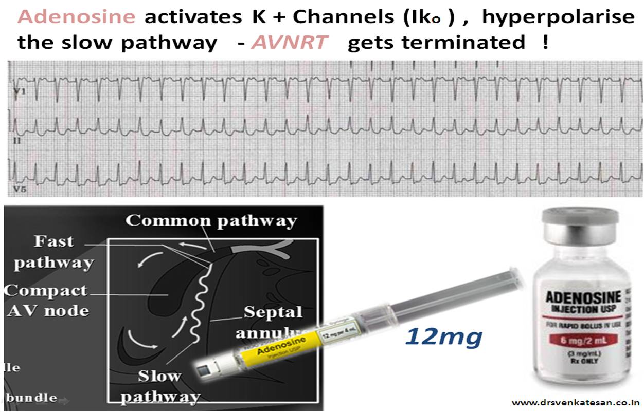
Adenosine is a  purine analogue. Acts by stimulating outward K+ channel  of AV nodal tissue, more specifically  in the posteriorly   located  slow pathway in the vicinity of  coronary sinus.

Another action of adenosine is inhibition of cAMP , which is similar to beta blocking action may also help in terminating the tachycardia.

Adenosine : A 10 second cardiac miracle

  • 12mg bolus is administered , preferably in a central vein (Not mandatory  though)
  • Termination is usually abrupt . Transient VPDs are observed during termination.
  • Transient flushing may occur.
  • If the patient is taking Aminophylline group of drugs (Which are adenosine antagonists) the AV nodal blocking action may be neutralised .

(It may be apt to recall  at this juncture ,  Aminophylline is used in sinus node dysfunction or AV block to increase heart rate )

Reference

A good one from Medscape http://www.medscape.com/viewarticle/585287_2

Coronary artery is the life line  of the  heart.The size ,  branches  and the course are  predetermined . Generally  it follows a  pattern ,  but still  it is   believed  every  human has a unique coronary finger print. (When retinal blood  vessels can do this . . .  why not the coronary  ? )

When the left main bifurcates it has to share its resources equally between the two  daughter vessel (Not exactly . . . LAD is  widow maker can’t be a daughter  !)

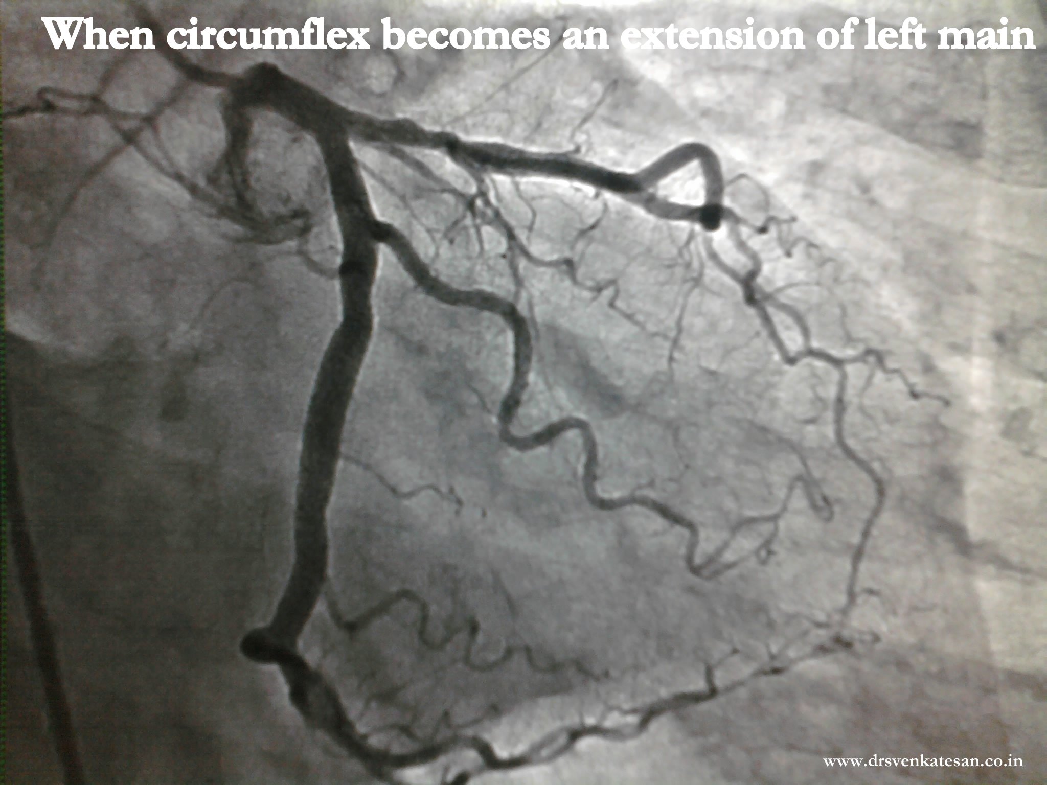
The logic would say LAD demands more as it has more  territorial commitment.But if LCX fights for  equality  share there is a  potential conflict here.But when LCX  is very aggressive and  demands  much more than it deserves the  issue becomes further complex

See how this LCX  gets bulk of the blood flow from Left main and no doubt this man came with  NSTEMI and  LAD region ischemia while his posterior circulation is comfortably  placed with  excess blood.

Note: The diameter of LCX even exceeds left main in certain segments.

One suggested formula (not validated ) is  diameter of   LCX +  LAD will be   at-least 150% of left main diameter .

1.5 x Left main diameter  =  (LAD + LCX diameter)

This amounts  to  50%  gain in  diameter   as it bifurcates .

Trifurcation  further increases the width conferring a hemodynamic advantage

What is the implication of unequal bifurcation  Left main ?

Hemodynamically there could be diversion  of flow into a larger orifice .But ultimately since  the blood flow is  determined  by the resistance arterioles at myocardial bed there  may not be any  practical significance . In pathological situations there could be a some impact  due to  stealing . Large LCX is more common with  left  dominant circulation  .

Coronary atherosclerosis  can  strike  an artery  with  variety  of   lesions.It can be  any of the following.

  • Focal
  • Ostial
  • Eccentric
  • Discrete
  • Diffuse
  • Tandem
  • Ectatic
  • Multiple

  Rarely  a coronary artery  is  blessed with  all  of the  above  characters ,     added  with homo and hetero  collaterals   . . .     resulting  in  Atherosclerotic    chaos  !


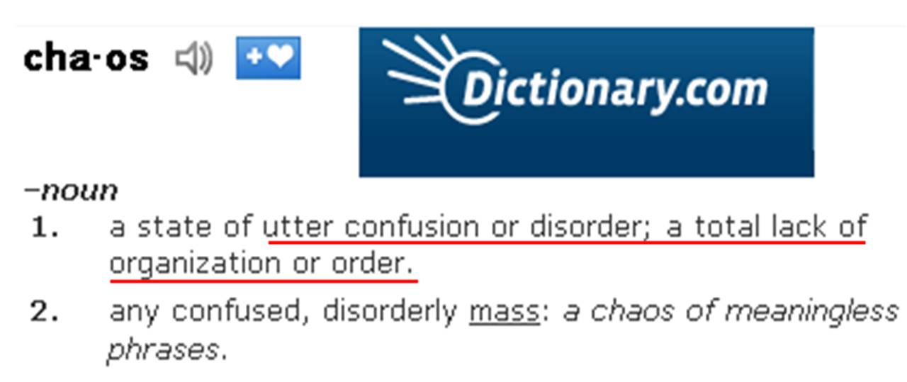
What is  chaos ?


Note one such vessel inflicted with chaos lesions

How to you report the above angiogram  ? What can be done ?

We  do not treat an angiogram. We need to know the clinical background. (This  is a  50 year old man with chronic stable angina )  He also had a  lesion  in LAD and   was advised CABG with grafts to LAD  and PDA.

Is PCI possible in such a vessel ?

Most would agree , it is a crime to think about  PCI in the above vessel. Still , few hard-core  interventional  cardiologists may vouch   for success  in this vessel !

Is there any alternative management other than CABG in this vessel ?

Leaving it alone can be an intelligent strategy . (If  LAD is normal  it  may be the  best possible management)  . But , CABG will remain a default choice. But , when a person is having such a rampant atherosclerosis  he is at high  risk  for pre- mature  graft disease as well. Hence  , intensive medical management will be the key  in  such patients irrespective  of  any revascularization procedure.

Coronary collateral circulation can be termed as one of the  mysterious  circulation in our body.Cardiologists generally do not  give much importance to it and some interventionists even ridicule it !  . But  ,  God has given it ,  with  a purpose. He adds a riddle though !  .Collaterals  grow  in  almost  every  individual  when   obstruction occurs gradually ( chronic coronary syndrome ) but only in  a few ,  it  will open up  during a real emergency like ACS !

How and why , only  few of us can  recruit  coronary collaterals   during   acute occlusion ?

God  blesses acute coronary collaterals only in selected few  , who  are on the right side of his good books .This can be  the other name for our  destiny !

Role of coronary collateral circulation  in acute coronary syndrome.

  • Limits  infarct size and volume
  • Promotes salvage
  • Converts q  MI to non q  MI
  • Prevents Unstable angina from becoming MI
  • Prevent primary VT and VF*

All  of the above can be vital  in saving a life  . Even as  we realise 30 % of STEMI do not even reach hospital  , it seems certain men and women with early collateral recruitment  will never  fail to reach the hospital alive

Is there a simple  method to identify  people who are blessed with acutely recruitable   collaterlas ?

I am afraid it is  almost equal to  asking   for a glimpse of GOD   !

Wait . . .  when we were on cath lab  few  days ago a  stunning  phenomenon happened  that could pave way for identifying  potential acute  collaterals  in any human being.  Follow this site  . . .the details will be posted !

 Hypertension  ranks  number one  in the risk for future  stroke . Surprisingly this is true  for ischemic  as well as  hemorrhagic strokes.

 What  causes  thrombosis or  rupture of small cerebral arterioles ?

 It is somewhat similar to coronary events . ( With one major exception,  coronary vesels  are   not prone for rupture ) .It is  believed   sudden spikes of   blood pressure  and the resultant endothelial injury are responsible. Atherosclerotic plaque fissure and inflammation  also  contribute. 

Is embolic stroke related to hypertension ?

The vast majority of embolic stroke are believed to  arise from heart .This belief is getting gradually eroded , as we now know aortic arch and carotid arteries vie for this honour . .(This was indirectly proved in AFFIRM trial  when rhythm control failed  to reduce the incidence of  stroke inpateints with AF ,   implying much of the strokes arise  in the upstream rather than within the cardiac chambers )  

Meanwhile , there is no controversy  in  SHT  promoting  both cardiac  and non cardiac embolus to brain

Systolic ,  Diastolic or Mean pressure   which is  important  in the genesis of stroke  ?

All parameters  are  important , but the   systolic blood pressure  is vested with more  vigour  to damage the  cerebral arterioles. The reason  systolic pressure is more important lies  in the  fact ,  it  can  attain  high pressure peaks instantly ,  unlike diastolic or pulse pressure which  slowly builds up. Further , systolic BP  carries  leading edge of the pressure  curve with high Dp/Dt and hits  the target  first !

At what pressure the cerebral artery becomes  uncomfortable ?

We do not know  the answer as yet , but any systolic pressure above 180 mmhg is a huge stress for the cerebral arterioles.The rapidity with which the BP  raises  (Dp/Dt) also becomes  important  . High blood pressure increases the shearing stress .It  interferes with nitric oxide synthesis and promotes endothelin release which precipitates  cerebro vascular event.

How do you identify people who are at risk for stroke ?

While  cardiac physicians are obsessed with exercise stress test to predict CAD  very  few  are worried about  stroke . In fact the same exercise stress test can be used to stratify stroke risk. The exercise induced systolic blood pressure  raise  is a useful risk stratifying  tool. This concept is there for more than a decade without reaching the clinical domain.

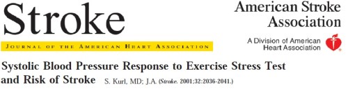
The following paper was  published in stroke journal (2001)  from the picturesque university of  Kupio Finland.(See below )  It is a wonderfully done study and throws great insight into the  new  emerging  science of  Intra cerebral hypertension .


The following can be summed up as risk factors for stroke during EST  (Derived from   various sources  and  . . .  with   liberal dose of personal  logic !)

  • Raise of 20 mmhg  SBP  at  2  minutes .
  • Increment of >  20mmhg in SBP any subsequent minute.   
  • Any  SBP  above 200mmhg during  EST
  • Failure to  reach baseline SBP  at 6 minutes recovery .
  • SBP  or DBP remaining high  even  after  the heart rate reaches baseline.


 Final message

For the kind attention  of all  cardiac physicians . . .  whenever you do an  EST for a cardiac indication ,  please spend the first  few  minutes  carefully ,and   look at the  blood pressure response . It is encouraged ,  to  specifically mention about the  behavior of  SBP  and write a remark about the propensity for  stroke in  every EST/TMT report .   Let us grow our brain  sense as well   . . .  for   the sake of our patients !

Thanks again  to Dr S.Kurl et all from Finland  for their  nice article which  stimulated  me to write  this post .

Reference

http://stroke.ahajournals.org/cgi/reprint/32/9/2036

http://heart.bmj.com/content/95/13/1072.abstract

Further queries

How common is stroke following a EST procedure ?  Can high blood pressure dislodge a carotid plaque during a stress test ?

The answers will be posted soon once I  get it . ( Of course you can do it if you know !)

Left bundle branch block (LBBB)   has a curious but important relationship with  STEMI . LBBB inflicts a dramatic change in qrs morphology   with  a diagonally  opposite   polarity of ventricular activation . This masks    the initial qrs vector  and  makes it a difficult task  to diagnose acute MI in this setting. The ST segment which is of primary importance  in STEMI is   lifted  up due to altered repolarization .

LBBB can be associated  with  STEMI in the following ways

  • Acute necrotic LBBB  with massive myocardial damage – Impending shock
  • Chronic LBBB with acute STEMI
  • Transient ischemic LBBB during STEMI
  • Rate dependent  LBBB (Usually tachycardic  ,  rarely bradycardic  )
  • STEMI in pacemaker rhythms

While every one of the above can be experienced ,  the most common diagnostic conundrum  occurs ,  when a patient   comes with acute  chest pain and LBBB . There has been many criterias  suggested to diagnose STEMI in the presence  of LBBB.

The criteria  proposed  by Sgarbosa  (A  GUSTO   off shoot )  in 1996  caught our imagination .One prime reason for this is ,  it came from the prestigious NEJM and Duke university combine. Suddenly this became the de- facto standard to diagnose  STEMI 

In the  past 15  years  ,  our experience in one of largest coronary  care units in India , we have   found this criteria   to have  little utility value  in STEMI and LBBB  . Most of the time  a correct diagnosis was made  by   simple clinical guessing .

Next to  clinical assessment, we found cardiac enzymes (Troponin and CPK ) were reliable in diagnosing  STEMI with LBBB.

Surprisingly ,echocardiography  was as unreliable as ECG .( The paradoxical  septal motion invariably confuses the already  confused  cardiology fellow who usually does the emergency echo  !) 

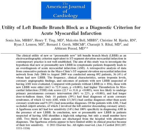
Even as our  CCU documentation was  far from satisfactory  , now this article from Mayo exactly reflect  our observation.

Sorry   Sgarbosa . . . the criteria was  based  on  sound observation and a  good  electrical principle  . . . still LBBB is able to beat   it convincingly ! ( Very low sensitivity !)

WPW syndrome remains as  a   fascinating ECG entity ,  ever since it was described by Wolf , Parkinson and White in the year 1930.It is  primarily a  disorder of cardiac embryology . Heart is an organ made up of  tissues from mesoderm and neuro ectoderm.The muscle which comes from mesenchyme has to be incorporated with specialized conducting system. This is a complex  process .It is determined by the bio-genetic forces. When errors happen in the embryonal  tissue  flow  congenital anomalies occur.

In  WPW this  error   happens  exclusively in the conduction  tissue movement  . Normally the specialized conducting system    pierces  the  entire  AV ring and connect atrium  and ventricle  .Later ,   it regresses in  all areas  except in the AV nodal zone  . When  It  fails  to regress ,  these  remnants of  conductive  tissue act as AV accessory tissue  and create electrical  short circuiting .This is the reason , all these pathways are located in the close vicinity of AV ring.

Accessory pathway shows   varying conduction velocity , but generally devoid of  decremental conduction properties .  The presence of such pathways make these individuals prone for variety of cardiac arrhythmias .It can range from  simple AVRT  to  malignant antidromic  AVRTs  that can end up in  VT /VF.

Resecting  these  pathways surgically was once popular.  Effective blocking  of  the pathways with  drugs  is a good option. Currently ,  it is possible to  locate and  ablate  most of these  pathways   successfully.

Even though there are many protocols to locate accessory pathway the one that is very popular is  simple   Type A and type B  WPW , which locates the pathway either in the  left  or  right  ventricle  respectively.

Huge data base  has been accumulated over the past 80 years  regarding WPW syndrome,  still   many questions are unanswered.  One of the important clinical issue is  multiple  accessory pathways , scattered  at  random  across the  tissue planes of atrium and ventricle  .

The other issue is intermittent pre-excitation and shuffling  of path ways during tachycardia  .

It is very rare to see a patient who manifests both Type A and type B pattern during sinus rhythm .Here is an  article from  unexpected  quarters  , Colombo Sri-Lanka in the year 1972  candidly  describes a patient with classical  combination  of  Type A and  B  WPW . It is great to see such an interesting  observation in the pre  EP/Echo era from a remote island nation.

Now , let us ponder over  these questions

    1. Can a pre-excitation  happen simultaneously in both right and left free wall pathway ?
    2. How will the ECG look like  when impulse travels over multiple pathway ?
    3. When dual pre-excitation combines   with  normal AV  conduction  ,     will  it not make  a  triple AV pathway ?
    4. How does a supra-ventricular impulse decide ,  which pathway it is going to travel  when confronted with a choice of  three or  four pathways ?
    5. How do you plan ablation for such a patient  ?