Archive for 2008
What is the greatest medical breakthrough since 1840?
Posted in cardiology-ethics, Uncategorized, tagged Add new tag, bmj, COMMON SENSE, ethics, INTERHEART on October 14, 2008| Leave a Comment »

What is the simplest possible guideline for doing coronary angiogram following acute myocardial infarction ?
Posted in Cardiology -Interventional -PCI, cardiology- coronary care, Infrequently asked questions in cardiology (iFAQs), tagged acc.aha, acc/aha guidelines for stemi, acute myocardial infarction, cath lab, conservative approach, coronary angiogram, drsvenkatesan, ethics, evidence based cardiology, excercise stress test, guidelines, interventional cardiologist, jacc, jama, lancet, nejm, nstemi, nuclear imaging, pci, pre discharge stress testing, pre discharge tmt, ptca, sestamibi, stemi, stent, streptokinase, sub maximal, thallium, thrombolysis on October 11, 2008| Leave a Comment »
Answer: Do coronary angiogram for all patients who had suffered from an acute myocardial infarction* ( Forget about all those mulitpage ACC/AHA guidelines !).
For an interventional cardiologist , it is often considered a crime to follow a conservative approach !

*Caution : This one line guideline is not based on scientific fact but reality based . Ideally one should identify high risk subsets among the patients who had an AMI .Patients who had complications during the MI get immediate CAG. Others need a focused LV function asessment , pre discharge sub maximal excercise stress test or perfusion studies .But this concept has been virtually replaced by pre discharge coronary angiogram for all , in many of the centres in the world.
Welcome to my website : www.drsvenkatesan.com
Posted in dr s venkatesan -Personal, tagged anna nagar, best cardiologist india, boiler plant high school, bphss, cardiological society of india, cardiologist, cardiologist india, cardiologist madras medical college, cardiologist tamilnadu, chennai, chennai cardiologist, coimbatore, coimbatre medical college, consultant cardiologist chennai, dr s venkatesan, dr venkatesan, famous, india, india cardiologist, india venkatesan, india's famous cardiologist, indian cardiologist, interventional cardiologist, kaniyalampatti, latha venkatesan, leading, madras, madras medical college, mani high school, on line heart care, online cardiologist, pudupatti, shreenila venkatesan, top, top indian cardiologist, top ten cardiologist india, venkatesan assistant professor of cardiology, venkatesan india, venkatesan madras, www.drsvenkatesan.com on October 10, 2008| 2 Comments »
Please click below to enter my web site

dr s venkatesan ,venkatesan india , india venkatesan , dr venkatesan, cardiologist india, india cardiologist, indian cardiologist, chennai cardiologist, venkatesan madras,cardiologist madras medical college, venkatesan assistant professor of cardiology, top indian cardiologist, top ten cardiologist india, best cardiologist india, online cardiologist, consultant cardiologist chennai, india’s famous cardiologist,cardiological society of india,
Tribute to my teachers: Professor T.K.Ganesan of coimbatore medical college.
Posted in dr s venkatesan -Personal, tagged coimbatore medical college, dr s venkatesan, dr t k ganesan, professor t.k ganesan on October 10, 2008| 1 Comment »
One of the greatest physicians of all time, I have come across , is my professor Dr.T.K.Ganesan from Coimbatore medical college.
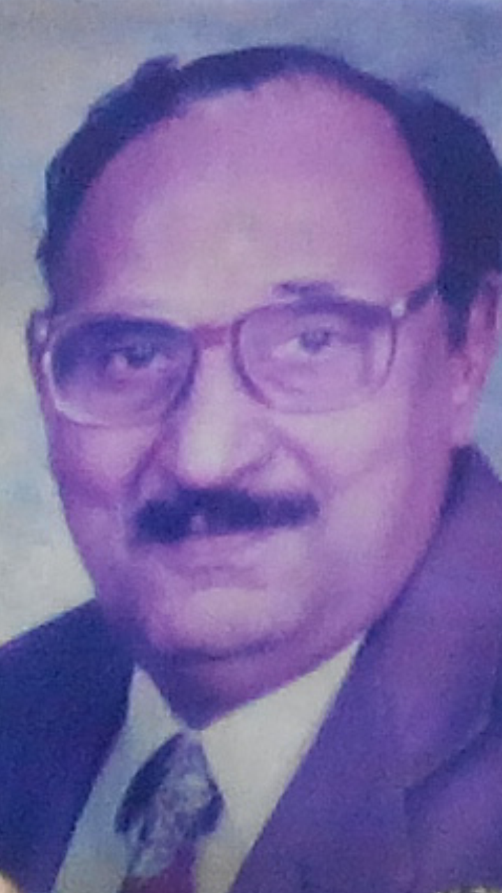
A man who taught medicine to generations of doctors.During those years (1980-1990) learning medicine was simple and also not contaminated with commerce . Dr TKG made it so lively .He infused passion in the subject.
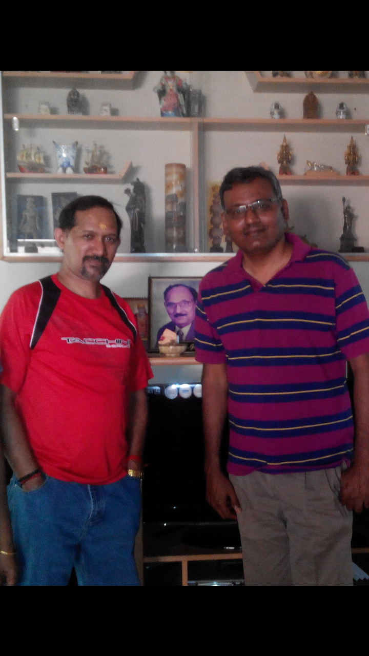
Myself and Dr K.A.Sambasivam (Son in law of DrTKG at his residence in Coimbatore )
* Dr K.A .Sambasivam was my class mate during both my under and post graduation . He is now a senior Interventional cardiologist in GKNM Hospital Coimbatore .
This post will be updated.
What do the myocardium need at times of ischemia ? Blood , oxygen, glucose or ATPs ?
Posted in Cardiology - Clinical, Cardiology -Interventional -PCI, cardiology -Therapeutics, cardiology- coronary care, Infrequently asked questions in cardiology (iFAQs), tagged acs, angina, atp, biochemical ischemia, cardiac failure, chronic stable angina, coronary, fatty acid, glucose, ischemia, ketoacid, metabolic modulation, mitochondrial energetics, myocardial energetics, ranalozine, trimetazidine, unstable angina on October 9, 2008| Leave a Comment »
During acute ischemia the most immediate requirement for the heart is
A.Blood
B.Oxygen
C.Glucose
D.High energy ATPs
E.Free fatty acid
Answer : A will be considered by most , as correct answer . A can provide B to E . But it is also a fact heart can survive without A.
Myocardium requires energy first ! it does not in fact bother about from where it is coming at the time of crises.It may be right if you restore the coronary blood flow all other components (B-E) are made available to the heart .
The heart can survive off the coronary circulation with only chemical support during cardiac surgery and also a during heart transplantation explanted donor heart survives on a ice box during transit and till it is transplanted into the recipient heart
But ironically we spend much of our energy and efforts in restoring blood flow.One need to spare a thought about the quality of blood also . This is especially important in the setting of ischmia where a metabolic centric approach will add further benefit.

Energy based approach to ischemia : Is it relevent ?
Heart is a fascinating mechano biological organ pumping millions of gallons of blood .Fuel for this is self generated on a continuous basis from the circulation blood .So the key to human survival is the coronary blood flow that supplies the fuel and nutrients to the heart. When this key supply line is under threat during acute coronary syndrome cardiologist have the only option of restoring the compromised blood supply by any means . But during chronic ischemia there is no urgency. There has always been an option of enriching the blood with energisers like ATPs, glucose, hemoglobin etc .Providing energy support to the failing heart has never captured the imagination of cardiac physicians until recently.Still most are skeptical about the concept of biochemical ischemia.
Click to download full PPT presentation
Metabolic manipulation of CAD( Will be available shortly)
How is the blood volume distributed in normal human body ?
Posted in Cardiology - Clinical, cardiology -Therapeutics, Infrequently asked questions in cardiology (iFAQs), tagged autonomic dysfunction, blood volume, bmj, capillary, cardiac output, cardiology, drsvenkatesan, effective circulatory volume, fludrocortisone, heart, Hemodynamics, lnacet, lvedp, nejm, ortho static hypotension, pcwp, physiology of circulation, pulmonary edema, syncope, venous circulation, venous insufficiency, venous pooling on October 9, 2008| Leave a Comment »
Humans have roughly 5 to 6 liters of blood at any given time in their body . Out of this*
50% (2500ml) is located in the systemic venous compartment. 18% is within the pulmonary circulation participating in the vital oxygenation 12% (500-600ml) is within the cardiac chambers. 8% is in the arterial tree of the body. 5% is within the capillaries. 2% is in the aorta.* Source : Best & Taylor Physiological basis of medical practice 1966, 8th edition
What is the implication of this predominantly venous distribution of blood at rest ?
- A competent venous tone is essential for the human beings to maintain the erect posture.
- Bulk of the cause of syncope in humans is due to peripheral mechanism like loss of vascular tone and resultant venous pooling.
- The concept of venous reservoir is so important in emergency situations like hypotension as simple elevation of legs is equivalent to infusing 500 -800 ml of intravenous saline .
- Similarly during acute left ventricular failure trunk elevation and legs dangling down can reduce the pulmonary congestion very significantly and reduce pulmonary capillary wedge pressure (LVEDP)
Autonomic dysfunction and venous insufficiency
Autonomic dysfunction and resultant orthostatic hypotension is directly related to venous reservoir dysfunction.Increasing effective circulatory volume by elastic stockings or administration of mineralocorticosteroids like fludrocortisone (.5mg/day ) can be useful in this condition
Can hypoglycemia cause angina ?
Posted in Cardiology - Clinical, cardiology- coronary care, Infrequently asked questions in cardiology (iFAQs), tagged acs, acute coronary syndrome, angina, cardiac arrhythmias, cardiology, diabetes mellites, drsvenkatesan, hypoglycemia, jama, lancet, nejm, tiggers on October 9, 2008| 1 Comment »
Glucose is the molecule of life ,burnt every second inside the body at the energy store house called mitochondria. Heart , the most active organ in the body gets bulk of it’s energy supply from fatty acids, glucose and a little from keto acids. Under anerobic conditions this energy substrates shifts towards glucose .
We are rarely inclined to think that heart can ever suffer from hypoglycemia ! But hypoglycemia can have distinct direct and indirect effects on heart. In fact indirect effects due to activation of adrenergic activation is more obvious.An episode of hypoglycemia can precipitate an arrhythmia . Glucose potassium insulin infusion


Final message
Hypoglycemia , can be a trigger of ACS .This aspect is poorly recognised and studied.
Is atrial fibrillation a benign arrhythmia ?
Posted in Cardiology - Electrophysiology -Pacemaker, Infrequently asked questions in cardiology (iFAQs), tagged atrial fibrillation, cardiac arrhythmia, cardiology, drsvenkatesan, ECG, electro physiology, heart rhythm, nejm, pace, ventricular fibrillation on October 7, 2008| 1 Comment »
Ventricular fibrillation is invariably fatal if not treated . When can atrial fibrillation be fatal ?

Atrial fibrillation is relatively a benign arrhythmia especially when it occurs in isolation with structurally normal heart.This is sometimes referred to lone atrial fibrillation . Even otherwise, atrial fibrillation is rarely fatal except in few situations.But AF commonly destabilises the patient who have baseline valvular or myocardial disease.(Post MI, dilated cardiomyopathy etc)
There are few situations where AF can be life threatening
- In patients with WPW syndrome*where , AF enters into a electrical short circuit , downhill to enter the ventricle and make it fire at the same rate as that of atria . ( ie 400-600) and result in ventricular fibrillation.Note , even here it is the VF that kills not , AF per se.
- AF in acute MI often precipitates LVF , but rarely fatal.
- In patients with critical aortic stenosis, or hypertrophic cardiomyopathy, sudden onset of AF can result in acute cardiac failure.
- AF is often a terminal event in primary pulmonary hypertension
While atrial fibrillation is less likely to cause death , it is a highly morbid arrhythmia .It is one of important cause of stroke in elderly as well as young !
What is the anatomical relationship between pulmonary artery and pulmonary veins ?
Posted in cardiology-Anatomy, Infrequently asked questions in cardiology (iFAQs), tagged pulmonary artery, pulmonary vein on October 6, 2008| Leave a Comment »
 Usually in the the vascular system both artery and vein go together .It is an irony in pulmonary circualtion these two never go together .Another paradox is that pulmonary artery carries the most deoxygenated blood and pulmonary vien carries the purest form of blood in the entrie body , probably God has kept them widely seperated as communication between them seriously affect the physiology.

What determines hemodynamic stability in ventricular tachycardia ?
Posted in Cardiology - Electrophysiology -Pacemaker, cardiology- coronary care, Infrequently asked questions in cardiology (iFAQs), tagged bmj, fasicular tachycardia, heart rhythm, Hemodynamics, ischemic vt, jama, lancet, lvot vt, myocardial VT, nrjm, pace, ventricular tachycardia, verapamil sensitive vt on October 3, 2008| Leave a Comment »
Ventricular tachycardia is considered as one of the most dangerous cardiac arrhythmia .Rather , it is the label VT that spreads more fear than the arrhythmia itself. It is a fact many patients with VT walk into hospital , still VT will always be a sinister arrhythmia as long as it carries a risk of degenerating into ventricular fibrillation.
What determines hemodynamic stability in VT ?
- Origin and location of VT
- The ventricular rate
- Presence or absence of AV dissociation
- Impact on mitral inflow pattern
- Associated left ventricular dysfunction or valvular heart disease.
- VT in the setting of acute coronary syndrome.(Ischemic VT)
- Inappropriate drug selection
Origin and location
VTs originating high up in the ventricle( High septal VT,Proximal VTs) have more organised ventricular contraction and they are more stable.Distal VT originating in the myocardium away from the conducting system has chaotic myocyte to myocyte conduction.These are very unstable.

The term fascicular VT is nothing but VTs originating in the His bundle and it’s branches( Can also be termed Septal VT ).These VTs are also stable and some of them respond well to calcium blockers indicating that they are very close to the AV junction and carry the properties of junctional tachycardia. QRS width gives a rough estimate about the location of VT. Narrower the VT higher it’s origin.( But remember even in VT , qrs can further widen on it’s way downhill !)
LV dysfunction.
This is probably the most important determinant of the outcome in VT. Patients with severe LV dysfunction (EF <30%) fare badly .Hence the land mark concepts from MADIT 1& 2 demanded ICDs in these patients.The most common clinical setting is dilated cardiomyopathy.SomE of them have bundle branch re entry(BBR).This particular VT can be stable for many hours.
Ventricular rate.
Usually VT has a rate between 120-200.Higher the rate of VT more the chances of instability .This rule is also not always true as fascicular VT can be well tolerated at high rates.So location of VT focus and LV dysfunction usually over rides the impact of ventricular rate.
Mitral inflow pattern
Proper left ventricular filling is the key to hemodynamic stability in VT. In proximal, septal,fascicular, LVOT VTs doppler studies suggest (ACC /AHA Type C evidence : Personal observations in CCU during VT) near normal preservation of bi modal filling of mitral valve inflow.In ischemic myocardial VT the mitral inflow profile is critically affected . There is no distinctive forward filling was observed .In fact at rapid rates a short pulsatile MR jets are noted instead.
Associated valvular diseases
It is obvious, aortic and mitral valve disorders can aggravate the hemodyanmic instability.
Final message
The clinical behavior of ventricular tachycardia is widely variable and dependent on multiple factors.
Associated LV dysfunction and structural heart disease ultimately determine the outcome.
Categories
-

-
The contents of the this blog is being published as Kindle E book , as per the request of many of the readers. Every article will continue to be open source in this site. Again I shall reiterate the book format is not aimed at any commercial intent. It is only to facilitate learning in a single book format Here is the link to book
https://amzn.in/d/euhL5vu Archives
- March 2026 (7)
- February 2026 (8)
- January 2026 (8)
- December 2025 (11)
- November 2025 (7)
- October 2025 (8)
- September 2025 (7)
- August 2025 (9)
- July 2025 (10)
- June 2025 (8)
- May 2025 (9)
- April 2025 (7)
- March 2025 (10)
- February 2025 (4)
- January 2025 (9)
- December 2024 (11)
- November 2024 (8)
- October 2024 (10)
- September 2024 (5)
- August 2024 (5)
- July 2024 (6)
- June 2024 (5)
- May 2024 (4)
- April 2024 (7)
- March 2024 (4)
- February 2024 (8)
- January 2024 (6)
- December 2023 (8)
- November 2023 (13)
- October 2023 (14)
- September 2023 (5)
- August 2023 (6)
- July 2023 (10)
- June 2023 (5)
- May 2023 (5)
- April 2023 (4)
- March 2023 (5)
- February 2023 (2)
- January 2023 (7)
- December 2022 (3)
- November 2022 (5)
- October 2022 (5)
- September 2022 (4)
- August 2022 (3)
- July 2022 (9)
- June 2022 (2)
- May 2022 (1)
- April 2022 (2)
- March 2022 (1)
- February 2022 (3)
- January 2022 (7)
- December 2021 (3)
- November 2021 (5)
- October 2021 (8)
- September 2021 (4)
- August 2021 (6)
- July 2021 (6)
- June 2021 (7)
- May 2021 (5)
- April 2021 (4)
- March 2021 (3)
- February 2021 (6)
- January 2021 (8)
- December 2020 (4)
- November 2020 (5)
- October 2020 (7)
- September 2020 (7)
- August 2020 (10)
- July 2020 (6)
- June 2020 (9)
- May 2020 (9)
- April 2020 (5)
- March 2020 (7)
- February 2020 (3)
- January 2020 (4)
- December 2019 (4)
- November 2019 (6)
- October 2019 (3)
- September 2019 (6)
- August 2019 (3)
- July 2019 (1)
- June 2019 (3)
- May 2019 (2)
- April 2019 (2)
- March 2019 (2)
- February 2019 (4)
- January 2019 (2)
- December 2018 (2)
- November 2018 (2)
- October 2018 (2)
- September 2018 (1)
- August 2018 (2)
- July 2018 (3)
- June 2018 (1)
- May 2018 (3)
- April 2018 (1)
- March 2018 (3)
- February 2018 (3)
- January 2018 (1)
- December 2017 (3)
- November 2017 (3)
- October 2017 (3)
- September 2017 (2)
- August 2017 (2)
- July 2017 (2)
- June 2017 (2)
- May 2017 (4)
- April 2017 (3)
- March 2017 (3)
- February 2017 (5)
- January 2017 (3)
- December 2016 (2)
- November 2016 (5)
- October 2016 (4)
- September 2016 (3)
- August 2016 (5)
- July 2016 (3)
- June 2016 (4)
- May 2016 (3)
- April 2016 (6)
- March 2016 (4)
- February 2016 (3)
- January 2016 (5)
- December 2015 (6)
- November 2015 (5)
- October 2015 (8)
- September 2015 (2)
- August 2015 (5)
- July 2015 (7)
- June 2015 (4)
- May 2015 (6)
- April 2015 (5)
- March 2015 (7)
- February 2015 (15)
- January 2015 (8)
- December 2014 (5)
- November 2014 (9)
- October 2014 (7)
- September 2014 (9)
- August 2014 (5)
- July 2014 (11)
- June 2014 (5)
- May 2014 (4)
- April 2014 (5)
- March 2014 (8)
- February 2014 (8)
- January 2014 (5)
- December 2013 (7)
- November 2013 (7)
- October 2013 (14)
- September 2013 (12)
- August 2013 (15)
- July 2013 (15)
- June 2013 (15)
- May 2013 (15)
- April 2013 (15)
- March 2013 (15)
- February 2013 (15)
- January 2013 (15)
- December 2012 (15)
- November 2012 (15)
- October 2012 (15)
- September 2012 (15)
- August 2012 (15)
- July 2012 (15)
- June 2012 (15)
- May 2012 (15)
- April 2012 (15)
- March 2012 (15)
- February 2012 (15)
- January 2012 (15)
- December 2011 (15)
- November 2011 (17)
- October 2011 (17)
- September 2011 (17)
- August 2011 (21)
- July 2011 (20)
- June 2011 (17)
- May 2011 (15)
- April 2011 (17)
- March 2011 (25)
- February 2011 (20)
- January 2011 (20)
- December 2010 (18)
- November 2010 (21)
- October 2010 (21)
- September 2010 (25)
- August 2010 (20)
- July 2010 (10)
- June 2010 (11)
- May 2010 (19)
- April 2010 (16)
- March 2010 (14)
- February 2010 (22)
- January 2010 (18)
- December 2009 (20)
- November 2009 (20)
- October 2009 (3)
- September 2009 (21)
- August 2009 (19)
- July 2009 (12)
- June 2009 (12)
- May 2009 (11)
- April 2009 (15)
- March 2009 (21)
- February 2009 (4)
- January 2009 (12)
- December 2008 (13)
- November 2008 (9)
- October 2008 (22)
- September 2008 (20)
- August 2008 (16)
- July 2008 (14)
- June 2008 (7)
Blog Stats
- 6,637,606 hits
Please give your feed back .
Click below to see who is watching this website live !

- This site will never aim for profit. Still ,this donation link is added at the request of few visitors who wanted to contribute and of-course that will help make it sustainable .

Please Note

