Feeds:
Posts
Comments

Archive for 2011

Prosthetic valve implantation has revolutionized the management of  valvular heart disease . The original concept valve  was a ball in a cage valve  , still considered as a  fascinating discovery.  It was conceived by the young Dr Starr and made by Engineer Edwards  .This was followed   by long hours of arguments,  debates and  experiments that ran into many months . The  silent corridors of  Oregon hospital Portland USA remain the only witness  to their hard work and motivation.  At last,  it happened , the first human valve was implanted in the year 1960. Since then . . . for nearly  50 years these valves  have done a seminal  job for the mankind.

With the advent of  disc valve and bi-leaflet valve in the  later decades of 20th century , we had to say a reluctant good-bye to this valve.

There is a  lingering question among many of the current generation cardiologists and surgeons why this valve became extinct ?

Starr and Edwards with their child !

We in India , are witnessing these old warrior inside the heart functioning for more than 30 years.From my institute of Madras medical college  which probably has inserted more Starr Edwards valve than any other  during the 1970s and 80s by Prof . Sadasivan , Solomon victor , and Vasudevan and others .

It is still a mystery why this valve lost its popularity and ultimately died a premature death.The modern hemodynamic  men  working from a theoretical labs thought  this valve was  hemodynamically  inferior. These Inferior valves worked  like a  power horse  inside the hearts  the poor Indian laborers  for over 30 years.

A Starr Edwards valve rocking inside the heart in mitral position
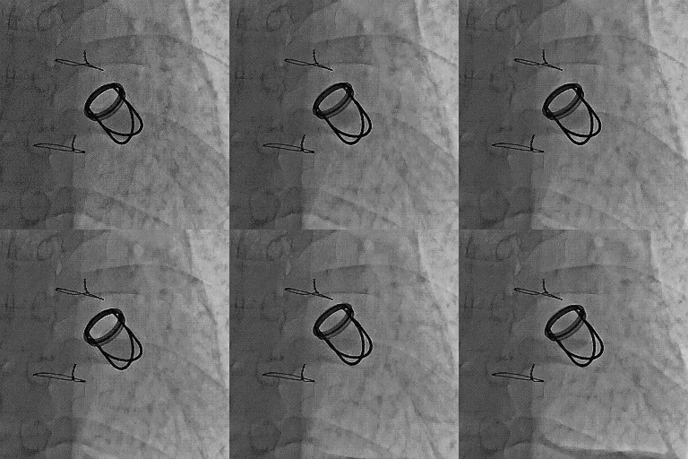
The cage which gives  a radial support* mimic  sub valvular apparatus, which none of the other valves can provide.

* Mitral  apparatus has 5 major  components. Annulus, leaflets, chordae, pap muscle, LV free wall.None of the artificial valves has all these components.  Though , we would love to have all of them technically it is simply not possible.  The metal cage of Starr Edwards  valve partially satisfies this  , as  it acts as a virtual sub valvular apparatus.Even though the cage has no contact with LV free wall, the mechano hydrolic  transduction of  LV forces to the annulus  is possible .

Further , the good hemodyanmics of this valve indicate , the cage ensures co axial blood  flow  across the mitral inflow throughout diastole. .Unlike the bi-leaflet valve ,  where the direction of  blood flow is determined by the quantum of leaflet excursion  in every beat . In bileaflet valves  each leaflet has independent determinants of valve  motion . In Starr Edwards valve the ball is the leaflet . In contrast to bi-leaflet valve , the contact area  of the  ball and the blood in Starr Edwards  is a smooth affair  and  ball makes sure  the LV forces are equally transmitted to it’s surface .

The superiority of bi-leaflet valves and disc valves  (Over ball and cage ) were  never proven convincingly in a randomized fashion . The other factor which pulled down this valve’s popularity was the supposedly high profile nature of this valve. LVOT tend to get narrowed in few undersized hearts.  This  can not be an  excuse , as no consistent  efforts were made to miniaturize this valve which is  distinctly possible.

Sudden deaths from  Starr Edwards valve  .

  • Almost unheard in our population.
  • The major reason  for the long durability of this valve is due to the  lack of  any metallic moving points .
  • Absence of hinge  in this  valve  confers  a huge mechanical  advantage with  no stress points.
  • A globe / or a ball  has  the universal hemodynamic advantage. This shape makes it difficult for thrombotic focus to stick and grow.

Final message

Science is considered as sacred as our religion Patients believe in us. We believe in science. A  good  durable valve  was  dumped from this world  for no good reason. If commerce is the  the main issue ( as many still believe it to be ! )  history will never  forgive those people who were  behind the murder of this innocent device.

Cardiologists and Cardio thoracic surgeons are equally culpable  for the pre- mature exit of this valve from human domain.  Why didn’t they protest ?  We  can get some solace  ,  if  only we can impress upon  the current valve manufacturers  to  give a fresh lease of life to this valve .

http://www.heartlungcirc.org/article/S1443-9506%2810%2900076-4/abstract

Read Full Post »

It is   surprising  to find  many   similarities between  our heart and the car .Both essentially carry out  mechanical function.  One carries the  life , while the  other beats ,  breaths life !

  • The car is the status symbol  of modern life  , while the heart is a life by itself .
  • The car has a 4 stroke engine  , while the heart has only two strokes – systole and diastole !
  • The car pumps petrol  , the heart does it with blood.
  • The car can afford to take rest at night in your garage   but  , your heart doesn’t.
  • Car  can be replaced  by a fresh one every 5 -10 years your heart can’t be.
  • In India  , it is  mandatory   for  the cars  to be  insured for  about  10 Lakh  rupees , while  the   poor hearts of our country men  are not even insured for a  single pie  !
  • A dent in your  new  Toyota  can give us   sleep less nights for many days .  A bruised and battered heart with tobacco and cholesterol is rarely  bothered about .

When the silent  screams of the heart  are not respected and heard ,   there is no other option left  for  it ,  to register its protest  , except with  a  heart attack   .This  can either be a  SOS call  or  a  call from  Heaven !

Final  message

Remember ,  the   heart breathes  your life ,  your car  doesn’t .Heart is   million times glorious than your car.

It is foolish to compare  the heart with a car . But let us  at least  learn to respect it  . . . like  our car.

Heart service station

  • Authorized heart service centers are few . Insist on genuine spare parts. Good  service engineers are becoming a rare breed.
  • Remember  both defective spare parts   and  dysfunctional service  engineers  can ruin your heart.
  • Do not allow your heart to  be explored and dismantled for flimsy reasons.
  • Never hand over your heart to strangers.
  • Do not-self indulge in 64 slices  of  fancy   shooting  of  your heart . Resist the temptation.
  • Finally do not ever go for unscheduled  free heart  service camps . That is the beginning of  trouble for your heart.

Some hearts  may  servicing alright , but realise , you are the master of the  service station .

If only you respect it , it  rarely requires to be sent to a  garage (cath lab )


Simple life style, adequate activity,   nutritious diet,  peaceful   sleep,  good work ethics ,respect to  fellow citizens  ,good-bye to  anger , helping the poor, a joyous family life , and finally  . . . less  visits to your physician  !  These  make  a  perfect , sure shot  recipe  for  living   100  glorious years !

Read Full Post »

Why is it .  .  .  so difficult ,  to  acquire  healthy   habits  and   good  behavior  in life ?

How to over come it ?

Answer :

No body can answer this question . . .Except yourself  !


Read Full Post »

We know,  electrical deaths constitute the bulk of sudden cardiac deaths in MI.  Mechanical deaths due to pump failure, muscle rupture , valve leak , also cause significant deaths   .(Surprisingly many of the mechanical deaths   may also   fulfill  the sudden death criteria !)

Free wall rupture is  invariably a fatal event. Papillary  muscle trunk  rupture  leads to severe LVF and unless intervened sure to result in fatality.

The ones who tear their interventricular septum  are some what blessed ! Here ,  the rupture does not result in instant death as there is  no loss of blood ,   instead , there is an  volume over load of right ventricle  followed by the  left ventricle  after a  few beats. Hypotension is the  rule. Even though this is a major complication there is something about  VSR which makes it unique.

Sudden giving way of IVS has  a decompressing effect on the ailing left ventricle.This many times  bring a  temporary relief to LV and if the patient survives the first few hour he is likely to stabilise  further . In fact , sudden deaths within 24hours after the onset of VSR is an exception.This defect always gives the cardiologists and surgeon some time to plan the management. We need to use this time judiciously.

The natural history is delicate . Five themes are possible

  1. Very unstable Instant death( Fortunately a  rare theme )
  2. Unstable – Deteriorating further
  3. Unstable to Stable * fit for discharge even without surgery
  4. Stable from the onset and  continue to be stable* .
  5. Stable to Unstable (Probably the most common theme )

* Pleasant themes occasionally witnessed !)

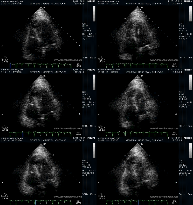
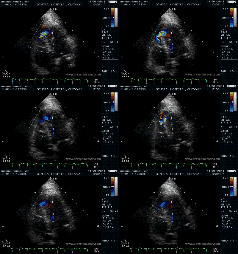
Here is 55 year old women came with extensive anterior MI with lower septal rupture.(She belonged to type 3 of the above scheme)

)

Note the septal rupture is visible even in 2D Echo


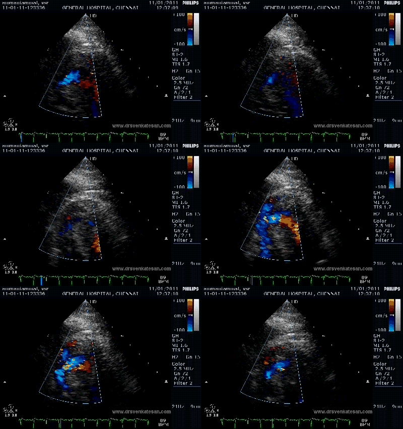
Color flow showing significant shunting from LV to RV.This shunt depends upon the LV contractile function, LVEDP and ofcourse the RV pressure


If there is severe RV dysfunction or bi ventricular dysfunction flow across the defect is inconspicuous.Brisk left to right shunting may be an indirect marker for good LV systolic function and absence of significant pulmonary hypertension.Both imply a better outcome.

The main determinant  of survival is the  underlying LV dysfunction and associated co morbidity(Renal function ) and complications .

Infero -posterior ruptures tend to be complex and  may have multiple irregular tracks  that makes it difficult to repair.

Investigations

Echo cardiogram is the mainstay .Serial echos should be done to assess the mechanical function and the progress of VSR.Hemodynamic monitoring may be done without injuring the patient .

Medical management

  • Often supportive , but  effective . Dobutamine infusion can maintain a life for few days.
  • Paradoxically , LV dysfunction and elevated LVEDP restricts volume overloading of VSD.
  • Associated MR, Arrhythmias  need to be taken care of .

Surgeons role

  • Very Vital.
  • Experience counts.(Individual as well as  Institutional )

Timing of surgery

Continues to be a controversy . Surgeons love to operate in a stable patient. But they need to realise , surgery is often needed to stabilise  many  patients. . The issue of tissue friability  is blown out of proportion in the literature .When a  life is  is at danger we can not worry about  friable tissues !

The rule of thumb could be

  • Operate as early as possible in unstable patient.
  • Post pone surgery in stable patient as late as possible ( Late here means . . .elective non emergent surgery )

Surgical options

  • Simple VSR closure without  knowing coronary anatomy
  • Simple VSR closure after knowing coronary anatomy
  • VSR closure with CABG ( total revascularization)
  • VSR closure with partial revascularization

In our experience  each of the above , has a role in a given patient depending upon the logistic , financial , social and even  the available expertise. (A good surgeon in bad Institution !)

Is coronary angiogram mandatory  before attempting to close VSR ?

Logically yes. If it is not available  just do not bother .  But, many times , when issue is saving lives , we can not afford to be too scientific , many lives have been saved by not following  such strict  protocols .A simple emergency  thoracotomy and closure of rupture site (Without even touching the LAD ) can be a distinct  and viable option in  a selected few .

Role of cardiologists

Contrary to the popular belief the role of cardiologists is minimal , except  to prepare  the patient and hand over to the surgeon.

Interventional approach to close  a VSR  is currently  be termed as an  adventurous option ! The VSRs  can assume unpredictable shapes  and the  tears can be multiple  in  different planes. The devices , catheters and  other hard ware are not specifically made to tackle these  issues  .An acquired VSR  should never be compared with congenital VSD.

Read Full Post »

Preamble

The much published TRANSFER -AMI study  has few important queries to ponder about.It was supposed to test the role of routine PCI following  thrombolysis. In other words it compared  rescue only strategy with routine strategy.The caveat is , even among  failed thrombolysis, the   rescue strategy has not convincingly proven superior to medical management  (if the time is lapsed ) as much of the damage is done .

In essence , Acute MI is  more about time management than drug or cath lab management

  1. Why the 67 % of  standard therapy cohort underwent PCI. Technically , you are supposed to transfer for rescue only if there is a  failed thrombolysis ?That is the standard approach , if  most of the cases are any way land up in cath lab , then you are trying to compare two similar groups .
  2. Why the rate of   failed thrombolyis with TNK-TPA in both arms not disclosed ?
  3. How can a 92% of study population be in class 1 Killip still considered to be high risk group ?
  4. Why the recurrent ischemia  was very vaguely  defined and still included and clubbed with primary end point along with deaths. If only recurrent ischemia was removed from primary end point . . .this study will straight away land in a regret bin.
  5. Why there were 6 additional deaths at 30 days  in routine early  PCI group ,  What was he cause of death ? Mind you these deaths have happened in a 92 %  Killip class  one cohort . Is it  not important ? The trend looks vitally   significant .We can not afford take refuge under a false  statistical roof .
  6. How many patients died or  developed MI  because of the early PCI in-spite of having  successful thrombolysis.This again could be vital . Complications during intervention  for a failed thrombolysis may be acceptable. While ,complications , when we try to  improve upon the already  successful thrombolysis is simply not acceptable .

Will the investigators share their experience ?

Finally

Why the title of the paper says it is about “Routine angioplasty” and  the conclusion emphasizes  it is indeed   “high risk subsets ofangioplasty” (While the study itself involves a 92 %  least risk Killip class 1 ) .  Why this double dose of confusion ?  (Is it deliberate  ! Which i think is unlikely )

NEJM please take note of this  . . .

All that glitters  are  not natural glitter . . .some are made to glitter !

Read Full Post »

NSTEMI is a  common clinical problems in CCU.

When we say  NSTEMI it can mean any of the following

  • NSTEMI with ST depression
  • NSTEMI with T  wave Inversion
  • NSTEMI with Biphasic T wave
  • NSTEMI with normal ECG
  • The irony   called STEMI evolving as  NSTEMI**

By default most of  us think ,  if it is NSTEMI  . . . there  must be ST depression. This thinking is  not logical but traditional. Still,   ST depression may be the  common presentation. NSTEMI with ST depression  has much worse outcome than other forms.

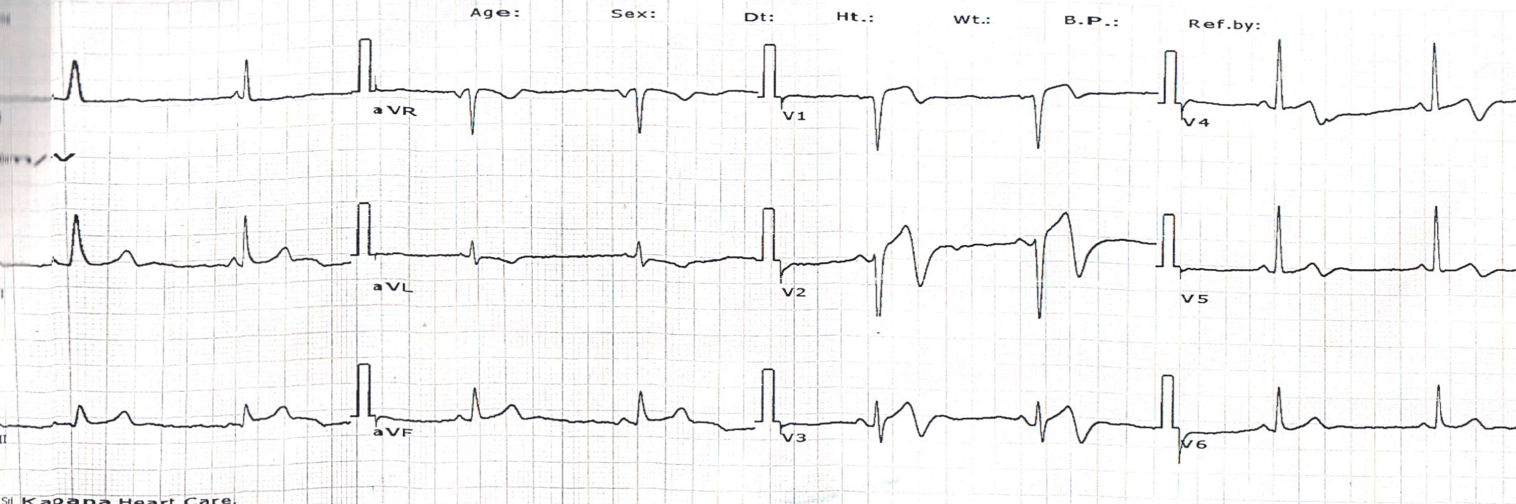
The following ECG is from a 45 year old man with a vague mid sternal  chest pain for 48 hours.

The unusual type of NSTEMI with Bi-phasic T waves

His echo showed wall motion defect in LCX territory .A diagnosis of NSTEMI was made.The predominant finding was biphasic T waves .

**One may wonder  why can’t we call this ECG as a  Classical STEMI ?

There is a 2mm  ST elevation ,  with a infarct as well  ? But , the point  here  is there is no business for T waves to get bi-phasic or inverted in the early hours  of  a  classical STEMI .

This  exactly has happened here. Hence we can not call  the above event as  STEMI . Instead it  is ,  STEMI   evolving into NSTEMI . So  a combination of  features of STEMI/NSTEMI occur together. The best description for above  entity is  STEMI in transition to Non Q MI

Read the related article in my site  Is the terminology of Non Q MI still relevant or obsolete ?

Read Full Post »

What happens to vegetation following  successful therapy ?

  1. It regresses almost completely  and become sterile
  2. It regresses about 50 % volume   but continue to harbor  live  viable bacteria
  3. Gets sterile   but  does not regress ,
  4. Vegetation vanishes completely .Gets dissolved circulation as micro particles.
  5. Appears slightly bulky.

Answer.

Each of the above statement can be true in different patients  at  / different times.  However No  1, is generally the dominant theme.

  • Most of the small vegetations disappear fully.
  • Large vegetations (>2 cm) almost never disappear fully .
  • Fungal vegetation is notoriously known  for a long haul battle
  • Systemic embolism is an important mode of  vegetation clearance from heart.
  • Size of vegetation is an independent indication for surgery .
  • Combination of vegetation with super added layer of  thrombus is common.The thrombus lyses in due course , mimicking thrombus regression.
  • Paradoxically healed vegetations may appear dense in  2 D echocadiography ,which may be wrongly interpreted as a growing vegetation.
  • The risk of recurrent vegetation formation remains till the raw area is completely endothelised.Hence antibiotics are given up to 4-6 weeks.

Persistent  culture negativity may be a  good index  for  successful management . But a negative blood culture  does not in any – way imply  absence of vegetation.

http://www.ncbi.nlm.nih.gov/sites/entrez?Db=pubmed&Cmd=Retrieve&list_uids=7985602&dopt=abstractplus

Read Full Post »

T waves attract less  attention in STEMI ,except for the  fact   tall T waves  implies   hyper acute phase of  STEMI.

What is the duration of hyper acute phase ?

  1. Few seconds
  2. Few minutes
  3. An hour
  4. Few hours
  5. Any of the above

Answer

No one exactly knows  .It can  be highly variable .  So , 5  could be  the correct answer .  

 * Most importantly  hyper acute phase  need not occur in all patients with STEMI as suggested in experimental models.

Some  observations in T wave behavior in STEMI

Mechanism of hyper acute  T waves

It is the pottsium channel dynamics.Transient intracellular hyperkalemia  is thought to be responsible.

T wave as marker of  reperfusion

Inverted T wave in precordial leads are a good marker of IRA patency  especially in LAD

Slowly evolving STEMI

This is relatively  new concept . STEMI with a prolonged hyper acute phase  ,  ie ,  T waves ” dilly dallying”  for hours or even few days have been recognised. (This was  refered  to pre-infarction angina in the past )

This sort of T wave behavior makes it difficult to diagnose STEMI.Enzymes will help , still  thrombolytic guidelines  demand us to wait till ST elevation to occur. This is  unfortunate .But as physicians we are  justified to thrombolyse tall T waves with a clinical ACS .The other simple solution is to shift the patient to cath lab to find what exactly is happening in the LAD ! 

Now , what is new about  T waves in STEMI ?

It is  the localizing value  in LAD infarct

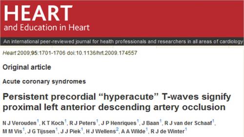
A tall persistent  hyper acute T wave  helps us to localise a LAD lesion .This paper from Netherlands ,  clearly  confirms this observation. The study was done from a primary PCI cohort,   a perfect setting to assess the  T wave behavior  in the early minutes /hours of  STEMI .

Other mysteries about T waves in STEMI

Does hyperacute T waves  occur in infero-posterior STEMI ?

I would believe it is very rare .Our CCU has not seen any tall T waves in inferior lead. Further analysis of the  data from the  above study could answer this question .

How often a  hyperacute T waves transform into NSTEMI ?

This again is not clear.Most of the hyper acute T will evolve as STEMI .But  , nothing prevents it to evolve as NSTEMI a well . After all , a hyper acute T   MI can  spontaneously lyse in a lucky few , ( Who has that critical  mass of natural  circulating TPA )  .If  these natural lytic forces are only partially successful , it may evolve into de nova NSTEMI.

Bi-phasic T waves in ACS.

A benign looking T waves with terminal negativity in precordial leads  can some times be a deadly marker of critical LAD disease.This has been notorious to cause deaths in young men which often correlates with the widow maker lesion in LAD.

What is a slowly evolving STEMI ?

Prolonged tall T wave phase  possibly   indicate , the myocardium is relatively resistant to hypoxic damage .

The most bizarre aspect in our understanding about ACS pathophysiology  is the concept of  time window , based on which , all our  ACS therapeutics revolve !

Does all myocardial   cells  have a same ischemic shelf  life ?  Can some patients  be  blessed with  resistant myocardial cells   when confronted with hypoxia or ischemia ?

                                 It is well-known  , in some hearts ,  the  muscles go for necrosis within  30 minutes of  ischemia,  while some hearts can not be infarcted even after 24 hours of occlusion .So , slowly evolving STEMI is a feature of  myocardial ischemic resistance .This is not  a new phenomenon as we have extensively studied about the concept   ischemic preconditioning .

We wonder there is something more to it . . .  the quantum of preconditioning  can be inherited .Further  , we are grossly ignorant about  the molecular secrets of  non ischemic metabolic  preconditioning  .

Final message

                         T waves attract less  attention in STEMI . Cardiologists are often tuned to look only the ST segment , after all ,  ACS  itself is classified based on  the behavior of this segment.(STEMI/NSTEMI) . We need to recognise ,there is a significant subset of ACS   affecting exclusively T waves.  Shall we call T elevation  MI ? ( TEMI )

Do not ignore T waves in STEMI. It has more hidden electrophysiological  treasures that  is waiting to be explored .

Read Full Post »

Great things happen in India nowadays  .Economy is growing    at  9% , the growth of  automobile industry is  fastest in the world. But in  scientific  research and development  it   has  been traditionally lagging behind . Things are set to change. The medical science , (If you want to call it as industry it is fine !)  especially  the imaging science is making rapid strides. The proliferation of  private and corporate hospitals and institutes   has helped us practice the latest .

Pulmonary atresia with VSD is a rare congenital disease where  there is partial or  total (chaotic  )pulmonary arterial blood supply .

When the pulmonary  artery becomes atretic , what will the lung do ?

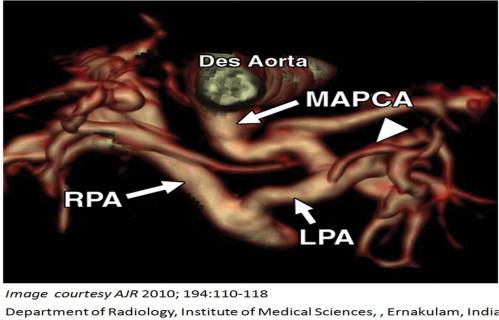
It has to get perfused somehow ! It tries to snatch the  blood from the aorta  in whatever  possible manner . Depending on the severity of pulmonary atresia  , there  can be  a total anomalous pulmonary  arterial supply (Type 4 ) .Here , few twigs directly originate  from Aorta, few from branches of  Aorta  and sometimes bilateral  PDA etc .These are collectively called major arotopulmonary collateral  (MAPCAS) .In fact differentiating a PDA from a MAPCA can be extremely difficult .(It has only academic purpose though !)

Hither to ,  visualising  the  MAPCAS was a  huge  task . Aortogram with selective cannulation of MAPCA  was  neccessary.Now,  with the advent of  MDCT we can get some stunning  images  of these collaterals non invasively.

Why is visualizing and delineating MAPCA anatomy important ?

This will facilitate the surgeon to plan  the  unification of pulmonary  arterial flow    and reenginnering the pulmonary circulation  (with or with  out  conduits)

Here is a rare  publication originating from India  in  American journal of radiology  . An  exclusive   article  with  CT scan  images of the  defect .

Amrita  Institute , from  the southern   Indian  state of  Kerala (  also known as God’s  own country )  is doing a phenomenal  cardiology  work  especially  in pediatric cardiology.

Three cheers to the team which published this  master piece . With the courtesy of   AJR the link to the article is placed here.

Read Full Post »

Rheumatic heart disease is rampant in India.  Advanced forms of mitral stenosis are  still common.

Critical mitral stenosis with LA clot formation is often seen.

But here is a  women in late twenties  presenting for the first time with syncope .

And what  you see inside is  not a fiction  . . .

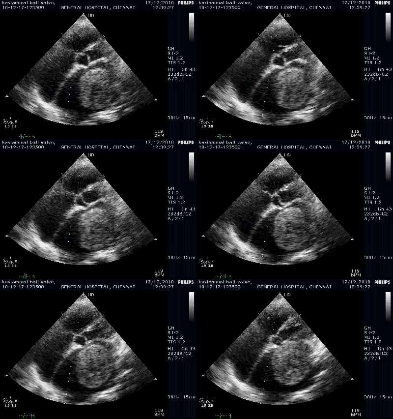
Left atrial clot occupying the whole cavity ! Where is the blood bound for left ventricle ?

4 chamber view

Luckily the clot is   so  big and MVO is less than 1 sq cm. It is highly unlikely the LA clot can negotiate the orifice.

Small fragments can dislodge .This patient developed syncope whenever she bends and lie down at a particular position.

What needs to be done in this patient  ?

Can it be lysed ? No ,Emergency surgery is required with concomitant mitral valvotomy or mitral valve replacement.

Is there a temporary aortic filter available to prevent systemic emboli from heart  ?

Distal protection devices are  available only temporarily in the coronaries and  carotids during interventional procedures.There is no aortic protection devices  for LA,LV clots in high risk patients .  When IVC filters  are used  block a potential pulmonary   clot why not aortic filters  for preventing systemic emboli  ?

Why we have not thought about  this  . . . is  surprising . May be intensive anti coagulation is as effective .

Read Full Post »

« Newer Posts - Older Posts »