Feeds:
Posts
Comments

Archive for the ‘Uncategorized’ Category

Research is the  guiding force for advancement in medical science.New drug and devices are developed every year that result in major break throughs.There need to be  a strong purpose and meaning for every research concepts in Medicine.The fundamental  principle in medical research  should be . . .

In basic science

It need to  generate a  new concept  that should help solve a existing  problem  or find an  eluding  answer ( Eg mechanism of an organ or cell function,  Genetic research  etc)

In clinical science

  1. An applied scientific concept  that can generate new treatment  modalities.
  2. Development of new diagnostic tools that can add new information to the existing imaging modalities.It should have either a cost or effectiveness advantages .
  3. Development of new drugs  or devices or surgical methods with  less side effects.
  4. Development of new drugs, which has a new additional benefit with less side effects.

A .What is Junk research or duplication of research

Most of the research methods testing  “non inferiority”  hypothesis can be considered junk  research (Note – Exceptions will be there. )

The most exploited concept in drug research  is  the concept of testing   non inferiority as it  allows comparing  new drugs with a proven old  drugs .Why should some one produce a drug which is likely to have a same effect  with  no additional benefit. Huge amount of time  , money  efforts  are wasted in  such  research

What is the need for 20 different ACE inhibitors  and 10 different ARBs when the basic structure  and function is almost similar ? Similarly Statins, analgesics, H2 receptor blockers, various forms of heparin  the list is end less . . .

There need to be a  strict vigil  on which research question the industry is trying to raise  and answer ?

Drug are released periodically  like movies with same old  screen play ( With a new  name  of course !) but with   a huge  cost.If any novel or music or art work or done in similar fashion there is  vibrant media to expose it, or  some times even legal action follows.  Alas . . . In medicine  it is free for all as  any one can legally plagiarise with  loopholes in plenty. Some times these drugs are hurriedly marketed as it has to generate the funds  before risk of it getting banned due to unforeseen side effects. In this sense some drugs exactly mimic a movie with good box office run for the initial few weeks.

A autonomous  world body ( Say WHO ) should supervise which research can be pursued in this world that can help betterment of human health . Currently tighter  regulations come into force only  during human testing .We need act  earlier   to prevent junk research in lab itself ! This can save huge resources .Some  may argue ,scientific work should not be curtailed  however futile they are !  I leave it your judgement .

B.Placebo compared superiority : This is another controversial concept  .This was a prerequisite  for a drug research in the earlier era .Now a drug need to be compared with the best current treatment! not with placebo.

Why superiority studies  are rarely designed by drug companies ?

Realise , how rare it would be to find  a study   where  a patented drug  is  compared  for superiority over another patented drug . Can you  guess the reason . . .  because they can do  superiority studies  easily over the placebos  !

Do we need to restrict drug development with similar structure and function?

The only argument can be in a global economy no body has a right to prevent any company  from  producing whatever they want  to  ! Survival of such  drugs  in this world is left to  market forces .The consequence  such approach could be disastrous ,  as  the  cheap  generic drugs with a  maximal  proven beneficial effect (Eg Hydrocholrthiazide in Hypertension ,  ) may not be manufactured at all in the near future . (Link to msf)

Is  the need for choices ,  an excuse ?

Like any commodity , people need choices so what is wrong  if a same drug is produced by 100 companies.Let the best survive !

Remember drugs can not be compared with other commodities. Even in other sectors , there are restrictions .( You can not allow 100 flights in a same sector or 10 mobile phone companies in a single city ,  however free the economy is ! )Every drug and device company should have a moral and legal obligation not to reduplicate the already proven concepts. Many tend to masquerade as new concept with a very subtle difference.

What is the favorite game  played by the drug companies ?

It is a too competitive market out there in world pharma industry. Drugs and device are developed at huge cost.They have to spend billions of dollors for R &D. They need money. The  manpower , time and cost involved are enormous. A drug or device has to pass on many acid tests and hurdles before releasing into market.Even after releasing there is a very good chances of it being  recalled  in the first few years as real human data start pouring.Apart from these many drugs and devices fail to take off at various levels. There can be a huge financial loss.(Eg Torcirapid HDL reducing agent).

So what is the solution ?

Drug companies involve in futile research that is imitating already proven concepts .And a drug which was approved for one indication by FDA is tested for many other indications as it becomes easier for increasing the market  share.This is can be  called as  the  “Phenomenon of  drugs  looking for a  new indication” .This  against  spirit of clinical research .

The classical example is  the attempt to  widen the  indication  for statins , amiodarone ,  ( Aortic stenosis   etc )Instead our energy  should be focused   into  developing  drug  for incurable  diseases

When can duplication of research  be beneficial ?

Scientists are working hard for a total artificial heart for years.Many teams are pursuing different methodology and concepts. Here the  competition is welcome. Like this there are so many labs  trying to create a solution for a genuine medical issue.

Disclaimer

This article has nothing against any pharma industry. It aims to reason out the futility in medical research which is to be addressed for the betterment of human health.

Read related topic in my blog.

Can a Aim of a study be wrong ?
Read Full Post »

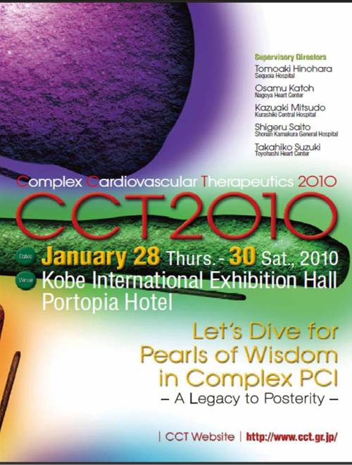
Annual workshops for interventional cardiologists has been  hugely popular events.They have  become the forum for all technological breakthroughs. Some of the popular ones are

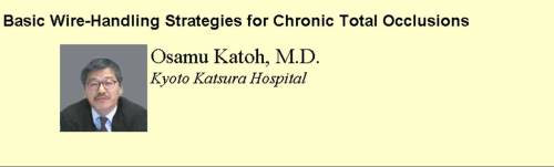
Japanese have gained a unique place in  complex cardiovascular  therapeutics interventions especially in chronic occlusions.

Landmark article for CTO crossing

cto chronic total occlusion  Katoh coronary angiogram

www.cct.gr.jp/2003/wirehand/index.html

www.cct.gr.jp  cto japan

How to reach Japan ?

Click below

CCT2010

Read Full Post »

Total coronary artery occlusion is a common finding in CAD  especially in chronic stable angina. Normal coronary blood flow is 5 % of cardiac output  that amounts to 250-300ml/mt.At an average  heart rate of  70/mt  , each  beat  injects  about 5cc blood into the coronary circulation.This is shared between two coronary arteries.  This means , only few CC (2-3cc) of blood enters  each coronary artery with each cardiac cycle .

When one of coronary artery is totally occluded what happens to the coronary

blood flow ?

A.Total coronary blood flow  can be be  maintained   normal  at rest  as it  forms  only about 5% of cardiac output  (or it is only  slightly reduced )

B. It is believed , the unobstructed coronary artery  could receive the blood meant for the contralateral coronary artery. This  possibly explains the increased coronary artery diameter in the non obstructed artery.

C. It’s nature’s wish ,  that the  contralateral  coronary artery  shall share  50% of  it’s  blood through  collaterals if available.

D.If collaterals are not formed it , the unobstructed coronary  artery  may be over perfused with double the amount  of blood flow.

E. Some times , the collaterals steal  much more than what  the  obstructed coronary artery  deserves and make the feeding coronary artery ischemic. This is many times observed in  total RCA occlusion with well formed  collaterals  from LAD/LCX.

F.The collateral flow  in CTO also depend on whether flow is directed from LAD system to RCA or from RCA -LAD system. The LAD is better placed to assist RCA than vice versa.This is for two reasons.1.LAD blood flow is higher than RCA so it can share it.2.The driving pressure is more  from LAD -RCA , as RCA can receive  blood flow even during diastole .

F.During exertion , the coronary hemodynamics become further complex.The collateral’s are traditionally thought to be less than adequate during times of exercise.But it is more of a perception than solid scientific data.This rule  may be applicable in only certain group of patients. We know CTO patients with very good exercise tolerance who have documented collateral’s.

G.Collaterals can be either  visible or invisible by CAG. The strength of collateral circulation is not in it’s visibility but it’s capacity to dilate and  respond to neuro humoral mediators at times of  demand.  Currently  , there is lot to be desired  regarding  our knowledge about  the physiology  of visible collaterals , no need to  mention about invisible collaterals !

Final message

The above statements  are based  on logics and observations .

Is it not a  irony  in cardiac literature ,  where  thousands of articles  are coming out every month  to tackle  totally occluded coronary artery(CTOs) ,  there is  very little data   regarding the coronary hemodynamics in chronic total occlusion .   How  does a patient with CTO can manage a active life with only one functioning  coronary artery ?

Read Full Post »

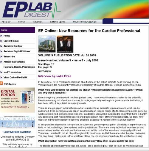
Thanks to wordpress.This  blog has caught the attention of some professional sites.

I was interviewed   by Jodie Elrod on behalf  of   EP lab digest July 09 Issue

dr s venkatesan www.drsvenkatesan.com

Read Full Post »

Contrast induced  nephropathy (CIN)  is potentially a serious problem. The following precautions are useful in the prevention of CIN.Patients with  serum creatinine>1.5mg carry a  progressive  risk .Diabetics and elderly are more prone.Protienuria is a added risk.

  • Adequate pre procedure hydration is a must . Normal saline (.45%NaCl) infused over 6 hours on the day prior  to flush the kidneys  of protiennaceous  substances.
  • Low protein diet in the days prior to procedure could be useful.
  • Ionic contrast to be  avoided.
  • Among noninionic  low osmolar , monomeric Iohexol may be avoided . Use of isoosmolar , dimeric Iodixanal has some advantage.
  • Oral antioxidant N-acetyl cysteine  600mg twice a day pre and post procedure along with .45% NaCl infusion  is found to be  useful.
  • Sodium bicorbonate infusion . Three  ampuoles of normal saline .9%  in one  litre normal saline infused 3ml/kg per hour started i hour prior to procedure and continued at 1ml/kg for 6 hours post procedure has a renoprotective effect.
  • There could be a role for combination of N -acetyl cystiene and sodium bicorbonate

Apart from the above measures the following general rules are vital

  1. Use minimal amount of dye .<30ml. Dye volume  is more important than the type.(50 ml of isoosmolar dye is more likey to cause CIN than a 30ml of ordinary dye !)
  2. Minimal views to delineate anatomy.
  3. Whenever possible utilise biplane angiography.
  4. Do not  give in to the  temptation of injecting a renal shot.( Although you could miss a renal artery stenosis (RAS), which is likely in these patients .Some may argue  for it ,  as  it gives us an opportunity to cure the RAS )
  5. Stage the  procedure and post it on different day if intervention is required.
  6. PCI for discrete straightforward lesions may be attempted
  7. Avoid complex PCI in renally compromised
  8. Review all the drugs for the potential renal offenders.
  9. Manage the diabetes , cardiac failure meticulously
  10. Have nephrologist always on a  standby mode

Post procedure follow up .

Hydration to continue

Follow up biochemistry

How often we require dialysis   in these patients  ?

It can be avoided in most if we have taken sufficient precaution. In severely compromised renal  function peri procedural dialysis is often used.

Finally , before doing a CAG in renally compromised patient always ask this  question and answer it genuinly . Is the CAG/PCI is really indicated in the given patient ?   Does it going to make a difference for the patient ‘s life ? If the answer is  – No – please avoid it !

Read Full Post »

Common causes

  • Left atrial appendage clots
  • Left atrial  clots
  • LV mural thrombus (Post MI, DCM)
  • Mitral aortic valve infective vegetations

Conditions that could be commoner than we think!

  • Aortic valve  calcific debri
  • Aortic arch atheromas
  • Paradoxical embolism through foramen ovale.*

If we consider incidence of patent foramen  ovale in general  population is up to 20%  the problem of paradoxical embolism could be really significant.PFO is a potential  right  to left channel of more than 5 square cm .

Unusual causes

  • Mitral annular  and posterior leaflet  calcification
  • Tumor embolus (Myxoma etc)
  • Prosthetic valve thrombus.

Cause never known and identified.

There are times a cardiac source can never be identiifed.This can very well happen , as a transient arrhytmia can trigger a thrombus formation and subsequent examination are totally normal .The incidence of such group can never be known !

A brief  account on cardiac embolus

Cardiac  embolus constitute an important cause for stroke or TIA. There are number of important conditions that can result in cardiac embolism. The embolus could be a  thrombus(95% of times ) , Vegetation, tumor, calcific debri, cholesterol , atheromatic particles , rarely chordal and subchordal structures following it’s rupture.The size of the embolus could vary between <.5mm to 1-2cm in diameter.The average size is 1cm . The clinical presentation depends upon the size, content of the emboli(Thrombus vs non thrombus) )  site of trapping, freshness of thrombus, natural lytic process.

The common  sites  of trapping is middle cerebral artery.The average diameter of MCA is around 2 mm.So one can imagine almost most of the cardiac emboli can not traverse it, and hence a  dense stroke .But , micro thromboemboli , can safely cross cerebral circulation.they usually present as TIA or a chronic lacunar infarcts and many times vascular dementias. Cardiac thrombus rarely gets struck within the carotids.This is especially common if there is associated critical carotid stenosis.The situation is a dire emergency.(Inspite of the fact there is circle of willis for

How do you investigate ?

A complete physical examination with well documented clinical history

A meticulous echocardiography with possibly a TEE (Transesophagel echo)  may  done .

Underlying disorder to be tackled.

When a emboli is released from the heart , what determines  it’s entry into carotid ?

The aortic ejection force is such that whatever particle that exit from the heart , tend to hit on the carotid first.Only if it fails to accept it , it is pushed across the aortic arch into the descending aorta.This result in peripheral embolism .Some times a emboli gets struck within the carotid .This  especially happens  if there is associated with critical  carotid obstruction .This can result in massive stroke and sudden neurological death( The threat is real  inspite of the presence of circle of willis which supposed to come to rescue in cases of sudden unilateral carotid obstruction)

What is the relationship between atrial fibrillation and cardiac emboli ?

For long ,the two conditions were thought to closely linked entities.AF slows blood flow across the atria, predispose to left atrial clots and possibly increased stroke.So vigorous means to restore sinus rhythm were attempted. But to our surprise, the incidence of stroke was not greatly reduced between optimal anticoagualtion and sinus rhythn restoration. This indicated many of the cardiac source of embolism could be distal to left atrium aortic arch atheromas, carotid etc (AFFIRM study)

Restoration of AF into SR can possibly prevent thrombus  formation  only in one chamber , while systemic anticoagualtion can prevent thromboembolism virtually any where from the high risk zones across the LA, LV,  Valves, Aorta, arch, carotids etc. So , it defies scientifc logic , to attempt AF restoration by all means (The exotic pulmonary vein isolation etc !)  to  eliminate one of the cause of stroke.

Read Full Post »

The valve replacement surgery is one of the great innovations in cardiac surgery. The common disorders that require mitral and aortic valve replacement are

  • Degenerative , calcific  aortic stenosis and regurgitation.
  • Rheumatic mitral, aortic valve disease.
  • Ischemic heart disease -Ischemic MR
  • Some cardiomyopathies

The mortality in valve replacement surgeries vary  between AVR, MVR, and DVR.

AVR – 2-5%

MVR 4-12%

DVR  6-15%

Source CTS.net

Determinants of outcome

General factors applicable for both valves

Elective vs Emergency

LV function

Associated CAD /CABG

Co morbid conditions

The following observations  can be  made  in valve replacement surgery

  • Mitral valve function is closely linked to LV function while Aortic valve  is not .
  • AVR  patients always do well than MVR in  the  immediate post operative period
  • Aortic stenosis patients do well than aortic regurgitation .
  • Mitral stenosis patient do well than mitral regurgitation
  • In   DVR  the excess mortality is due to  the addition of MV , not by  AVR .

Aortic valve replacement has better post operative outcome when compared to mitral valve replacement ,Why ?

Aortic valve has only two components namely a  annulus  and leaflets. The prosthetic  aortic valve  replaces both these natural components . Mitral valve has 6 components , prosthetic mitral valve has only two components . Hence  , any prosthetic mitral valve is far inferior to natural mitral valve . The  pap muscle, chordae, and LV muscle fail to assist the artificial  mitral valve.  So , between AVR and MVR   AVR is far perfect  prosthetic surgery and  the hemodynamics   mimic as closely to the natural valve.

Why aortic stenosis patients do better than aortic regurgitation ?

Aortic stenosis  results in severe  LV outflow obstruction .The LV struggles to pump across the obstruction.So , once it is relieved by a prosthetic valve , there is great relief for LV .We know the the aortic valve orifice becomes <1cm2 in critical AS . (Like a pin hole !) .Prosthetic aortic valve at least doubles or triples this orifice and the LV enjoys this sudden relief  and  becomes active or even hyperactive in immediate post operative phase , later  it    settles to a near normal LV function. It has been observed even very severe LV dysfunction associated with aortic stenosis recovers well .

What happens  in AVR done for  dominant or isolated  aortic regurgitation ?

Here the situation  is dramatically opposite.The purpose of  prosthetic aortic valve is  reduce the  aortic valve orifice .

In AR ,  the  left ventricle  is  used to eject  the blood  with ease  across LVOT   without  much  resistance  ,  only to find part of the blood returning  back into the chamber . In  the next beat it does the same and  the cycle   continues for ever .This in due course , dilates the LV  and increases  wall stress and afterload.  LV dysfunction follows .This  takes long time to set in.That’s why chronic asymptomatic AR patients  do so well and they do not require surgery until after the onset of LV dysfunction .(End systolic LV >55mm)

After the aortic valve replacement , the LV suddenly finds  the newly introduced prosthetic valve  a hindrance !. As all artificial  valves  have  less than the natural orifice. LV   takes some time to adapt to the new environment . The EF initially may slightly fall and recovers later.

If pre- operative LV dysfunction was significant the immediate post operative period can be critical.As even a slight fall in EF can result in prolonged hypotension.Many of these   pateint may  require  prolonged inotropic  support.

What are the differences between MVR done for mitral stenosis and MVR done for mitral regurgitation ?

Here again ,  the same principles apply.The Mitral stenosis patients do well following MVR than MR patients.This is because of two reasons .  MR patients  have dilated LV  and may also have associated impaired LV function .A chronic MR is  some   what  a stress reliever  for the LV  ,  as  with every contraction it can decompress a little   bit  . It is an important hemodynamic  fact  ie  ,  presence of   even a  trivial  MR helps the LV to tackle the  it,s  afterload  easily by increasing the dp/dt and also the EF.

So when we introduce a a fully competent prosthetic mitral valve all of a sudden the LV again struggles for some time.

Final message

MVR  patients has less favorable  clinical outcome than AVR .

Coming  soon

How  different is the anticoagulation  protocol difference between AVR  and  MVR ?

Read Full Post »

No one can deny ,  there is a huge revascularisation  dilemma  between CABG and PCI  in patients with CAD. This is especially  prevalent in multivessel disease in chronic coronary syndromes.

In acute STEMI , CABG is never considered as a primary revascularisation  procedure.There should be strong reason for this !  Few studies , suggested a role for CABG in acute MI if it is done within3- 6 hours .But it became very clear , by and large CABG for acute STEMI is contraindicated . This especially applicable when q waves are formed.

Reasons.

Performing a complex surgery  on a   blood vessel subtending a  dead  , irritable ,myocardium is dangerous. Even a graft for non IRA vessel has no great benefit in the acute setting. The mortality of CABG in the first 48hours of MI  can be up to 15%.  Primary PCI  opens up the IRA without the hazards of major surgery

Issues  for  CABG in STEMI

Failed thrombolysis :  Rescue PCI  could be useful provided it is also performed within the same time window . In most situations  there is nothing called  “rescue CABG ” 

Some would believe Left main and critical TVD is an  indication for an emergency  CABG. Yes , CABG may be indicated  in this setting , but even here it may be delayed for a week if there is no ongoing ischemia , angina or hemodynamic instability.

Still  , there is a  definite role for CABG in STEMI in the following situations.

  • Mechanical complication- VSR/MR/Free wall rupture
  • Cardiogenic shock
  • Failed  and complicated primary PCI.( Note : Simple failure to open a IRA is not an indication for CABG , there need to be a life threatening situation ! )

 Coming soon

Routine  CABG  is  generally  dangerous and contraindicated   for STEMI ,  while it  is  a great ,  life saving surgery  in  most of  the  refractory  NSTEMI : How ?

Read Full Post »

Amiodarone has brought a major change in the medical management of ventricular arrhythmias over  the last  few decades. It is  a powerful antiarrhytmic drug , with all class 1 -4 action  (of vaugan williams classification.) It  has   sodium , pottasium, calcium and beta blocking properties.  Hence there is no surprise, amiodarone is  aptly called a broadspectrum anti arrhythmic as it  acts  on many of the cardiac receptors .

Of course , the major action is thought to be pottasium channel blocking effect(Class 3) that prolongs the action potential duration and refractory period resulting in termination of many re entrant arrhythmias.  While , amiodarone by structure resembles thyroid hormone, takes a long time to reach the steady state plasma levels  .Oral amiodarone takes  up to a week time to exert it’s action.

If amiodarone is a slow acting drug , is it not  surprising  , Why  IV amiodarone is given in the emergency managment of  VT ?

Typically , there is difference between the mechanism of  action between  oral and IV amiodarone

The class 3 property of amiodarone ,  is a late observation following oral adminstration. QT prolongation rarely  occurs  following bolus iv amiodarone . So , VT  terminating effect is thought to be some thing , other than class 3 action. Many believe the combined beta and calcium channel blocking effect could be responsible for rapid reversion of ventricular arrhythmias. The sodium channel blocking action with fast kineticks may also contribute.

Final message.

While amiodarone is a  prototype class 3 anti arrhythmic drug,  it’s  VT terminating property  may  be ,   attibutible to other class action. mean while  , the Class 3 action is responsible for long term prevention of VT/VF .

Read Full Post »

Exercise  stress test ( Also called treadmill test ) is an important investigation  not only in patients  with suspected  CAD  but also in  established CAD . In the former  group ,  it helps us to exclude CAD in patients with chest  pain and in the later group ,  it helps us to assess  functional capacity , risk stratification and to detect any  additional ( New or residual ) ischemia.

Stress test being a physiological test , has a huge  advantage of assessing the adequacy of myocardial blood flow without even  knowing the coronary anatomy , while Coronary angiogram (CAG)   has a zero physiological value* in spite of   excellent assessment of the coronary anatomy !

It is an irony , in the assessment of angina we are expected to assess the physiological adequacy of myocardial blood flow ,  we have kept coronary angiogram as a gold standard  over and above the much  neglected  physiological stress test.

Of course, the limitation of stress test is that ,  it has only 75%  specificity(  to rule out CAD ) and about 80% sensitivity (To detect CAD ) .In simple terms  stress test is likely to miss  20% times to miss a CAD  in patients with CAD  and 25% of times falsely diagnose CAD  in patients without CAD.

In the above statistics  ,  coronary angiogram was considered   gold standard . The problem with this data is that , CAG is not the real gold standard ,but it was  nominated  as a gold standard . We now know normal coronary angiogram is not equivalent  to  normal coronary arteries and vice versa.

While both test have limitations , it is logical to believe CAG has an edge over stress test since it visualises the anatomy. But ,  once an obstruction is demonstrated by CAG, stress test scores over in assessing the physiological impact of the lesion.

Is a 70% LAD lesion significant or not ?

Stress test will give vital information to answer this question.If this patient performs 10-12Met exercise without symptoms it means , the obstruction is not impeding the flow even during stress. He may do well with medical therapy.

What does a positive stress *mean for the patient and for the physician ?

(* A false positive EST in LVH, anemia, baseline ST shifts are included in discussion )
  • A positive stress test  with or without angina at low workload <5 METS  indicates very significant obstructive CAD either in left main , or proximal LAD/LCX. They should get immediate CAG.
  • A positive stress test at load  5-10METS  is again significant and patients should get early CAG
  • A positive stress test with angina at good work load >10-12 mets  would indicate insignificant or minimally obstructive  CAD.
  • A positive stress test at  the peak of exercise  at good work load > 10-12METS without angina could indicate a false positive or very minimal CAD.

For the physician , the proper way  of interpretation  should be , the fact that a person performs 10-12  METS  indicate the myoacardial blood flow  would  be  more than adequate in most life situations. Knowing the coronary anatomy serves no purpose here, as no revascularisation will be attempted even if he is going to have a significant CAD ( Which again , is also highly unlikely ) .He should be managed with appropriate lifestyle (Diet, activity, relaxation )  anti anginal drugs,  aspirin , good lipid control and plaque stabilisation with statins .

Can a  patient with critical left main  or proximal LAD  perform >10METS in exercise stress test ?

No , large clinical experience (Also refered to Class C evidence  by ACC/AHA!) indicate no patient with critical  left main or equivalent disease  can perform 10 METS  excercise

While  ,  EST may be less hyped investigation, but it is the  only  noninvasive test , ( that too , simple and  cheap ) that can rule out * a significant left main  or equivalent almost   100%  correctly .

Now that,   the results of COURAGE  and BARI 2D have clearly indicated medical therapy is best form of management  in chronic  CAD , ( except in severe obstructive CAD in vital locations)  a  positive EST  at > 10-12Mets  , has absolutely no indication* to for doing a CAG.

*Some would advocate a policy of  doing a  CAG as a baseline investigation in all patients with positive EST  to know the coronary anatomy and will not proceed onto revascularisation if there is insignificant lesions.

Further ,  real life experience has taught us , routine  CAG in these patients

  1. Increases patient anxiety as he is given a report with a diagram of obstructed heart vessels
  2. Leads to multiple cardiac consultations
  3. Divergence of opinions
  4. Finally end up in  the likely hood of a inappropriate  revascularisation for a  insignificant distal CAD.

Final message

Every patient,  who has positive stress test  , ( Please note , it could  even be  true positive  )  need not undergo CAG .  Most  interventional cardiologists could  feel  otherwise , but one should also  remember ,  There is one  more role  for the interventional cardiologist ie  , to intervene when inappropriate interventions are done to their patients.

//

Read Full Post »

« Newer Posts - Older Posts »