Feeds:
Posts
Comments

Posts Tagged ‘nstemi’

                                     Hypertension is considered a major cardiovascular risk factor.Hypertension  can have multiple physiological and pathological effects on heart . The common response to  raised arterial pressure is the hypertrophy of the left ventricle ( LVH). This can increase the risk of heart failure in few ( Mainly diastolic failure)  It is a leading cause for stroke  and   less often a  coronary event.

What links Hypertension and  coronary artery disease

                                           Coronary artery disease is almost synonymous with atherosclerosis. There is no separate entity called hypertensive coronary artery disease. But HT can accelerate the process of atherosclerosis. It is widely understood, hypertension can cause  physical endothelial damage and functional impairment of endothelial function.The physical damage ie enothelial disruption , or erosion is a very uncommon phenomenon . So currently  there is sufficient clinical experience  HT is considered dangerous for coronary artery only if it is with the  company of diabetes and hyperlipidemia. (This will seem controversial as it is against the findings of iconic Framingham trial!)

What the medical community refers to hypertension , may not be really so inside  for the coronary arteries.

                                             The relationship between brachial cuff blood pressure and the intra coronary pressure has very little linear relationship. So one should recognise it is the intra coronary hypertension that has a immediate impact on the coronary events. Now only , we are beginning to understand the complexities  of the relationship between HT and CAD. If we analyse a series of individuals HT per se is not a very serious risk factor for CAD* , but it is a number one risk factor for stroke. 

Why HT in isolation  often result in stroke , rather than a MI ?

While HT  is notoriously common to result  intracerebral hemorrhage, the same HT  would not cause  intramyocardial bleeds . Why ?

What is protecting the myocardium against this complication ?

                                      The exact mechanism  is not clear.Acute surges of blood pressure can increase the risk of stroke many times  but  rarely precipitate  a coronary event(  But may cause a LVF) . The reasons could be the coronary endothelial shearing stress is less than the cerebral blood vessels.Both cerebral and coronary circulation has  auto regulatory mechanism . The coronary auto regulation is more robust in that it does not allow  intra coronary pressures to reach critical levels .There is no clinically relevant intra myocardial hemorrhage reported  even during malignant hypertension.

*But a  high intra coronary pressure can sometimes  result in spontaneous coronary dissection and plaque fissure .Lipid mediated injury is vey much facilitated in a high pressure environment.

Has Controlling blood pressure  to optimal levels  , reduced the overall CAD morbidity and mortality ?

                    The answer is yes, ( But not an emphatic yes ! ) Some studies had been equivocal. It is very difficult to say , how much benefit is attributable to BP reduction  per se  and   how much is attributable to indirect effect on atherosclerosis prevention.

Hypertension during ACS

                            High blood pressure during an episode of unstable angina or STEMI can increase the myocardial oxygen demand and worsen the ischemia. It requires optimal control with nitroglycerine ( Preferably ) or beta blocker and ACE inhibitors.Even though HT is commonly associated  with ACS,  one can not be sure the ACS is preciptated by HT. Many times the sympathetic surge during an ACS keeps the blood pressure high.It is a common experience the blood pressure suddenly dropping to normal or hypotensive levels once the pain and anxiety is controlled.

Hypertension during thrombolysis

                           High blood pressure is a relative contraindication for thrombolysis.It need to be emphasised here, It is the  the fear of stroke that make  it contraindicated .The heart can tolerate  thrombolytic agents delivered at high BP .In fact logically ,  hemodynamically and also  practically it is obseved , thrombolytic agents administered at relatively high blood pressure (140-160 systolic) has better thrombolysis than a patient who is lysed at 100mmhg.

                       The coronary pressure head which contain the thrombolytic agent (streptokinase and others ) need to have pressure jet effect on the thrombus.So the  mean coronary perfusion pressure becomes  a critical determinant of success of thrombolysis.

                            It is a paradox of sorts , very high blood pressures are a relative contraindication for thrombolysis and at the same time normal pressure patients fare less well to thrombolysis.

 Final  message

                        Hypertension continues to be a major cardiovascular risk factor.It has direct and indirect effects on the heart.Generally HT is more of a risk factor for stroke than CAD.A slightly high BP ( Just around the  upper limits of normal or just above it ) has a hemodynamic advantage during thrombolysis.(Class C evidence )

Read Full Post »

                                Acute coronary syndrome (ACS) is currently classified as STEMI and NSTEMI.This classification came into vogue  primarily to  triage patients for thrombolysis eligibility , as ST elevation is the  only criteria for thrombolysis.The  earlier term  non q MI  is largely used  to denote the  present day NSTEMI. In the past q  MI was referring to transmural MI non q MI  to non transmural  pathologically.(Of course , now we know  the relationship between q waves and transmurality is not good )

So when can we still use term non q MI ?

These terminologies of STEMI and NSTEMI are made on admission  at the emergency room.  ACS being a dynamic entity these  patients can  have rapidly changing  ST shifts , from depression to elevation and vice versa. Fresh T wave changes can also occur .Q waves  may or may not develop ,  depending upon the damage sustained to the myocardium and the efficacy of thrombolysis / PCI. So it should be emphasised here STEMI,  NSTEMI ,  q  MI ,  non q MI are the  descriptions of the  same group of patients in different time frames. The common mode of  evolution  of  STEMI  is  to q MI and NSTEMI  into non q MI. Cross overs can occur.


 The problem here is NSTEMI getting converted into STEMI  is quiet common and has no nomenclature issues . But  when   STEMI down grades  into NSTEMI  there is apparent  nomenclature incompatibility .This category of  patients have  no other labelling option other than “A STEMI evolving into non q MI”. Because one can’t label  STEMI  evolving into NSTEMI as  many of  them  will  have a residual ST elevation as well.

What is the final message ?

The term non q MI is still relevant and is used at discharge , in a patient with STEMI when he or she evolves without a q wave .In the setting of unstable angina , NSTEMI has largely replaced  the term  non q MI either on admission or at discharge.

Before I close

                 The important point to remember here  is NSTEMI getting converted into STEMI  is an adverse outcome and  in fact, it is  a complication and the patient should get an immediate  thrombolysis or PCI , while a STEMI getting converted into non Q MI is generally a  major therapeutic success.( Effective salvaging and preventing q waves )

//

Read Full Post »

Non ST elevation Myocardial infarction  (NSTEMI) is a major mode of presentation of acute coronary syndrome.

Patients present with clinical unstable angina and  elevated cardiac enzymes or troponin.

ECG features can be any of the following.

1.ST depression (70-80%)

2.T wave inversion(10-20%)

3.Both ST depression and T wave inversion

4Post MI NSTEMI  -ECG changes variable ( Ironically ,even a residual  ST elevation may be present)

5.Normal ECG.

                                     Bulk of the NSTEMI belong to ST depression group.NSTEMI with purely T wave inversion is less common but occurs mainly in perioperative settings, pre existing CAD.NSTEMI with normal ECG is very rare  but can occur.

Read Full Post »


 Rescue thrombolysis in acute   myocardial   Infarction  

 *Venkatesan sangareddi ,Madras medical college,Chennai.India


   Back ground  Failed thrombolysisin acute myocardial infarction occurs in 30-40% of patients. The incidence of progressive pathological remodelling and cardiac failure is high in these patients. The approach to the patient with failed thrombolysis is generally considered to be catheter based and the outcome is not clear. Bleeding can be troublesome in patients, taken for interventional procedures in the immediate post thrombolytic state. The option of repeat thrombolysis has not been studied widely and is not popular among cardiologists.

Methods:We present our experience with six patients (Age 42-56, M-6, F-0) who were thrombolysed for failed first thrombolysis. All had anterior MI and had received either urokinse or streptokinase (between four to nine hours) after the onset of chest pain. All of them had persistent ST elevation, angina not responsive to maximal doses of IV NTG and beta blockers. The initial thrombolysis was deemed to have failed. Repeat thrombolysis with streptokinase (15 lakhs) was given between 16 and 24 th hour. The clinical outcome following the second thrombolysis was rewarding. It relieved the angina, ST segment elevation came down by 50% and coronary angiogram done at 2-4 weeks showed complete IRA patency in four out of six patients. The factors responsible for failed thrombolysis is complex and multifactorial. A logical explanation from the fundamentals of clinical pharmacology would suggest that a common cause of failure of any drug is due to a inadequate first dose.

Conclusion :We conclude that repeat (Rescue) thrombolysis can be an effective medical intervention for failed thrombolysis in AMI.

Personal perspective                  

                             Repeat  thrombolysis for failed ( initial ) thrombolysis  is still   considered  a  fantasy treatment  by most of the cardiologists !  The utility and efficacy of this modality of  treatment (Rescue thrombolyis ) , will never be known to humanity , as planning  such a  study , in a large population  would  promptly be  called unethical by the modern day cardiologists.

                     While a cathlab based cardiologist  take on the lesion head on with multiple attempts  , it is an irony , poor  thrombolytic agents are given only one shot  and if failed in the first attempt,  it is doomed to be a  failure for ever.Currently,  the incidence of  failed thromolysis could be up to a whooping 50 %  .There has not been much scientific initiative  to enhance the efficacy of these drugs.

                            Common sense and logic would suggest it  is the  inadequate first dose ,  improper delivery , pharmacokinetics is   the major cause of failure of action of  a drug in clinical therapeutics.

If the first  dose is not working ,  always think about another  incremental dose if found safe to administer.

Can we increase the dose of thrombolytic agents  as we like ? Will it not increase the bleeding risk to dangerous levels ?

This is a clinical trial  question.

  • In patients with prosthetic valve thrombosis and acute pulmonary embolism we have safety data of administering of  1 lakh units for an hour for up to 48 hours.

Can  the same regimen be tried in STEMI if the initial thrombolysis has  failed  and emergency intervention is not possible  ?

Logic would say yes . Unfortunately we can’t go with logic alone in medicine .We need scientific data ( with or without logic ! ).But now ,  as we realise common sense is also a integral part of therapeutics  It is called as level 3 evidence / expert consensus by AHA/ACC .

Applying  mind , to all relevant issues ,  continuous streptokinase infusion 1 lakh/hour for 24-48 hours in patients with failed thrombolysis can indeed be an option,  especially when the patient is sinking and  no immediate catheter based intervention  possible .This study question is open to all researchers , and may be tested in a scientific setting if feasible.

Read Full Post »

Answer: Do  coronary angiogram  for all patients  who had suffered from an acute myocardial infarction* ( Forget about all those mulitpage ACC/AHA  guidelines !).

For an  interventional cardiologist ,  it is often  considered a crime to  follow a conservative  approach !

*Caution : This one line guideline is not based on scientific fact  but reality based . Ideally one should identify  high risk subsets among the patients who had an AMI .Patients who had complications during the MI get immediate CAG. Others need  a focused LV function asessment ,  pre discharge  sub maximal excercise stress test or perfusion studies .But this concept has been  virtually replaced by pre discharge coronary angiogram for all ,  in many  of the centres in the world.

Read Full Post »

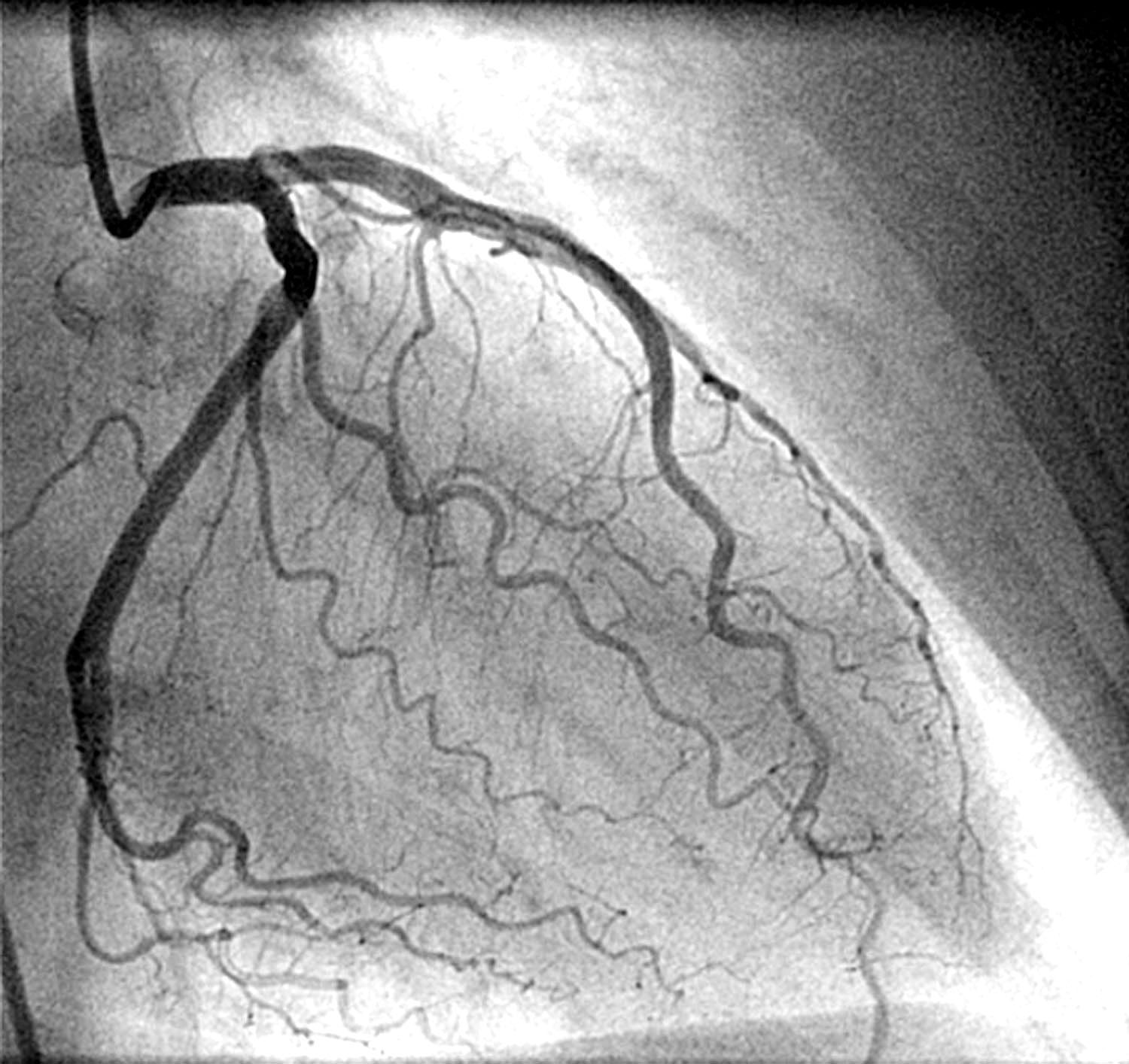
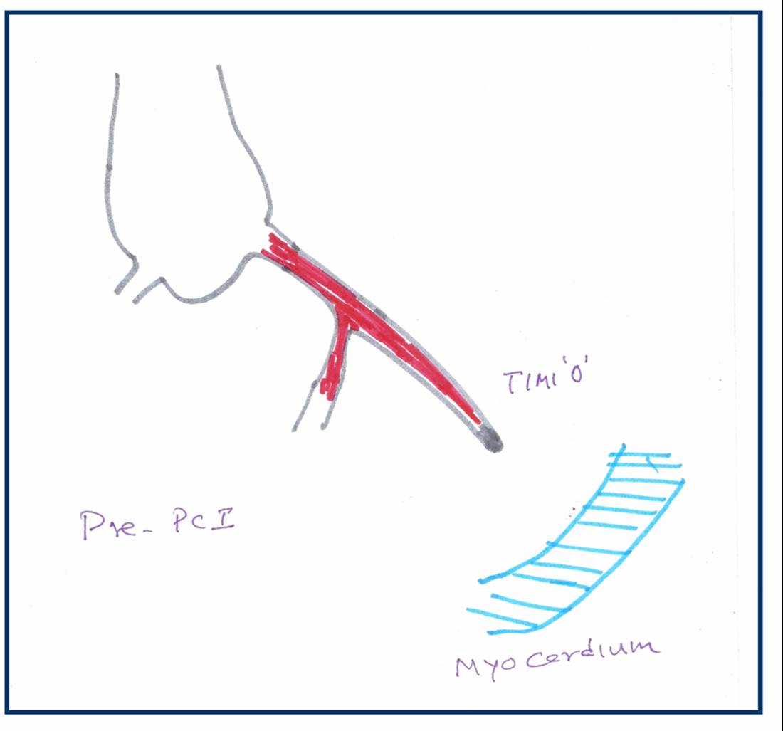
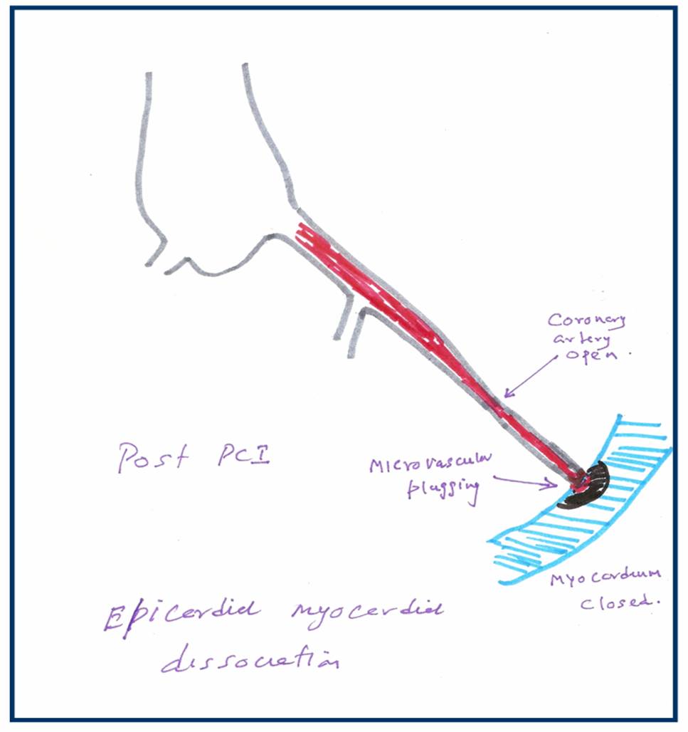
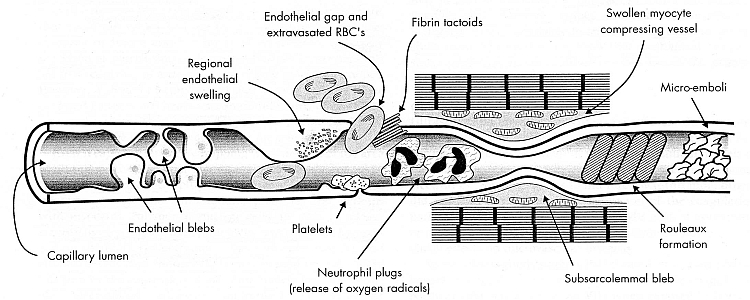
No reflow is the terminology used primarily in cath labs where, even  after a successful opening and stenting  of a coronary artery the coronary blood flow is not  restored to myocardium . The point to be emphazised here is blood do cross  successfully the site of  the obstruction but fails to enter the muscle segment  to which the coronary artery is supplying. So the paradoxical situation of artery  being open but the  myocardium is closed to receive  blood flow  happens . This is termed as no -reflow.  Actually it is a  misnomer , and  ideally it should be called “no flow” because  normal distal flow  does not  occur (After PCI)  in the first instance  to get interrupted  later on  and be labeled as  no re-flow.  .The only positive effect of PCI in these situation is blood flow would have improved by few centimeters ie till it reaches  but falls short of myocardium . In fact no reflow , can be termed as  glorified and concealed  terminology  for  PCI failure . It needs urgent action . No reflow is also called as myocardial epicardial dissociation.

Mechanism of no reflow.

Curious case of open coronary artery and closed myocardium !

Coronary  microvascular plugging  is mainly  due to thrombus and atheromatous debri , myocardial  edema , microvascular spasm may also contribute.

Where can it occur ?

  • First described in cath lab, especially following primary angioplasty.
  • It can very  well happen following thrombolysis in STEMI.
  • Can occur in venous grafts.

How do you recognise no reflow?

In cath lab it will be self evident from the check angiogram. Some times it is less obvious and may  require, myocardial  blush score, TIMI frame  count, contrast echocardiography, PET scan etc. In post MI a very simple method to recognise this entity could be the observation of persistent ST elevation in ECG .

Treatment.

Extremely difficult. Almost every coronary vasodilator has been tried.(Nitrates, nicorandil, calcium blockers, etc).Success is less than 30%.  High pressure flushing with saline inside the coronary artery is advocated by some.Others believe it’s dangerous to do it. So prevention is the key. Avoid doing PCI in complex, thrombotic lesions. Use thrombus suction device like export catheter(Medtronic). Distal protective devices are double edged devices , useful only in experienced hands.

Unanswered question

What is the size of the particle (thrombotic and atheromatous  debri)  the   coronary microcirculation safely handle and push it into the coronary venous circulation and the coronary sinus for disposal ?

If we can lyse the thrombus into micro particles by some mechanism and make it traverse the coronary circulation this complication of microvascular  plugging can be treated and prevented .

What is the final message ?

  • No reflow is relatively common condition during emergency PCI done for ACS patients
  • More common in complex thrombotic lesions.
  • Can also  occur in STEMI
  • Treatment is often vexing . In fact the treatment of this condition is so difficult , it can be termed  almost synonymously with “Failed PCI” if flow is not restored.
  • Successful treatment of no- reflow  means not momentry restoration of  myocardial flow  by mechanical and pharmacological modalities ,but to maintain sustained myocardial   perfusion. This we realise, as patients who have had a no reflow during  a PCI, do not perform as well in the follow up  .
  • So prevention is the key.

Read Full Post »

Differential response of thrombolysis between left and right coronary system

  • Thrombolysis is the specific treatment for acute myocardial infarction. ( Privileged few , get primary PCI))
  • Failed thrombolysis occurs in significant number of patients ( 30-40%).
  • Persistent ST elevation  120 minutes after thrombolysis is best indicator of failed thrombolysis.
  • It has been a consistent observation  failed  thromolysis  is more frequent in anterior   or LAD myocardial infarction.

In a simple study we have documented  patients  with inferior MI  rarely had persistent ST elevation and thrombolysis  was   successful in vast majority  of  patients  ( Except in few patients associated lateral MI)


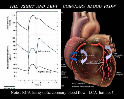
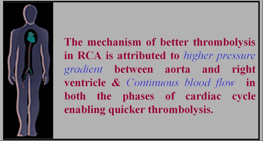
The mechanism of better thrombolysis in right coronary artery  is simple.The success of thrombolysis , apart from early time window , is directly correlated with pressure head  and the duration of contact between the thrombolytic agent and the thrombus. In right coronary circulation the  blood flow is continuous ,  occurs  both in systole and diastole that facilitates the maximum delivery of the thrombolytic agent . Further there is a favorable  pressure gradient  across RV myocardium  as the transmural occluding pressure across RV is considerably less then LV myocardium.

This paper was presented in the  “Annual cardiological society of India scientific sessions”

at Chennai, Tamil Nadu.India December 2000

Click to down load PPT full presentation

Read Full Post »

This is a 15-year-old post about LVH, written in 2008. Few of my colleagues, now agree with this, still hesitate to oblige in the open, suggesting it is too good to be true! Re-posting it for your own assessment. Surprised, why cardiology community didn’t consider this observation worthy to pursue.

Advantages of Left ventricular hypertrophy (LVH)

Left ventricular hypertrophy is one of the most common clinical cardiac entity.It is recognised either by ECG or echocardiography.LVH has a unique place in cardiology as it can imply a  grossly pathological state or  a marker of healthy heart as in physiological hypertrophy in athletes.

Logic would suggest, in this era of  stem cells and  nano medicine ,  every muscle fibre in ventricle is worth in gold !. So when the nature provides an  extra reserve of myocardium in the form of LVH one should welcome it , if otherwise not harmful.

Is LVH due to systemic hypertension benign ?

Not really, LVH has been shown to be an independent cardiac risk factor. (The famous Framingham study)Further LVH can result in diastolic dysfunction and the risk of cardiac failure increases.

But in spite of these observations, an  astute clinician with considerable experience will appreciate , patients with LVH fare better during an acute coronary syndrome !

This has been a consistent clinical observation . (Shall we call it as class C . ACC /AHA evidence ? )

Is LVH  an asset during ACS ?

  • A hypertrophied heart takes ischemic injury very easy , it doesn’t really hurt much . Another possibility is that in  LVH myocytes are relatively resistant to hypoxia .
  • Patients with LVH rarely show  significant wall motion defect following an STEMI.This is probably because the full thickness transmural necrosis is almost never possible even if extensive MI occurs.
  • This is also reflected in ECG  as these patients   rarely develop q waves in  following STEMI .
  • Persistent ST elevation and failed thrombolysis is very uncommon in pateints with LVH.
  • LVH provides  a relative immunity against development of cardiogenic shock . It requires 40% of LV mass destruction to produce cardiogenic shock.This can rarely happen in LVH. In a  long term analysis we have found none of the patient with LVH developed cardiogenic shock following STEMI.
  • LVH patients  are also protected against development of free wall rupture.

 Concluding message

                   “Lack of published evidence is the weakest evidence to dismiss a true myth”

LVH , either pathological or physiological, has a hitherto unreported beneficial effect.It acts as a myocardial reserve and helps limit the impact of STEMI.


Read Full Post »

              Intra coronary thrombosis is the sine qua non of acute coronary syndrome ( Both STEMI and NSTEMI.) But thrombolysis is the specific therapy in STEMI and is contraindicated in NSTEMI/UA.
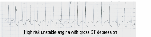
Why is this apparent paradox ? What is basic differnce between UA and AMI ?

In STEMI there is a sudden & total occlusion of a coronary artery usually by a thrombus with or without a plaque .The immediate aim is to open up the blood vessel . Every minute is important as myocardium undergoes  a continuous process ischemic necrosis. So thrombolysis (or more specifically fibrinolysis should be attempted immediately) .The other option is primary angioplasty,  which will not be discussed here.

The thrombus in STEMI  is RBC &  fibrin rich and often called a red clot. Number of fibrinolytic agents like streptokinase, Tissue palsminogen activator,(TPA) Reteplace, Tenekteplace etc have been tested and  form the cornerstone of STEMI management.The untoward effect of stroke  during  thrombolysis  is well recognised , but usully the risk benefit ratio favors thrombolyis in most situations except in very elderly and previous history of stroke or bleeding disorder.
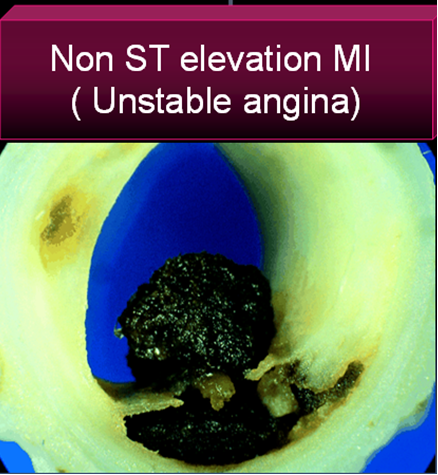
Unstable angina is a  close companion of STEMI . Many times it precedes STEMI often called preinfarction angina. During this phase blood flow in the coronary artery  becomes sluggish gradually,and patients develop  angina at rest .But unlike STEMI there is never a total occlusion and myocardium  is viable but ischemic,  and emergency salvaging of myocardium is not a therapeutic aim but prevention of MI becomes an aim. It is a paradox of sorts ,  even though thrombus is present in  UA ,  It has been learnt by experience thrombolytic agents are not useful in preventing an MI .


Why  thrombolysis is not useful in UA ?

1.In unstable angina  mechanical obstruction in the form of plaque fissure/rupture is more common than completely occluding thrombus. So lysis becomes less important.

2. Even if the thrombus is present , it is often intra plaque  or intra lesional and the  luminal  projection of thrombus is reduced  and hence thromolytic agents have limited area to act.

3.Further in UA/NSTEMI since it is a slow and gradual occlusion (Unlike sudden & total occlusion in STEMI) the platelets  get marginalised and trapped within the plaque .Hence in UA  thrombus is predominantly  white  . Often, a central platelet core  is  seen over which fibrin clot may also be  formed.

4.All available  thrombolytic agents act basically as a fibrinolytic agents,  and   so it finds   difficult to lyse the platelet rich clot.There is also a small risk of these agents lysing the fibrin cap and exposing underlying platelet  core and trigger a fresh thrombus.This has been documented in many trials( TIMI 3b to be specific) So if we thrombolyse in UA , there could be a risk of recurrent ACS episodes in the post thrombolytic phase.

5. UA is a semi emergency where  there is no race against time to salvage myocardium .Administering a  stroke prone thrombolytic agent tilts the risk benefit ratio against it.

6. Among UA, there is a significant group of secondary /perioperative UA   due to increased demand situations. Here there is absolutely no role for any thromolytic agents,  the  simple reason is , there is  no thrombus to get lysed. 

7.Many of the UA patient have multivessel CAD and might require surgical revascualarisation directly .


So fibrinolytic  agents are contraindicated in UA so what is the next step ?

The emergence of  intensive and aggressive platelet-lytic agents.

A combination of aspirin, clopidogrel, heparin, glycoprotien 2b 3a antagonist formed the major therapeutic protocol in these patients.Even though these are called antiplalet agents some of them  like 2b/3a antagonist eptifibatide, tirofiban, and many times even heparin has a potential to dissolve a thrombus. So technically one can call these agents  as thrombolytic agents.

What are the unresolved issues

                                       Even though clinical trials have convincingly shown thrombolytic agents  have no use in UA .There is a nagging belief  THAT  there could  be group of patients  with UA , still might benefit from thrombolysis as total occlusions have been documented  in some cases with UA.This is  especially true in peri-infarction unstable angina (Pre & post) as there is a fluctuation  between total and subtotal occlusions ) .But bed side recognition of this population is very difficult.

Many would consider this issue as redundant now,  since  most of  these patients  are taken up for emergency revascularisations

Read Full Post »
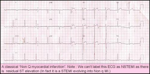
Recurrent myocardial infarction following an ACS is a fairly common clinical problem. Many times this is not recognised because it is difficult to establish the diagnosis.

The issues relevant here is

When does the first infarct (Index infact) process end ? and when the second infarct process start ?

Can the first infarct be a STEMI and the reinfarct be NSTEMI ? ( Dual acute coronary syndrome )

The only way to confirm a diagnosis of reinfarction is to document raising titres of cardiac enzymes and second peaking of CPK MB . New fresh ST elevation after a succesful thrombolysis is also a useful sign. But ST elevation in a q lead simply reflects a wall motion defect . So it requires enzymes to confirm it.

When there is tachycardia the ST segments tend to elevate following MI.

Other confounders are Infarct expansion and infarct extension .

These are macropathological entities almost impossible to dignose with surface ECG. What we diagnose as re-infarction could be an infact a infarct expansion.The modern terminology for infarct expansion is ventricle remodeling .The extreme remodeling results in ventricular aneurysm .Adverse acute ventricular remodeling can closely mimic a reinfarction .

What is clinical relevance of diagnosing reinfarction ?

Nothing great !

In modern day cardiology it is not a bother whether the infarct is expanding, extending or reinfarcting !All one has to do in a patient with chest pain ,showing a fresh ST elevation following STEMI is to take him/her to cath lab .

The only issue here one has to remember there are mechanical cause also for ST elevation following STEMI .

Dr.S.Venkatesan,Madras medical college, Chennai.

Read Full Post »

« Newer Posts - Older Posts »