Feeds:
Posts
Comments

Posts Tagged ‘nstemi’

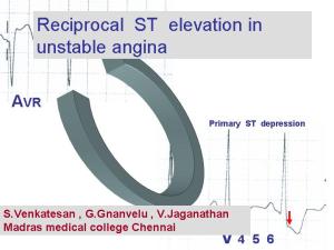
Is reciprocal ST  segment changes  occur  only in STEMI ? Can it occur in UA/NSTEMI ?   


                      Even   after   100 years of electro cardiology   the electrophysiological mechanism of ST elevation in STEMI  and ST depression in Unstable  angina is   still in the hypothetical stages. One popular theory   says that the   current of injury   as we see   as  ST  segment elevation  in surface ECG  is  actually   an illusion. It’s   apparently due to   constant negative current    pushing down the   rest of ECG segments. Ironically   the concept   of  reciprocal ST depression in patients who have  ST elevation is well debated  for over 3 decades and  is considered  a  settled issue. It   probably   represents , a  purely electrical phenomenon where the  tail end  of the  lead   picks up the opposite vector. Even   as   conflicts   continue to confront the basic electro physiological   concepts management    strategies   of   acute coronary syndromes is witnessing   great strides.
Aim
                                     We   hypothesized   if   ST depression occurs as response to ST elevation it’s logic to expect strong ST depressive forces should  possibly elevate The ST segments in the reciprocal leads .
In fact  we have seen this phenomenon in three distinct  clinical situations. 
1) ST   elevation   in posterior leads: Patients who present   with isolated   ST depression in V1,  V2 , V3  and  ST elevation in posterior chest leads V7, V8 .These patients were initially thought to have isolated posterior MI. But later the cardiac enzymes were found to be normal   indicating no myocardial necrosis   echo evaluation revealed wall motion defects in anterior segments rather than in posterior segments. CAG revealed critical   LAD disease .  This we believe a pure reciprocal ST elevation in the posterior leads to  a  ST depressive forces in anterior leads.
 2) Inferior  ST   elevation   with ST depression   in   V4- V6 : Few  patients who present with  infero    lateral STEMI   later  do not  evolve into  Q  MI but as a NSTEMI .The initial ST elevation  was found  be transient and  disappeared  much earlier,  while the  ST depression  lateral leads persisted.
 3) ST elevation in AVR   in high risk unstable angina :As   already reported in the literature,  we have seen  ST  elevation in AVR  in patients with  high risk unstable angina. This was   more often observed when there is > 3mm ST depression in V4-V6. The AVR  ST elevation  possibly   represents   the  reciprocal  vector.
    Conclusion
                                            ST elevation in certain   specific leads in   some of the patients with ACS,   could   be   a pure reciprocal   electrical phenomenon   to   dominant ST depressive forces in Opposite leads .  And hence   ST elevation in the surface ECG during early hours of ACS   should be interpreted more cautiously. The   sanctity   assocociated with ST segment   elevation   could  be  opened   for debate. 
 
                                     To down load full PPT  click on  the slide
//
Read Full Post »

                    Lateral myocardial infarction is not a common site when compared to anterior and inferior MI. But the lateral MI has some unique features, since it involves free wall of the ventricle.The laplace law mediated wall stress is more as the dyskinetic segments bulge with a long radius .Due to this,  lateral MI has a distinctly high risk for free wall rupture .Further pericardial rub is more common in thse patients.Ischemic mitral regurgitation and vulnerability to LVF is also more prevalent if the lateral wall is involved.  Generally lateral MI pateints have a turbulent and complicated course than a simple inferior or anteroseptal MI.

The angiographic correlation of  lateral MI is rarely reported in literature.

The following leisons are commonly observed.

1. Proximal  LAD with large D1  involvement

2. Isolated large D1/D2 disese

3.A left dominat LCX  with large OM1 disease

4.Large ramus disease

5.LAD total and RCA to LAD/D1 collateral pattern

Final message

If we encounter a lateral MI either alone or in combination with inferior/  posterior MI , it is better to manage these patients  aggresively with early triaging for CAG and revascularisation.

Read Full Post »

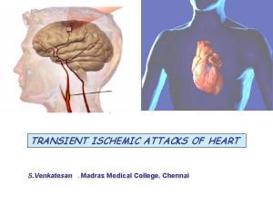
Transient ischemic attacks are not exclusive to cerebral circulation.

Many such episodes can occur in coronary circulation also .

TIA of heart PPT presentation

Click here to download  tia-of-heart

Read Full Post »

Thousands of dissections happen in cath labs  all over the world every day  very rarely it is painful . The answer is not clear. Both have rich vasa nervorum. Aortic dissection  involves media and smooth muscle . Coronary dissection may also be a  equally painful  , probably we are not recognising it ! or we attribute   all  chest pain in ACS  to ischemia .

Deep dissections into the smooth muscle should be painful.  Type c nerve fibers carry pain signals from heart

Answers welcome.

Read Full Post »

Plaque fissure ,rupture and subsequent thrombois is the hallmark of acute coronary syndrome . Are these events painful ? We always attribute any chest pain in an ACS patient to ischemia of myocardium.Is that always true? Coronary artery also has a rich vasa nervorum that could be activated by plaque disruption.

Why  we need an answer to this question ?

We are triaging patients for early invasive apporach based on chestpain .

Many patients may be subjected to revascularisation process for an non ischemic coronary pain !

Read Full Post »

How is LAD angina differnt from RCA angina ?

Can we localise the “Angina related artery ”  from the  the type of chest pain ?

Patients with stable  angia  many times have  multivessel CAD. There has been some correlation with radiation of anginal pain and the culprit artery.If the angina spreads to jaw or neck it is possibleit might indicate RCA(RIGHT coronary angina) but rarely it indicates LAD/LCX lesions. if the angina radiates to left shoulder it virtually ruels out a RCA disease

Source .Braunwald 1992 Edition

Dr.S.Venkatesan ., Madras medical college. Chennai.

Read Full Post »

Aspirin confusion spreads to clopidogrel !

It all started with 75 mg clopidogrel in CURE study  and others.

It went up to 150, 300, 600, and in some centres 900 mg.

No body knows how much clopidogrel optimally inhibits the platelet.

Aspirin had the same story three decades ago. It started from 40mg went up to 1200mg

and finally settled at 162mgs.

Why this confusion?

 It is because there is no simple platletlet function tests available in bedside.

and also the wide safety margin of this drug.At what level  clopidogrel  is unsafe

is also not clear !

Answers are expected soon .


Read Full Post »

                                   ACS   is the  most common cardiac emergency .  Management of STEMI is relatively straight forward.  The  only decision that to be taken is the  modality of reperfusion. (Primary PCI   or thrombolysis.) There is no need to risk stratify  STEMI on arrival. All STEMI patients are considered high risk on admission. Whereas  NSTEMI consists of  a heterogeneous  population. They need to be   triaged into low intermediate  or high risk categorizes on arrival.There is two management  approaches for unstable angina .All high risk UA should enter early invasive strategy . And low risk and intermediate risk group will get early conservative management. 

                                       The principle of management of  UA differ from STEMI in a fundamental way , as there is no issue of myocardial salvage in UA .The primary aim is to provide relief from pain and prevent an MI. So in the strict sense there is no time window in unstable angina /NSTEMI.


                                       But it is generally considered 48 hours is the time limit for an early invasive approach.If the patient has crossed this time there is apparently no great difference in outcome for conservative and invasive approach. 

Read Full Post »

2b -3a antagonists have revolutinised ACS management .

But the irony is Reo pro is approved for use only  inside cath lab or on the way to cath lab ! when PCI is done . 

If PCI with stenting is planned,  then subsequently cancelled due to  minimal coronary lesion or spontaneous reperfusion  what will be the effect of Abxicimab on outcome ?

Message 1

Abxicimab (Reopro,Faximab)

Useful only if PCI and stenting is done.

Dont use it for regular managment of UA/NSTEMI

Read Full Post »

« Newer Posts
