Feeds:
Posts
Comments

Archive for September, 2008

                                       Left atrium is the posterior most chamber of the heart.  It is almost a mid line structure.  The normal size of left atrium is about 4 / 4 cm. Normal left atrial volume is 46ml in men and 38 ml in women .(Atrial volume in a normal adult population by two-dimensional echocardiography Y Wang, Chest, Vol 86, 595-601.)  Left atrium  is not an easy chamber to identify in the  X ray chest as it does not form  the cardiac border.( Except a small circumference of left atrial appendage.(LAA)

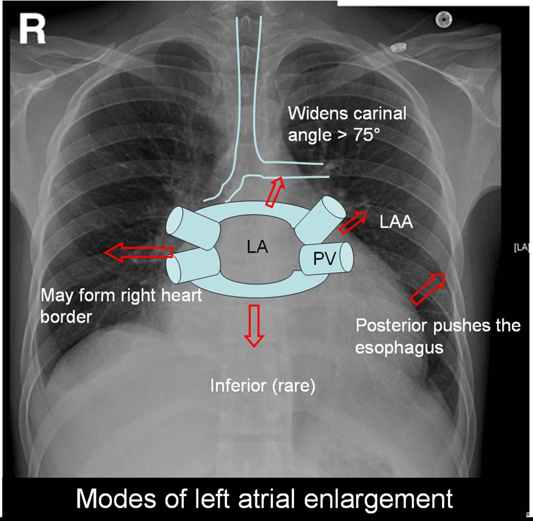
Left atrium can enlarge in multiple directions.Generally it dilates in the path of least resistance.


  • It is believed left atrial appendage  enlargement occur early .  LAA enlargemnet seen as a fullness beneath the pulmonary artery shadow. It may be the earliest finding of LAE in X ray. ( This may appear as straight left heart border , as in classical  mitral stenosis where MPA is also enlarged). The LAA enlargement is not necessarily in  in proportion  with LAE.
  • LA could  also enlarge posteriorly by pushing the esophagus towards the spine.This is visible only in barium swallow.
  • Then LA can enlarge either to left or right ( Usually towards right) and  reach the right heart border or over shoot it and form the right heart border by itself.This occurs very late in the course.
  • The other direction  LA goes on to enlarge is superiorly. When LA enlarges superiorly it hits on the left main  bronchus and lifts it.This is measured by the widened subcarinal angle which is normally less than 75 degrees.
  • LA can enlarge anteriorly  sometimes , but it is resisted by right ventricle but rarely right ventricle yields to the LA push and produce a left parasternal lift which could be mistaken  for RV enlargement.
  • Inferior enlargement can not happen in a significant way as it is limited by the AV groove and strong fibrous skeleton. 

With the advent of echocardiography X ray assessment of LA is redundant .(Academic value and in fellows training programs).The upper limit of normal LA size is around 4.5cm.

LA enlargement is commonly seen in

  • Rheumatic mitral stenosis, regurgitation. Gross enlargement up to 10 cms are common.
  • Hypertensive heart disese.
  • Cardiomyopathy, especially restrictive where both atria enlarge.

In all these conditions if  atrial fibrillation occurs  LA size increases further.

Read Full Post »

Chest pain is one  of the commonest presenting symptom  in any  hospital both as  an emergency  or non emergency. Reaching an accurate diagnosis is very important. The main  purpose of evaluation of chest pain is to recognise it as cardiac or non cardiac origin . Cardiac chest pain almost always means ischemic chest pain . That is called angina. (Of course there are few important causes for non ischemic cardiac chest pain which Will be discussed later).

Standard features of typical angina.

Chest pain which falls short of typical features are called atypical chest pain . Some recommend at least three typical features to label it as angina.
After the clinical examination patients  should be categorised in one of the following .

  • Typical angina
  • Atypical chest pain
  • Non cardiac chest pain** Non cardiac chest pain is not a diagnosis. Any physician (or a specialist)  should take some effort to localise it. (Muscle, nerve , pleura , anxiety  etc) . But  generally once these patients are ruled out of cardiac pain  they become less special and are simply referred back to their  family physician, only to return back  with  another cardiac  pseudo-emergency  in a different hospital .

    Why we are diagnosing atypical chest pain liberally ?

    Currently   more number of  patients as well as  the physicians  are   aware of the looming epidemic of CAD. The other major reason is the  lack of application of mind during  foirst clinical appraisal  and examination. Many of the patients with non cardiac chest pain  (Muscle, nerve , pleura )  are termed as atypical chest pain. Though some of the popular texts use atypical  chest pain  and non cardiac chest pain interchangeably , it is not  correct to do so. For example don’t ever label a  patient with chest pain with chest wall tenderness as atypical chest pain and order a cardiac work up .It  is a poor model to  emulate , that consumes time and resources!.Instead they should be diagnosed a confident non cardiac chest pain and dealt properly.

Once a patient is diagnosed  atypical chest pain what’s next ?

They should get a  complete physical examination,ECG, and  undergo exercise stress test.   In the  screening of CAD , angina can be termed a hard sign,  atypical chest pain is a soft sign,  resting ECG is surprisingly  a soft sign again (unless you record it during chest pain). Exercise stress testing is  the ideal  investigation in evaluation of  chestpain.( 70-80% accuracy). This can be improved upon by Thallium, SPECT, stress echo etc. As of now coronary angiogram is considered the ultimate gold standard (Not pure gold !) to rule out  CAD.

It is also worthwhile to remember non anginal  chest pain can also be an emergency and life threatening

  • Pulmonary embolism
  • Pneumothorax
  • Thoracic tumors
  • Aortic aneurysm (Dissection and non dissection)  The list is not  exclusive

Final message

What do we really mean by  atypical chest pain ?

In reality we don’t mean any thing !

When a  cardiac  physician is confused or rather , unable to  rule out angina , at the same time he is not confident of calling it as non cardiac chest pain,  he has the luxury of using this terminology . It is obvious  this terminology  should  minimally  be used.  Once diagnosed  these patients  can’t carry on with this tag  for long. They should be reinvestigated , (Right from history  and clinical ex) .They should either enter the cardiac work up  protocol  or  a non cardiac source for pain should be fixed  immediately.

Read Full Post »

                                                   Infective endocarditis is a serious clinical cardiac problem. The disease has evolved over many decades and now we are witnessing the  most virulent forms of the disease . Infection of heart , can occur in a native healthy valve, native diseased valve, or a prosthetic valve. Further, IE can occur either as  an acute (usually non diseased valve) , or sub acute form (usually in diseased valve).The changing microbial pattern has made this entity very complex. The vigorous   treatment protocols are available for IE. Still  the  prognosis and outcome with medical management is  dismal even in best centers.So the role of surgery in IE has increased over the years.We propose here,  a radically different approach to the problem.

 Traditionally there is a set of criteria for surgery in IE  :  These include

  •  Abscess formation
  •  Worsening valve lesion
  •  Refractory cardiac failure
  •  Persistent fever even after  2 weeks of  appropriate and adequate anti microbial therapy .
  •  Vegetation of more than 10mm size.
  •  Failed medical treatment

(The list is not exclusive)

In any large tertiary  hospital  series, if you  apply the above rule  more than 50 % of all patients with IE will be the candidates for  immediate surgery.

In the remaining 50% the mortality in medical management is very high. The reason being,  the  medical treatment is often prolonged over weeks. Many  of the complications occur  during the course of medical treatment.The common ones are abscess formation, embolic episodes, renal failure etc.Once a complication set in we call it as failed medical treatment and ask our surgical colleagues  to operate.By this time patient’s  general condition  deteriorates and either the surgeon refuses to take  up the case or  patient dies on the table.

So the key point  is , failure of medical treatment  is so common , it is simply not acceptable  to delay  the surgery in these patients as  majority of  them are  doomed to  fail  the trial of medical therapy.

What is the incidence of failed medical management, how to recognise it ? what is the impact of recognising it late ?

  • Failed medical therapy is around 60-70%  even in best centers.
  • Failed medical patients  constitute the greatest  surgical risk .
  • So it is proposed all IE patients should be triaged  early and the  dominant theme should be surgery (Commonly valve replacement, or valve repair)   .
  • If there is large vegetation surgery may be done for the sole purpose of physical removal of the vegetation*.

Final message

In Infective endocarditis experience has taught us, surgery  should be the default management protocol and medical therapy should be offered  to selected few who don’t require surgery.This is especially true in preexisting  rheumatic valve disease.

*The fundamental principle of management of infectious diseases, state that when there is a  resistant focus of infection .Always  remove the focus whenever possible.


Read Full Post »

This is a 15-year-old post about LVH, written in 2008. Few of my colleagues, now agree with this, still hesitate to oblige in the open, suggesting it is too good to be true! Re-posting it for your own assessment. Surprised, why cardiology community didn’t consider this observation worthy to pursue.

Advantages of Left ventricular hypertrophy (LVH)

Left ventricular hypertrophy is one of the most common clinical cardiac entity.It is recognised either by ECG or echocardiography.LVH has a unique place in cardiology as it can imply a  grossly pathological state or  a marker of healthy heart as in physiological hypertrophy in athletes.

Logic would suggest, in this era of  stem cells and  nano medicine ,  every muscle fibre in ventricle is worth in gold !. So when the nature provides an  extra reserve of myocardium in the form of LVH one should welcome it , if otherwise not harmful.

Is LVH due to systemic hypertension benign ?

Not really, LVH has been shown to be an independent cardiac risk factor. (The famous Framingham study)Further LVH can result in diastolic dysfunction and the risk of cardiac failure increases.

But in spite of these observations, an  astute clinician with considerable experience will appreciate , patients with LVH fare better during an acute coronary syndrome !

This has been a consistent clinical observation . (Shall we call it as class C . ACC /AHA evidence ? )

Is LVH  an asset during ACS ?

  • A hypertrophied heart takes ischemic injury very easy , it doesn’t really hurt much . Another possibility is that in  LVH myocytes are relatively resistant to hypoxia .
  • Patients with LVH rarely show  significant wall motion defect following an STEMI.This is probably because the full thickness transmural necrosis is almost never possible even if extensive MI occurs.
  • This is also reflected in ECG  as these patients   rarely develop q waves in  following STEMI .
  • Persistent ST elevation and failed thrombolysis is very uncommon in pateints with LVH.
  • LVH provides  a relative immunity against development of cardiogenic shock . It requires 40% of LV mass destruction to produce cardiogenic shock.This can rarely happen in LVH. In a  long term analysis we have found none of the patient with LVH developed cardiogenic shock following STEMI.
  • LVH patients  are also protected against development of free wall rupture.

 Concluding message

                   “Lack of published evidence is the weakest evidence to dismiss a true myth”

LVH , either pathological or physiological, has a hitherto unreported beneficial effect.It acts as a myocardial reserve and helps limit the impact of STEMI.


Read Full Post »

              Intra coronary thrombosis is the sine qua non of acute coronary syndrome ( Both STEMI and NSTEMI.) But thrombolysis is the specific therapy in STEMI and is contraindicated in NSTEMI/UA.

Why is this apparent paradox ? What is basic differnce between UA and AMI ?

In STEMI there is a sudden & total occlusion of a coronary artery usually by a thrombus with or without a plaque .The immediate aim is to open up the blood vessel . Every minute is important as myocardium undergoes  a continuous process ischemic necrosis. So thrombolysis (or more specifically fibrinolysis should be attempted immediately) .The other option is primary angioplasty,  which will not be discussed here.

The thrombus in STEMI  is RBC &  fibrin rich and often called a red clot. Number of fibrinolytic agents like streptokinase, Tissue palsminogen activator,(TPA) Reteplace, Tenekteplace etc have been tested and  form the cornerstone of STEMI management.The untoward effect of stroke  during  thrombolysis  is well recognised , but usully the risk benefit ratio favors thrombolyis in most situations except in very elderly and previous history of stroke or bleeding disorder.

Unstable angina is a  close companion of STEMI . Many times it precedes STEMI often called preinfarction angina. During this phase blood flow in the coronary artery  becomes sluggish gradually,and patients develop  angina at rest .But unlike STEMI there is never a total occlusion and myocardium  is viable but ischemic,  and emergency salvaging of myocardium is not a therapeutic aim but prevention of MI becomes an aim. It is a paradox of sorts ,  even though thrombus is present in  UA ,  It has been learnt by experience thrombolytic agents are not useful in preventing an MI .


Why  thrombolysis is not useful in UA ?

1.In unstable angina  mechanical obstruction in the form of plaque fissure/rupture is more common than completely occluding thrombus. So lysis becomes less important.

2. Even if the thrombus is present , it is often intra plaque  or intra lesional and the  luminal  projection of thrombus is reduced  and hence thromolytic agents have limited area to act.

3.Further in UA/NSTEMI since it is a slow and gradual occlusion (Unlike sudden & total occlusion in STEMI) the platelets  get marginalised and trapped within the plaque .Hence in UA  thrombus is predominantly  white  . Often, a central platelet core  is  seen over which fibrin clot may also be  formed.

4.All available  thrombolytic agents act basically as a fibrinolytic agents,  and   so it finds   difficult to lyse the platelet rich clot.There is also a small risk of these agents lysing the fibrin cap and exposing underlying platelet  core and trigger a fresh thrombus.This has been documented in many trials( TIMI 3b to be specific) So if we thrombolyse in UA , there could be a risk of recurrent ACS episodes in the post thrombolytic phase.

5. UA is a semi emergency where  there is no race against time to salvage myocardium .Administering a  stroke prone thrombolytic agent tilts the risk benefit ratio against it.

6. Among UA, there is a significant group of secondary /perioperative UA   due to increased demand situations. Here there is absolutely no role for any thromolytic agents,  the  simple reason is , there is  no thrombus to get lysed. 

7.Many of the UA patient have multivessel CAD and might require surgical revascualarisation directly .


So fibrinolytic  agents are contraindicated in UA so what is the next step ?

The emergence of  intensive and aggressive platelet-lytic agents.

A combination of aspirin, clopidogrel, heparin, glycoprotien 2b 3a antagonist formed the major therapeutic protocol in these patients.Even though these are called antiplalet agents some of them  like 2b/3a antagonist eptifibatide, tirofiban, and many times even heparin has a potential to dissolve a thrombus. So technically one can call these agents  as thrombolytic agents.

What are the unresolved issues

                                       Even though clinical trials have convincingly shown thrombolytic agents  have no use in UA .There is a nagging belief  THAT  there could  be group of patients  with UA , still might benefit from thrombolysis as total occlusions have been documented  in some cases with UA.This is  especially true in peri-infarction unstable angina (Pre & post) as there is a fluctuation  between total and subtotal occlusions ) .But bed side recognition of this population is very difficult.

Many would consider this issue as redundant now,  since  most of  these patients  are taken up for emergency revascularisations

Read Full Post »

Diuretics are the most commonly prescribed anti hypertensive agents.Thiazide diuretics which  was introduced many decades ago ,  lost popularity  in recent years ,  again got a second life after  the publication of ALLHAT trial recently. Now diuretics has become the  the drug of first choice in almost any hypertension unless any specific contraindication.

The most commonly used thaizide is hydrochlorthiazide ,and chlorthalidone.The blood pressure reducing effect is so consistent and smooth , all the currently popular molecules like ACE inhibitors and ARBs come with a combination with thiazide. While every one is clear diuretics are  effective anti hypertensive agent How it does is not clear.

How does a diuretic reduce blood pressure?

A. The exact mechanism is not clear. May not be uniformly effective in all patients  with HT.

B . Salt sensitive HT respond well to diuretics.

C. Volume correction /free water clearance might be a factor

D. Direct effect on vascular smoth muscle documented.The sodium transporter is blocked and  hence   calcium : sodium exchange is prevented .This depletes  intracellular calcium  in vascular smooth muscle cells .Less calcium for actin myosin interaction and hence vasodilatation

E. Thiazides combine well with all other antihypertensive drugs (ACEI, ARB, Beata blockers, calcium blockers)

F. Loop diuretics  like frusemide  can never be  a good antihypertensive agent.

Read Full Post »


Chronic renal failure and CAD are common companions.Severe CAD  in patients with renal failure  pose an imposing task on the treating physicians.CABG  and kidney transplantation  both are major interventions.When a patient  requires both the decision making becomes much more difficult.

The possible  choices are

A. Do CABG first follow it with renal transplant .

B. Do  renal transplant first follow it with CABG.

C. Do CABG first  and  defer transplant &  advice life long dilaysis

D.Do  renal transplant and offer medical management / PCI for CAD if feasible.

E.Simultaneous CABG & renal transplant is a remote possibility .

F.In terminally ill , combined cardiac and renal transplantation is the ultimate option. (Possible in very few centres in the world)   

G.In severe co-morbid condtions avoid both and support life. Success is not in completing   the procdeures but in providing useful life !

Among the options the most prefered worldwide is option no 1. This has a caveat. If angina is dominant  CABG should precede transplant. If cardiac failure is dominant the issue need further scrutiny.

Given a situation ( DCM & End stage renal disese) , your patient could  undergo only one procedure,  which will you prefer ?

              This again is highly emprical but logic could still be applied. Never do  CABG with a sole  aim of improving severe LV dysfunction in ischemic DCM .It happens only in  journal articles & major clincal trials!.Of course mitral valve correction and LV reduction surgery might help.But in a patient with  renal failure prolonging the CABG on table time , with add on surgery is highly risky. So it would be logical to think intensively  for  postponement of  the CABG in a patient with class 4 cardiac failure and renal failure. Do only the transplant .

 What is the impact of end stage renal failure  on LV dysfunction ?

 End stage renal failure has a great adverse impact on LV function. Many times it is reversible.We will never ever know, if you do a CABG first on them. So always think twice or even thrice before voting  on this vital issue . Correction of renal impairment can improve the cardiac status dramatically in some.

Read Full Post »

CURRENT   CARDIOLOGY  PRACTICE: EVIDENCE  OR  EXPERIENCE  BASED ?    AN  ANALYSIS OF  ACC/AHA  GUIDELINES.

S. Venkatesan,  Madras Medical College. Chennai


If  a  major therapeutic procedure is adviced based on simply by experience or expert opinion  how can we say evidence based medicine is practiced !


                                    Evidence based cardiology  is  the  buzz word  in global cardiovascular  health care  organizations. All diagnostic  and therapeutic  interventions are  undergoing  rigorous randomized  trials  for  proof of  efficacy  and  safety. ACC/AHA   have published  management guidelines and it  has been accepted  as de-facto standard of clinical cardiology practice world wide.  In these guidelines  class  1  indication  is defined as Conditions for which there is evidence for and/or general agreement that the procedure is useful and effective. These indications are supported by three levels of evidence.(A,B,C) .It has been observed,   many of the recommendations  in  class 1  were supported by only level  C  evidence. (Expert consensus or  agreement  ). We  analysed how much of todays guidelines is  agreement based  and  how much is evidence based. The  latest  practice  guidelines  of  ACC/AHA   for  Acute myocardial infarction , Unstable Angina and Non–ST-Segment Elevation Myocardial Infarction , chronic  stable angina  ,coronary angiography  were analysed. The  no  of  class 1  indications  were counted  in each set of guidelines  and  each  of the indication were  sub grouped with reference to the  levels of  evidence  to which it was supported. There  were a total  of 210  class 1  indications.


Class  1

Level A

Class   1

Level  B

Class  1

Level  C

P value

1A vs 1C

AMI(54)

7

25

22

<.0001

UA  (66)

11

26

29

<.0001

CSA(59)

8

29

22

<.0001

CAG(31)

3

12

16

<.0001

Total(210)

29(13.9%)

92(43.8%)

89(42.4%)

<.001

 13.9%   of class 1  indications were based on  level  A evidence.  42.4%  of class 1 indication were based  on Level C  ( agreement  of experts).Though evidence based cardiology   is   considered  to  define  the  standards in  Cardiology  practice  in reality  we lack evidence in most of the situations. 

                                       We  conclude  that  consensus or  agreement  based cardiology  practice is the dominant theme in current   ACC/AHA 

Read Full Post »

There are numerous complex grading for bifurcation lesions available.

The one proposed by Medina is simple and most useful.

In this grading three segments

  • Proximal main vessel
  • Distal main vessel
  • Branch vessel

Are given a code 0, and 1 if  lesion is present or absent .

This grading gives simple and fast method to label a bifurcatiuon lesion and to asssess the response to PCI. The only issue here is the individual  lesions are not graded , for example branch vessel ostium just involved about 20 % is not addressed . Further TIMI flow in these vessels may also be incorporated

How medina grading can be used to assess effectiveness of

angioplasty  ?

A patient with 1.1.1  after the treatment should revert back to 0.0.0.  if converted into 0.0.(.5) may indicate a residual side branch lesion  .5 shall indicate 50% residual lesion, .3 , 30% etc


What is the best management strategy for bifurcation lesions?

The topic has been discussed extensively for over a decade in various forums.

Though the lesions and intervention techniques  appear complex the basic concept is simple.

Following is the 8 point algorithm

1. Assess the bifurcation lesion accurately.

2. Apply the general rule and ask the first question whether PCI is neccessary at all ? if decided for PCI

3. Stent the main vessel.Protect the side branch.  

4. Dilate the side branch with a balloon.(KIss or through the struts) 

5. Very rarely,  if the side vessel is more significant and large  stent it and balloon the main vessel.

6. Use drug eluting stents with caution .

7. Resist the temptation of using two stents unless the situation demands and is absolutely required.

8. Never attempt to do bifurcation angioplasty during ACS as apart of primary angioplasty.( Unless you’re extremely competent, even then aim of primary PCI is to salvage myocarium quickly , not to provide TIMI 3 flow in non IRA vessel.)

Dr.S.Venkatesan.Madras medical college.Chennai.

Read Full Post »

Edema is considered the cardinal feature of cardiac failure for decades. It is one of the minor criteria of  the Framingham protocol  to diagnose cardiac failure.

The mechanism of edema in cardiac failure is multifactorial . The major mechanism attributed is raised venous pressure. This is transmitted backwards into the circulation and resultant increase in hydrostatic pressure in the venous end of capillaries results  in edema .

Even though this is considered as the  dominant mechanism for decades , it may not be true. Apparent reduction in cardiac output (Effective circulatory volume) and resultant reduction in renal blood flow triggers renal ischemic response and RASS activation (Rennin angiotensin aldosterone system). This is a powerful stimulus for sodium and water retention . This could be the major mechanism of cardiac edema. While there is clearcut explanation for the mechanism of edema in cardiac failure the explanation for lack of edema in many is not available in literature.

Does the activation of neuroendocrine happen in every one with cardiac failure ?

We think so ! but it may not be. Further,  activation of RASS trigger a counter hormone  response mediated by atrial natriuretic peptide  that promotes active water loss  from  the distal tublues which could prevent edema formation in many. So there is a distinct group of patients with cardiac failure who rarely develop edema .

In which type of cardiac failure edema is common and gross?

  • Predominatly right sided failure.
  • Corpulmonale
  • Tricuspid valve disese
  • Constrictive pericarditis

Read Full Post »

« Newer Posts